|
Neonatal Cholestasis
Neonatal cholestasis refers to elevated levels of conjugated bilirubin identified in newborn infants within the first few months of life. Conjugated hyperbilirubinemia is clinically defined as >20% of total serum bilirubin or conjugated bilirubin concentration greater than 1.0 mg/dL regardless of total serum bilirubin concentration. The differential diagnosis for neonatal cholestasis can vary extensively. However, the underlying disease pathology is caused by improper transport and/or defects in excretion of bile from hepatocytes leading to an accumulation of conjugated bilirubin in the body. Generally, symptoms associated with neonatal cholestasis can vary based on the underlying cause of the disease. However, most infants affected will present with jaundice, scleral icterus, failure to thrive, acholic or pale stools, and dark urine. Epidemiology Neonatal cholestasis can present in newborn infants within the first few months of life. The incidence of neonatal cholestasis i ... [...More Info...] [...Related Items...] OR: [Wikipedia] [Google] [Baidu] |
Bilirubin
Bilirubin (BR) (adopted from German, originally bili—bile—plus ruber—red—from Latin) is a red-orange compound that occurs in the normcomponent of the straw-yellow color in urine. Another breakdown product, stercobilin, causes the brown color of feces. Although bilirubin is usually found in animals rather than plants, at least one plant species, '' Strelitzia nicolai'', is known to contain the pigment. Structure Bilirubin consists of an open-chain tetrapyrrole. It is formed by oxidative cleavage of a porphyrin in heme, which affords biliverdin. Biliverdin is reduced to bilirubin. After conjugation with glucuronic acid, bilirubin is water-soluble and can be excreted. Bilirubin is structurally similar to the pigment phycobilin used by certain algae to capture light energy, and to the pigment phytochrome used by plants to sense light. All of these contain an open chain of four pyrrolic rings. Like these other pigments, some of the double-bonds in bilirubin isomer ... [...More Info...] [...Related Items...] OR: [Wikipedia] [Google] [Baidu] |
Peroxisomal Disorders
A peroxisome () is a membrane-bound organelle, a type of microbody, found in the cytoplasm of virtually all eukaryotic cells. Peroxisomes are oxidative organelles. Frequently, molecular oxygen serves as a co-substrate, from which hydrogen peroxide (H2O2) is then formed. Peroxisomes owe their name to hydrogen peroxide-generating and scavenging activities. They perform key roles in lipid metabolism and the reduction of reactive oxygen species. Peroxisomes are involved in the catabolism of very long chain fatty acids, branched chain fatty acids, bile acid intermediates (in the liver), D-amino acids, and polyamines. Peroxisomes also play a role in the biosynthesis of plasmalogens: ether phospholipids critical for the normal function of mammalian brains and lungs. Peroxisomes contain approximately 10% of the total activity of two enzymes (Glucose-6-phosphate dehydrogenase and 6-Phosphogluconate dehydrogenase) in the pentose phosphate pathway, which is important for energy meta ... [...More Info...] [...Related Items...] OR: [Wikipedia] [Google] [Baidu] |
Choledochal Cysts
Choledochal cysts (a.k.a. bile duct cyst) are congenital conditions involving cystic dilatation of bile ducts. They are uncommon in western countries but not as rare in East Asian nations like Japan and China. Signs and symptoms Most patients have symptoms in the first year of life. It is rare for symptoms to be undetected until adulthood, and usually adults have associated complications. The classic triad of intermittent abdominal pain, jaundice, and a right upper quadrant abdominal mass is found only in minority of patients. In infants, choledochal cysts usually lead to obstruction of the bile ducts and retention of bile. This leads to jaundice and an enlarged liver. If the obstruction is not relieved, permanent damage may occur to the liver - scarring and cirrhosis - with the signs of portal hypertension (obstruction to the flow of blood through the liver) and ascites (fluid accumulation in the abdomen). There is an increased risk of cancer in the wall of the cyst. In olde ... [...More Info...] [...Related Items...] OR: [Wikipedia] [Google] [Baidu] |
Common Bile Duct
The common bile duct (also bile duct) is a part of the biliary tract. It is formed by the union of the common hepatic duct and cystic duct. It ends by uniting with the pancreatic duct to form the ampulla of Vater (hepatopancreatic ampulla). Its sphincter the sphincter of Oddi, enables the regulation of bile flow. Anatomy The bile duct is some 6–8 cm long, and normally up to 8 mm in diameter. Its proximal supraduodenal part is situated within the free edge of the lesser omentum. Its middle retroduodenal part is oriented inferiorly and right-ward, and is situated posterior to the first part of the duodenum, and anterior to the inferior vena cava. Its distal paraduodenal part is oriented still more right-ward, is accommodated by a groove upon (sometimes a channel within) the posterior aspect of the head of the pancreas, and is situated anterior to the right renal vein. The bile duct terminates by uniting with the pancreatic duct (at an angle of about 60°) t ... [...More Info...] [...Related Items...] OR: [Wikipedia] [Google] [Baidu] |
Cystic Duct
The cystic duct is the duct that (typically) joins the gallbladder and the common hepatic duct; the union of the cystic duct and common hepatic duct forms the bile duct (formerly known as the common bile duct). Its length varies. Anatomy The cystic duct typically measures (sources differ) 2–4 cm/2–3 cm in length (though its length has been known to range from 0.5 cm to 9 cm), and 2–3 mm in diameter. It is often tortuous. It is the distal continuation of the neck of the gallbladder, from where it is directed inferoposteriorly and to the left/medially (this occurs in half of individuals). It typically terminates by uniting with the common hepatic duct to form the bile duct (usually anterior to the right hepatic artery). It usually joins the common bile duct from the right lateral side (forming an oblique angle between the two), and at such a distance that the bile duct is twice as long as the common hepatic duct. It often fuses with the common ... [...More Info...] [...Related Items...] OR: [Wikipedia] [Google] [Baidu] |
Common Hepatic Duct
The common hepatic duct is the first part of the biliary tract. It joins the cystic duct coming from the gallbladder to form the common bile duct. Structure The common hepatic duct is the first part of the biliary tract. It is formed by the union of the right hepatic duct (which drains bile from the right functional lobe of the liver) and the left hepatic duct (which drains bile from the left functional lobe of the liver). The duct is about 3 cm long. The common hepatic duct is about 6 mm in diameter in adults, with some variation.Gray's Anatomy, 39th ed, p. 1228 Termination The common hepatic duct typically unites with the cystic duct some 1–2 cm superior to the duodenum and anterior to the right hepatic artery, with the cystic duct approaching the common hepatic duct from the right. Relations The right branch of the hepatic artery proper usually passes posterior to the duct, but may rarely pass anterior to it instead. Histology The inner surface ... [...More Info...] [...Related Items...] OR: [Wikipedia] [Google] [Baidu] |
Panhypopituitarism
Hypopituitarism is the decreased (''hypo'') secretion of one or more of the eight hormones normally produced by the pituitary gland at the base of the brain. If there is decreased secretion of one specific pituitary hormone, the condition is known as selective hypopituitarism. If there is decreased secretion of most or all pituitary hormones, the term panhypopituitarism (''pan'' meaning "all") is used. The signs and symptoms of hypopituitarism vary, depending on which hormones are under-secreted and on the underlying cause of the abnormality. The diagnosis of hypopituitarism is made by blood tests, but often specific scans and other investigations are needed to find the underlying cause, such as tumors of the pituitary, and the ideal treatment. Most hormones controlled by the secretions of the pituitary can be replaced by tablets or injections. Hypopituitarism is a rare disease, but may be significantly under-diagnosed in people with previous traumatic brain injury. The first de ... [...More Info...] [...Related Items...] OR: [Wikipedia] [Google] [Baidu] |
Hypothyroidism
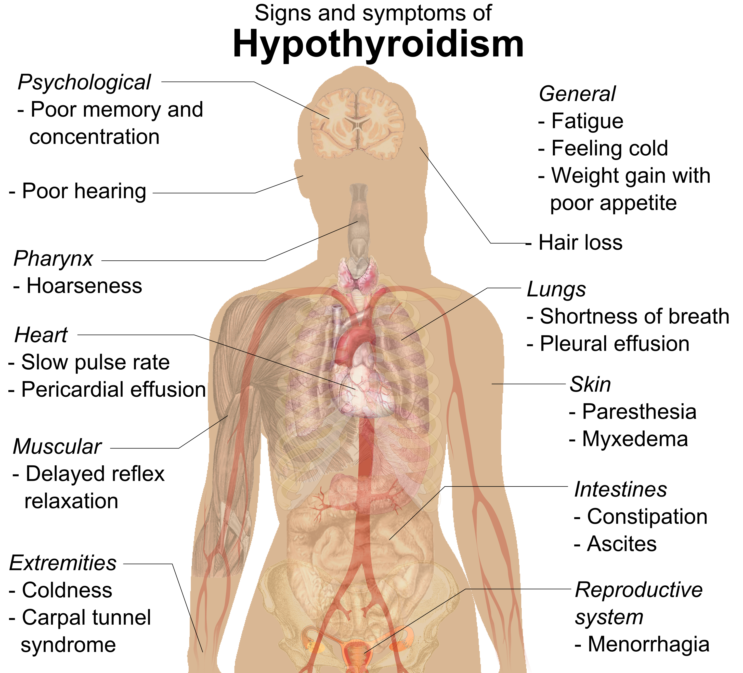
Hypothyroidism is an endocrine disease in which the thyroid gland does not produce enough thyroid hormones. It can cause a number of symptoms, such as cold intolerance, poor ability to tolerate cold, fatigue, extreme fatigue, muscle aches, constipation, slow heart rate, Depression (mood), depression, and weight gain. Occasionally there may be swelling of the front part of the neck due to goiter. Untreated cases of hypothyroidism during pregnancy can lead to delays in child development, growth and intellectual development in the baby or congenital iodine deficiency syndrome. Worldwide, iodine deficiency, too little iodine in the diet is the most common cause of hypothyroidism. Hashimoto's thyroiditis, an autoimmune disease where the body's immune system reacts to the thyroid gland, is the most common cause of hypothyroidism in countries with sufficient dietary iodine. Less common causes include previous treatment with iodine-131, radioactive iodine, injury to the hypothalamus ... [...More Info...] [...Related Items...] OR: [Wikipedia] [Google] [Baidu] |
Syphilis
Syphilis () is a sexually transmitted infection caused by the bacterium ''Treponema pallidum'' subspecies ''pallidum''. The signs and symptoms depend on the stage it presents: primary, secondary, latent syphilis, latent or tertiary. The primary stage classically presents with a single chancre (a firm, painless, non-itchy Ulcer_(dermatology), skin ulceration usually between 1 cm and 2 cm in diameter), though there may be multiple sores. In secondary syphilis, a diffuse rash occurs, which frequently involves the palms of the hands and soles of the feet. There may also be sores in the mouth or vagina. Latent syphilis has no symptoms and can last years. In tertiary syphilis, there are Gumma (pathology), gummas (soft, non-cancerous growths), neurological problems, or heart symptoms. Syphilis has been known as "The Great Imitator, the great imitator", because it may cause symptoms similar to many other diseases. Syphilis is most commonly spread through human sexual activi ... [...More Info...] [...Related Items...] OR: [Wikipedia] [Google] [Baidu] |
Tuberculosis
Tuberculosis (TB), also known colloquially as the "white death", or historically as consumption, is a contagious disease usually caused by ''Mycobacterium tuberculosis'' (MTB) bacteria. Tuberculosis generally affects the lungs, but it can also affect other parts of the body. Most infections show no symptoms, in which case it is known as inactive or latent tuberculosis. A small proportion of latent infections progress to active disease that, if left untreated, can be fatal. Typical symptoms of active TB are chronic cough with hemoptysis, blood-containing sputum, mucus, fever, night sweats, and weight loss. Infection of other organs can cause a wide range of symptoms. Tuberculosis is Human-to-human transmission, spread from one person to the next Airborne disease, through the air when people who have active TB in their lungs cough, spit, speak, or sneeze. People with latent TB do not spread the disease. A latent infection is more likely to become active in those with weakened I ... [...More Info...] [...Related Items...] OR: [Wikipedia] [Google] [Baidu] |
Echovirus
Echovirus is a polyphyletic group of viruses associated with enteric disease in humans. The name is derived from "enteric cytopathic human orphan virus". These viruses were originally not associated with disease, but many have since been identified as disease-causing agents. The term "echovirus" was used in the scientific names of numerous species, but all echoviruses are now recognized as strains of various species, most of which are in the family ''Picornaviridae''. List of echoviruses Thirty-four echoviruses are known: * Human echoviruses 1–7, 9, 11–21, 24–27, and 29–33 are strains of the species '' Enterovirus B'' of the genus ''Enterovirus''. * Human echovirus 8 was shown to be identical to Human echovirus 1 and was abolished as a species. * Human echovirus 10 was reclassified as a strain of the species ''Reovirus type 1'', currently named ''Mammalian orthoreovirus'' of the genus ''Orthoreovirus'', which belongs to the family ''Reoviridae''. As such, Human echovirus ... [...More Info...] [...Related Items...] OR: [Wikipedia] [Google] [Baidu] |
Adenovirus
Adenoviruses (members of the family ''Adenoviridae'') are medium-sized (90–100 nm), nonenveloped (without an outer lipid bilayer) viruses with an icosahedral nucleocapsid containing a double-stranded DNA genome. Their name derives from their initial isolation from human adenoids in 1953. They have a broad range of vertebrate hosts; in humans, more than 50 distinct adenoviral serotypes have been found to cause a wide range of illnesses, from mild respiratory infections in young children (the common cold) to life-threatening multi-organ disease in people with a weakened immune system. Virology Classification This family contains the following genera: * '' Aviadenovirus'' * '' Barthadenovirus'' * '' Ichtadenovirus'' * ''Mastadenovirus'' (including all human adenoviruses) * '' Siadenovirus'' * '' Testadenovirus'' Diversity In humans, currently there are 88 human adenoviruses (HAdVs) in seven species (Human adenovirus A to G): * A: 12, 18, 31 * B: 3, 7, 11, 14, ... [...More Info...] [...Related Items...] OR: [Wikipedia] [Google] [Baidu] |


