|
Nasal Concha
In anatomy, a nasal concha (; : conchae; ; Latin for 'shell'), also called a nasal turbinate or turbinal, is a long, narrow, curled shelf of bone tissue, bone that protrudes into the breathing passage of the nose in humans and various other animals. The conchae are shaped like an elongated seashell, which gave them their name (Latin ''concha'' from Greek ''κόγχη''). A concha is any of the scrolled spongy bones of the nasal cavity, nasal passages in vertebrates.''Anatomy of the Human Body'' Gray, Henry (1918) The Nasal Cavity. In humans, the conchae divide the nasal airway into four groove-like air passages, and are responsible for forcing inhaled air to flow in a steady, regular pattern around the largest possible surface area of nasal mucosa. As a cilium, ciliated mucous membrane with shallow blood supply, the nasal mucosa ... [...More Info...] [...Related Items...] OR: [Wikipedia] [Google] [Baidu] |
Ethmoid Bone
The ethmoid bone (; from ) is an unpaired bone in the skull that separates the nasal cavity from the brain. It is located at the roof of the nose, between the two orbits. The cubical (cube-shaped) bone is lightweight due to a spongy construction. The ethmoid bone is one of the bones that make up the orbit of the eye. Structure The ethmoid bone is an anterior cranial bone located between the eyes. It contributes to the medial wall of the orbit, the nasal cavity, and the nasal septum. The ethmoid has three parts: cribriform plate, ethmoidal labyrinth, and perpendicular plate. The cribriform plate forms the roof of the nasal cavity and also contributes to formation of the anterior cranial fossa, the ethmoidal labyrinth consists of a large mass on either side of the perpendicular plate, and the perpendicular plate forms the superior two-thirds of the nasal septum. Between the orbital plate and the nasal conchae are the ethmoidal sinuses or ethmoidal air cells, which are a var ... [...More Info...] [...Related Items...] OR: [Wikipedia] [Google] [Baidu] |
Pseudostratified Epithelium
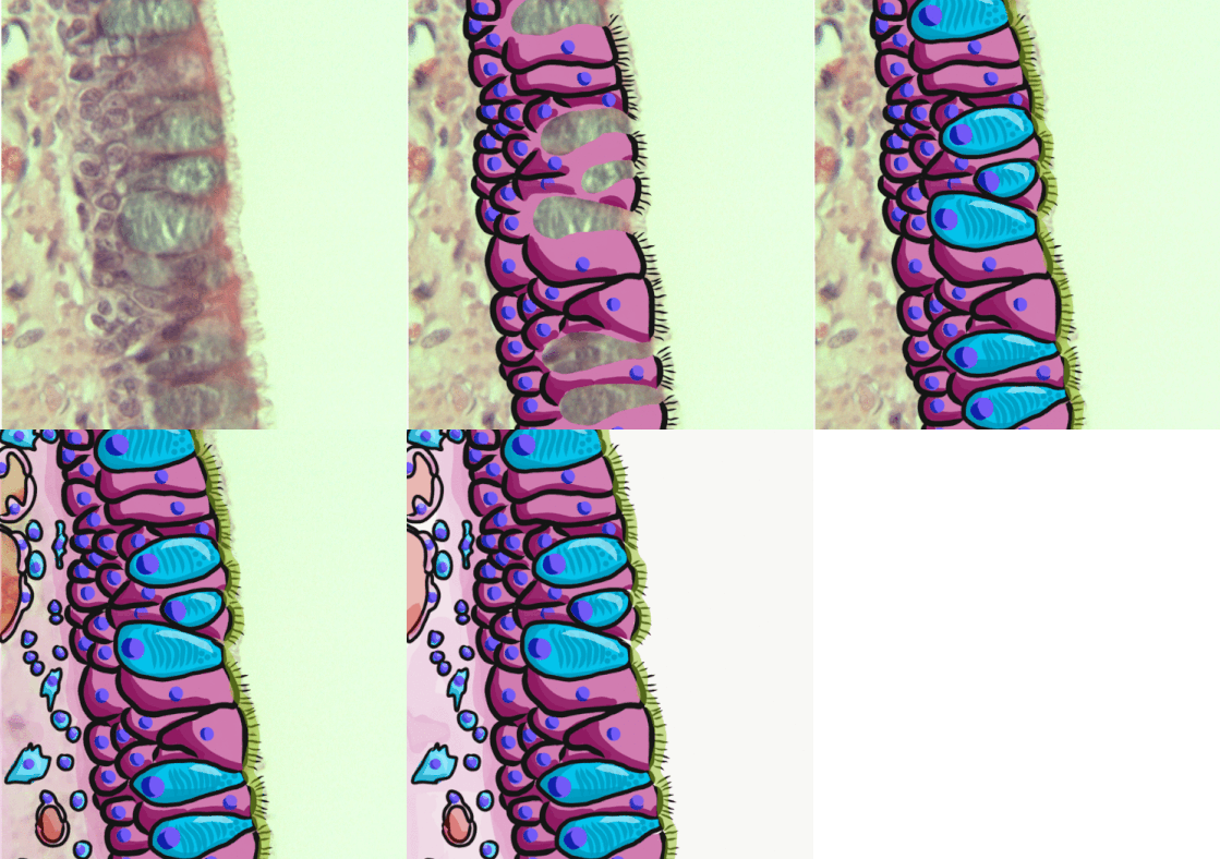
Pseudostratified columnar epithelium is a type of epithelium that, though comprising only a single layer of cells, has its cell nuclei positioned in a manner suggestive of stratified columnar epithelium. A stratified epithelium rarely occurs as squamous or cuboidal. The term ''pseudostratified'' is derived from the appearance of this epithelium in the section which conveys the erroneous (''pseudo'' means almost or approaching) impression that there is more than one layer of cells, when in fact this is a true simple epithelium since all the cells rest on the basement membrane. The nuclei of these cells, however, are disposed at different levels, thus creating the illusion of cellular stratification. All cells are not of equal size and not all cells extend to the luminal/apical surface; such cells are capable of cell division providing replacements for cells lost or damaged. Pseudostratified epithelia function in secretion or absorption. If a specimen looks stratified but has c ... [...More Info...] [...Related Items...] OR: [Wikipedia] [Google] [Baidu] |
Frontal Process Of Maxilla
The frontal process of the maxilla is a strong plate, which projects upward, medialward, and backward from the maxilla, forming part of the lateral boundary of the nose. Its ''lateral surface'' is smooth, continuous with the anterior surface of the body, and gives attachment to the quadratus labii superioris, the orbicularis oculi, and the medial palpebral ligament. Its ''medial surface'' forms part of the lateral wall of the nasal cavity; at its upper part is a rough, uneven area, which articulates with the ethmoid, closing in the anterior ethmoidal cells; below this is an oblique ridge, the ethmoidal crest, the posterior end of which articulates with the middle nasal concha, while the anterior part is termed the agger nasi; the crest forms the upper limit of the atrium of the middle meatus. The ''upper border'' articulates with the frontal bone and the ''anterior'' with the nasal; the ''posterior border'' is thick, and hollowed into a groove, which is continuous below wit ... [...More Info...] [...Related Items...] OR: [Wikipedia] [Google] [Baidu] |
Cribriform Plate
In mammalian anatomy, the cribriform plate (Latin for lit. '' sieve-shaped''), horizontal lamina or lamina cribrosa is part of the ethmoid bone. It is received into the ethmoidal notch of the frontal bone and roofs in the nasal cavities. It supports the olfactory bulb, and is perforated by olfactory foramina for the passage of the olfactory nerves to the roof of the nasal cavity to convey smell to the brain. The foramina at the medial part of the groove allow the passage of the nerves to the upper part of the nasal septum while the foramina at the lateral part transmit the nerves to the superior nasal concha. A fractured cribriform plate can result in olfactory dysfunction, septal hematoma, cerebrospinal fluid rhinorrhoea (CSF rhinorrhoea), and possibly infection which can lead to meningitis. CSF rhinorrhoea (clear fluid leaking from the nose) is very serious and considered a medical emergency. Aging can cause the openings in the cribriform plate to close, pinching olf ... [...More Info...] [...Related Items...] OR: [Wikipedia] [Google] [Baidu] |
Middle Nasal Concha
The medial surface of the labyrinth of ethmoid consists of a thin lamella, which descends from the under surface of the cribriform plate, and ends below in a free, convoluted margin, the middle nasal concha (middle nasal turbinate). It is rough, and marked above by numerous grooves, directed nearly vertically downward from the cribriform plate; they lodge branches of the olfactory nerves, which are distributed to the mucous membrane covering the superior nasal concha. The middle turbinates insert anteriorly into the frontal process of the maxilla and posteriorly into the perpendicular plate of the palatine bone. There are three mutually perpendicular segments of the middle turbinate: from proximal to distal, there is the horizontal segment ( axial plane), the basal lamella (coronal plane), and the vertical segment (sagittal plane). Additional images File:Illu nose nasal cavities.jpg, Nose and nasal cavities File:Gray152.png, Ethmoid bone from the right side. File:Gray19 ... [...More Info...] [...Related Items...] OR: [Wikipedia] [Google] [Baidu] |
Sphenoid Sinus
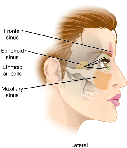
The sphenoid sinus is a paired paranasal sinus in the body of the sphenoid bone. It is one pair of the four paired paranasal sinuses.Illustrated Anatomy of the Head and Neck, Fehrenbach and Herring, Elsevier, 2012, page 64 The two sphenoid sinuses are separated from each other by a septum. Each sphenoid sinus communicates with the nasal cavity via the opening of sphenoidal sinus. The two sphenoid sinuses vary in size and shape, and are usually asymmetrical. Structure On average, a sphenoid sinus measures 2.2 cm vertical height, 2 cm in transverse breadth; and 2.2 cm antero-posterior depth. Each spehoid sinus is in the body of sphenoid bone, just under the sella turcica. The sphenoid sinuses are separated from each other medially by the septum of sphenoidal sinuses, which is usually asymmetrical. An opening of sphenoidal sinus forms a passage between each sphenoidal sinus and the nasal cavity. Posteriorly, an opening of sphenoidal sinus opens into the sphenoidal ... [...More Info...] [...Related Items...] OR: [Wikipedia] [Google] [Baidu] |
Olfactory Bulb
The olfactory bulb (Latin: ''bulbus olfactorius'') is a neural structure of the vertebrate forebrain involved in olfaction, the sense of smell. It sends olfactory information to be further processed in the amygdala, the orbitofrontal cortex (OFC) and the hippocampus where it plays a role in emotion, memory and learning. The bulb is divided into two distinct structures: the main olfactory bulb and the accessory olfactory bulb. The main olfactory bulb connects to the amygdala via the piriform cortex of the primary olfactory cortex and directly projects from the main olfactory bulb to specific amygdala areas. The accessory olfactory bulb resides on the dorsal-posterior region of the main olfactory bulb and forms a parallel pathway. Destruction of the olfactory bulb results in ipsilateral anosmia, while irritative lesions of the uncus can result in olfactory and gustatory hallucinations. Structure In most vertebrates, the olfactory bulb is the most rostral (forward) part ... [...More Info...] [...Related Items...] OR: [Wikipedia] [Google] [Baidu] |
Superior Nasal Concha
The superior nasal concha is a small, curved plate of bone representing a medial bony process of the labyrinth of the ethmoid bone. The superior nasal concha forms the roof of the superior nasal meatus. Anatomy Anatomical relations The superior nasal concha is situated posterosuperiorly to the middle nasal concha. It forms the superior boundary of the superior nasal meatus. Superior to the superior nasal concha is the sphenoethmoidal recess where the sphenoid sinus communicates with the nasal cavity; the sphenoethmoidal recess is interposed between the superior nasal concha, and (the anterior aspect of) the body of sphenoid bone. The sphenoid sinus ostium exists medial to the superior turbinate. See also * Nasal concha In anatomy, a nasal concha (; : conchae; ; Latin for 'shell'), also called a nasal turbinate or turbinal, is a long, narrow, curled shelf of bone tissue, bone that protrudes into the breathing passage of the nose in humans and various other anim ... Add ... [...More Info...] [...Related Items...] OR: [Wikipedia] [Google] [Baidu] |
Nasal Septum
The nasal septum () separates the left and right airways of the Human nose, nasal cavity, dividing the two nostrils. It is Depression (kinesiology), depressed by the depressor septi nasi muscle. Structure The fleshy external end of the nasal septum is called the Human nose#Cartilages, columella or columella nasi, and is made up of cartilage and soft tissue. The nasal septum contains bone and hyaline cartilage. It is normally about 2 mm thick. The nasal septum is composed of four structures: * Maxillary bone (the crest) * Perpendicular plate of ethmoid bone * Septal nasal cartilage (ie, quandrangular cartilage) * Vomer bone The lowest part of the septum is a narrow strip of bone that projects from the maxilla and the palatine bones, and is the length of the septum. This strip of bone is called the maxillary crest; it articulates in front with the septal nasal cartilage, and at the back with the vomer. The maxillary crest is described in the anatomy of the nasal septum as h ... [...More Info...] [...Related Items...] OR: [Wikipedia] [Google] [Baidu] |
Human Anatomical Terms
Anatomical terminology is a specialized system of terms used by anatomists, zoologists, and health professionals, such as doctors, surgeons, and pharmacists, to describe the structures and functions of the body. This terminology incorporates a range of unique terms, prefixes, and suffixes derived primarily from Ancient Greek and Latin. While these terms can be challenging for those unfamiliar with them, they provide a level of precision that reduces ambiguity and minimizes the risk of errors. Because anatomical terminology is not commonly used in everyday language, its meanings are less likely to evolve or be misinterpreted. For example, everyday language can lead to confusion in descriptions: the phrase "a scar above the wrist" could refer to a location several inches away from the hand, possibly on the forearm, or it could be at the base of the hand, either on the palm or dorsal (back) side. By using precise anatomical terms, such as "proximal," "distal," "palmar," or "do ... [...More Info...] [...Related Items...] OR: [Wikipedia] [Google] [Baidu] |
Glandular
A gland is a cell or an organ in an animal's body that produces and secretes different substances that the organism needs, either into the bloodstream or into a body cavity or outer surface. A gland may also function to remove unwanted substances such as urine from the body. There are two types of gland, each with a different method of secretion. Endocrine glands are ductless and secrete their products, hormones, directly into interstitial spaces to be taken up into the bloodstream. Exocrine glands secrete their products through a duct into a body cavity or outer surface. Glands are mostly composed of epithelial tissue, and typically have a supporting framework of connective tissue, and a capsule. Structure Development Every gland is formed by an ingrowth from an epithelial surface. This ingrowth may in the beginning possess a tubular structure, but in other instances glands may start as a solid column of cells which subsequently becomes tubulated. As growth proceeds, the ... [...More Info...] [...Related Items...] OR: [Wikipedia] [Google] [Baidu] |
Blood Vessel
Blood vessels are the tubular structures of a circulatory system that transport blood throughout many Animal, animals’ bodies. Blood vessels transport blood cells, nutrients, and oxygen to most of the Tissue (biology), tissues of a Body (biology), body. They also take waste and carbon dioxide away from the tissues. Some tissues such as cartilage, epithelium, and the lens (anatomy), lens and cornea of the eye are not supplied with blood vessels and are termed ''avascular''. There are five types of blood vessels: the arteries, which carry the blood away from the heart; the arterioles; the capillaries, where the exchange of water and chemicals between the blood and the tissues occurs; the venules; and the veins, which carry blood from the capillaries back towards the heart. The word ''vascular'', is derived from the Latin ''vas'', meaning ''vessel'', and is mostly used in relation to blood vessels. Etymology * artery – late Middle English; from Latin ''arteria'', from Gree ... [...More Info...] [...Related Items...] OR: [Wikipedia] [Google] [Baidu] |


