|
Naegleria Fowleri
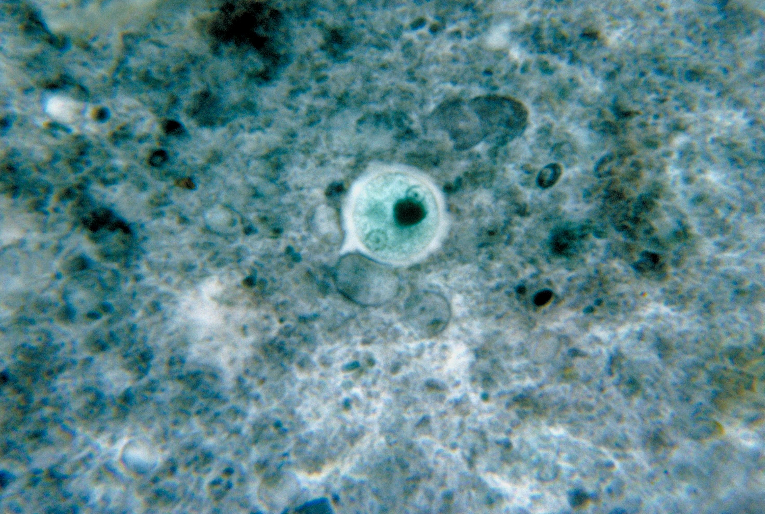
''Naegleria fowleri'', also known as the brain-eating amoeba, is a species of the genus ''Naegleria''. It belongs to the phylum Percolozoa and is classified as an amoeboflagellate Excavata, excavate, an organism capable of behaving as both an amoeba and a flagellate. This free-living microorganism primarily feeds on bacteria, but can become pathogenic in humans, causing an extremely rare, sudden, severe, and almost always fatal brain infection known as naegleriasis or primary amoebic meningoencephalitis (PAM). It is typically found in warm freshwater bodies such as lakes, rivers, hot springs, warm water discharge from industrial or power plants, geothermal well water, and poorly maintained or minimally Water chlorination, chlorinated swimming pools with residual chlorine levels under 0.5 g/m3, water heaters, soil, and pipes connected to tap water. It can exist in either an amoeboid or temporary flagellate stage. Etymology The organism was named after Malcolm Fowler, an ... [...More Info...] [...Related Items...] OR: [Wikipedia] [Google] [Baidu] |
Naegleria
''Naegleria'' is a genus consisting of 47 described species of protozoa often found in warm aquatic environments as well as soil habitats worldwide. It has three life cycle forms: the amoeboid stage, the cyst stage, and the flagellated stage, and has been routinely studied for its ease in change from amoeboid to flagellated stages. The ''Naegleria'' genera became famous when ''Naegleria fowleri'', the causative agent of the usually fatal human and animal disease primary amoebic meningoencephalitis (PAM), was discovered in 1965. Most species in the genus, however, are incapable of causing disease. Etymology The genus ''Naegleria'' is named after the German protozoologist, Kurt Nägler. History In 1899, Franz Schardinger discovered an amoeba that had the ability to transform into a flagellated stage. He named the organism ''Amoeba gruberi'', which was later changed to the genus ''Naegleria'' in 1912 by Alexeieff. Before 1970, the genus was generally used as a model organism ... [...More Info...] [...Related Items...] OR: [Wikipedia] [Google] [Baidu] |
Olfactory Receptor Neuron
An olfactory receptor neuron (ORN), also called an olfactory sensory neuron (OSN), is a sensory neuron within the olfactory system. Structure Humans have between 10 and 20 million olfactory receptor neurons (ORNs). In vertebrates, ORNs are Bipolar neuron, bipolar neurons with dendrites facing the external surface of the cribriform plate with axons that pass through the cribriform foramina with terminal end at olfactory bulbs. The ORNs are located in the olfactory epithelium in the nasal cavity. The cell bodies of the ORNs are distributed among the wikt:stratified, stratified layers of the olfactory epithelium. Many tiny hair-like non-motile cilia protrude from the olfactory receptor cell's dendrites. The dendrites extend to the olfactory epithelial surface and each ends in a dendritic knob from which around 20 to 35 cilia protrude. The cilia have a length of up to 100 micrometres and with the cilia from other dendrites form a meshwork in the olfactory mucus. The surface of ... [...More Info...] [...Related Items...] OR: [Wikipedia] [Google] [Baidu] |
Axons
An axon (from Greek ἄξων ''áxōn'', axis) or nerve fiber (or nerve fibre: see spelling differences) is a long, slender projection of a nerve cell, or neuron, in vertebrates, that typically conducts electrical impulses known as action potentials away from the nerve cell body. The function of the axon is to transmit information to different neurons, muscles, and glands. In certain sensory neurons ( pseudounipolar neurons), such as those for touch and warmth, the axons are called afferent nerve fibers and the electrical impulse travels along these from the periphery to the cell body and from the cell body to the spinal cord along another branch of the same axon. Axon dysfunction can be the cause of many inherited and acquired neurological disorders that affect both the peripheral and central neurons. Nerve fibers are classed into three types group A nerve fibers, group B nerve fibers, and group C nerve fibers. Groups A and B are myelinated, and group C are unmyelina ... [...More Info...] [...Related Items...] OR: [Wikipedia] [Google] [Baidu] |
Olfactory Epithelium
The olfactory epithelium is a specialized epithelium, epithelial tissue inside the nasal cavity that is involved in olfaction, smell. In humans, it measures and lies on the roof of the nasal cavity about above and behind the nostrils. The olfactory epithelium is the part of the olfactory system directly responsible for detecting odors. Structure Olfactory epithelium consists of four distinct cell types: * Olfactory receptor neuron, Olfactory sensory neurons * Supporting cells * Basal cells * Brush cells Olfactory sensory neurons The olfactory receptor neurons are sensory neurons of the olfactory epithelium. They are bipolar neurons and their apical poles express odorant receptors on cilium, non-motile cilia at the ends of the dendritic knob, which extend out into the airspace to interact with odorants. Odorant receptors bind odorants in the airspace, which are made soluble by the serous secretions from olfactory glands located in the lamina propria of the mucosa.Ross, MH, '' ... [...More Info...] [...Related Items...] OR: [Wikipedia] [Google] [Baidu] |
Replicate (biology)
In the biological sciences, replicates are an experimental units that are treated identically. Replicates are an essential component of experimental design because they provide an estimate of between sample error. Without replicates, scientists are unable to assess whether observed treatment effects are due to the experimental manipulation or due to random error. There are also analytical replicates which is when an exact copy of a sample is analyzed, such as a cell, organism or molecule, using exactly the same procedure. This is done in order to check for analytical error. In the absence of this type of error replicates should yield the same result. However, analytical replicates are not independent and cannot be used in tests of the hypothesis because they are still the same sample. See also * Self-replication * Fold change Fold change is a measure describing how much a quantity changes between an original and a subsequent measurement. It is defined as the ratio between the ... [...More Info...] [...Related Items...] OR: [Wikipedia] [Google] [Baidu] |
Ostiole
An ''ostiole'' is a small hole or opening through which algae or fungi release their mature spores. The word is a diminutive of wikt:ostium, "ostium", "opening". The term is also used in higher plants, for example to denote the opening of the involuted syconium (fig inflorescence) through which fig wasps enter to Pollination, pollinate and breed. The species pharamacosycea have an arrangement interlocking pattern but there is an exception because of insipdia because it is partly cover the ostiole. On the adaxial side of the bracts is made out of cubic cells, that has a staining reactions and contain phenolic compounds. Sometimes a stomatal aperture is called an "ostiole"."Synergistic Pectin Degradation and Guard Cell Pressurization Underlie Stomatal Pore Formation", See also *Ostium (other) References Castro-Cárdenas, N., Vázquez-Santana, S., Teixeira, S. P., & Ibarra-Manríquez, G. (2023). Correction to: The roles of the ostiole in the fig-fig wasp mutu ... [...More Info...] [...Related Items...] OR: [Wikipedia] [Google] [Baidu] |
Cell Nucleus
The cell nucleus (; : nuclei) is a membrane-bound organelle found in eukaryote, eukaryotic cell (biology), cells. Eukaryotic cells usually have a single nucleus, but a few cell types, such as mammalian red blood cells, have #Anucleated_cells, no nuclei, and a few others including osteoclasts have Multinucleate, many. The main structures making up the nucleus are the nuclear envelope, a double membrane that encloses the entire organelle and isolates its contents from the cellular cytoplasm; and the nuclear matrix, a network within the nucleus that adds mechanical support. The cell nucleus contains nearly all of the cell's genome. Nuclear DNA is often organized into multiple chromosomes – long strands of DNA dotted with various proteins, such as histones, that protect and organize the DNA. The genes within these chromosomes are Nuclear organization, structured in such a way to promote cell function. The nucleus maintains the integrity of genes and controls the activities of the ... [...More Info...] [...Related Items...] OR: [Wikipedia] [Google] [Baidu] |
Microbial Cyst
A microbial cyst is a resting or dormant stage of a microorganism, that can be thought of as a state of suspended animation in which the metabolic processes of the cell are slowed and the cell ceases all activities like feeding and locomotion. Many groups of single-celled, microscopic organisms, or microbes, possess the ability to enter this dormant state. Encystment, the process of cyst formation, can function as a method for dispersal and as a way for an organism to survive in unfavorable environmental conditions. These two functions can be combined when a microbe needs to be able to survive harsh conditions between habitable environments (such as between hosts) in order to disperse. Cysts can also be sites for nuclear reorganization and cell division, and in parasitic species they are often the infectious stage between hosts. When the encysted microbe reaches an environment favorable to its growth and survival, the cyst wall breaks down by a process known as excystation. E ... [...More Info...] [...Related Items...] OR: [Wikipedia] [Google] [Baidu] |
Cerebrospinal Fluid
Cerebrospinal fluid (CSF) is a clear, colorless Extracellular fluid#Transcellular fluid, transcellular body fluid found within the meninges, meningeal tissue that surrounds the vertebrate brain and spinal cord, and in the ventricular system, ventricles of the brain. CSF is mostly produced by specialized Ependyma, ependymal cells in the choroid plexuses of the ventricles of the brain, and absorbed in the arachnoid granulations. It is also produced by ependymal cells in the lining of the ventricles. In humans, there is about 125 mL of CSF at any one time, and about 500 mL is generated every day. CSF acts as a shock absorber, cushion or buffer, providing basic mechanical and immune system, immunological protection to the brain inside the Human skull, skull. CSF also serves a vital function in the cerebral autoregulation of cerebral blood flow. CSF occupies the subarachnoid space (between the arachnoid mater and the pia mater) and the ventricular system around and inside t ... [...More Info...] [...Related Items...] OR: [Wikipedia] [Google] [Baidu] |
Biflagellate
A flagellate is a cell or organism with one or more whip-like appendages called flagella. The word ''flagellate'' also describes a particular construction (or level of organization) characteristic of many prokaryotes and eukaryotes and their means of motion. The term presently does not imply any specific relationship or classification of the organisms that possess flagella. However, several derivations of the term "flagellate" (such as "dinoflagellate" and "choanoflagellate") are more formally characterized. Form and behavior Flagella in eukaryotes are supported by microtubules in a characteristic arrangement, with nine fused pairs surrounding two central singlets. These arise from a basal body. In some flagellates, flagella direct food into a cytostome or mouth, where food is ingested. Flagella role in classifying eukaryotes. Among protoctists and microscopic animals, a flagellate is an organism with one or more flagella. Some cells in other animals may be flagellate, f ... [...More Info...] [...Related Items...] OR: [Wikipedia] [Google] [Baidu] |
Trophozoite
A trophozoite (G. ''trope'', nourishment + ''zoon'', animal) is the activated, feeding stage in the life cycle of certain protozoa such as malaria-causing ''Plasmodium falciparum'' and those of the ''Giardia'' group. The complementary form of the trophozoite state is the thick-walled microbial cyst, cyst form. They are often different from the cyst stage, which is a protective, dormant form of the protozoa. Trophozoites are often found in the host's body fluids and tissues and in many cases, they are the form of the protozoan that causes disease in the host. In the protozoan, ''Entamoeba histolytica'' it invades the intestinal mucosa of its host, causing dysentery, which aid in the trophozoites traveling to the liver and leading to the production of hepatic abscesses. Life cycle stages ''Plasmodium falciparium'' The causative organism of malaria is a protozoan, ''Plasmodium falciparium'', that is carried by the female Anopheles mosquito, ''Anopheles'' mosquito. Malaria is reco ... [...More Info...] [...Related Items...] OR: [Wikipedia] [Google] [Baidu] |


