|
Myocyte
A muscle cell, also known as a myocyte, is a mature contractile Cell (biology), cell in the muscle of an animal. In humans and other vertebrates there are three types: skeletal muscle, skeletal, smooth muscle, smooth, and Cardiac muscle, cardiac (cardiomyocytes). A skeletal muscle cell is long and threadlike with multinucleated, many nuclei and is called a ''muscle fiber''. Muscle cells develop from embryonic precursor cells called myoblasts. Skeletal muscle cells form by cell fusion, fusion of myoblasts to produce multinucleated cells (syncytium, syncytia) in a process known as myogenesis. Skeletal muscle cells and cardiac muscle cells both contain myofibrils and sarcomeres and form a striated muscle tissue. Cardiac muscle cells form the cardiac muscle in the walls of the heart chambers, and have a single central Cell nucleus, nucleus. Cardiac muscle cells are joined to neighboring cells by intercalated discs, and when joined in a visible unit they are described as a ''cardiac m ... [...More Info...] [...Related Items...] OR: [Wikipedia] [Google] [Baidu] |
Cardiac Muscle
Cardiac muscle (also called heart muscle or myocardium) is one of three types of vertebrate muscle tissues, the others being skeletal muscle and smooth muscle. It is an involuntary, striated muscle that constitutes the main tissue of the wall of the heart. The cardiac muscle (myocardium) forms a thick middle layer between the outer layer of the heart wall (the pericardium) and the inner layer (the endocardium), with blood supplied via the coronary circulation. It is composed of individual cardiac muscle cells joined by intercalated discs, and encased by collagen fibers and other substances that form the extracellular matrix. Cardiac muscle contracts in a similar manner to skeletal muscle, although with some important differences. Electrical stimulation in the form of a cardiac action potential triggers the release of calcium from the cell's internal calcium store, the sarcoplasmic reticulum. The rise in calcium causes the cell's myofilaments to slide past each other i ... [...More Info...] [...Related Items...] OR: [Wikipedia] [Google] [Baidu] |
Skeletal Muscle
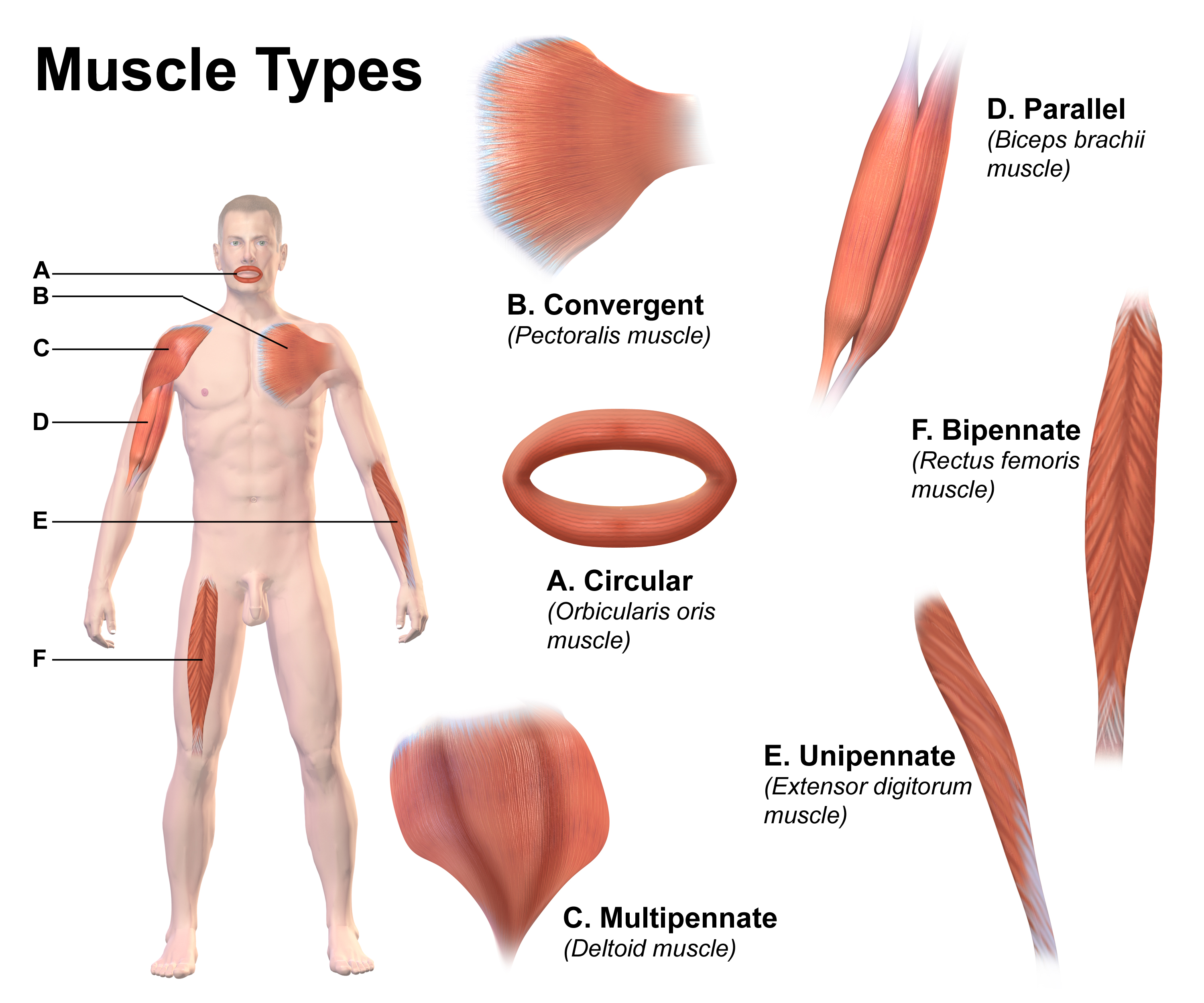
Skeletal muscle (commonly referred to as muscle) is one of the three types of vertebrate muscle tissue, the others being cardiac muscle and smooth muscle. They are part of the somatic nervous system, voluntary muscular system and typically are attached by tendons to bones of a skeleton. The skeletal muscle cells are much longer than in the other types of muscle tissue, and are also known as ''muscle fibers''. The tissue of a skeletal muscle is striated muscle tissue, striated – having a striped appearance due to the arrangement of the sarcomeres. A skeletal muscle contains multiple muscle fascicle, fascicles – bundles of muscle fibers. Each individual fiber and each muscle is surrounded by a type of connective tissue layer of fascia. Muscle fibers are formed from the cell fusion, fusion of developmental myoblasts in a process known as myogenesis resulting in long multinucleated cells. In these cells, the cell nucleus, nuclei, termed ''myonuclei'', are located along the inside ... [...More Info...] [...Related Items...] OR: [Wikipedia] [Google] [Baidu] |
Syncytium
A syncytium (; : syncytia; from Greek: σύν ''syn'' "together" and κύτος ''kytos'' "box, i.e. cell") or symplasm is a multinucleate cell that can result from multiple cell fusions of uninuclear cells (i.e., cells with a single nucleus), in contrast to a coenocyte, which can result from multiple nuclear divisions without accompanying cytokinesis. The muscle cell that makes up animal skeletal muscle is a classic example of a syncytium cell. The term may also refer to cells interconnected by specialized membranes with gap junctions, as seen in the heart muscle cells and certain smooth muscle cells, which are synchronized electrically in an action potential. The field of embryogenesis uses the word ''syncytium'' to refer to the coenocytic blastoderm embryos of invertebrates, such as ''Drosophila melanogaster''. Physiological examples Protists In protists, syncytia can be found in some rhizarians (e.g., chlorarachniophytes, plasmodiophorids, haplosporidians) and acellul ... [...More Info...] [...Related Items...] OR: [Wikipedia] [Google] [Baidu] |
Sarcoplasm
Sarcoplasm is the cytoplasm of a muscle cell. It is comparable to the cytoplasm of other cells, but it contains unusually large amounts of glycogen (a polymer of glucose), myoglobin, a red-colored protein necessary for binding oxygen molecules that diffuse into muscle fibers, and mitochondria.Roberts, Michael D.; Haun, Cody T.; Vann, Christopher G.; Osburn, Shelby C.; Young, Kaelin C. (2020). "Sarcoplasmic Hypertrophy in Skeletal Muscle: A Scientific "Unicorn" or Resistance Training Adaptation?". Frontiers in Physiology. 11. . . . . The calcium ion concentration in sarcoplasm is also a special element of the muscle fiber; it is the means by which muscle contractions take place and are regulated. The sarcoplasm plays a critical role in muscle contraction as an increase in Ca2+ concentration in the sarcoplasm begins the process of filament sliding. The decrease in Ca2+ in the sarcoplasm subsequently ceases filament sliding.Shahinpoor, Mohsen (2013). Muscular Biomimicry. Elsevier. pp. ... [...More Info...] [...Related Items...] OR: [Wikipedia] [Google] [Baidu] |
Cytoplasm
The cytoplasm describes all the material within a eukaryotic or prokaryotic cell, enclosed by the cell membrane, including the organelles and excluding the nucleus in eukaryotic cells. The material inside the nucleus of a eukaryotic cell and contained within the nuclear membrane is termed the nucleoplasm. The main components of the cytoplasm are the cytosol (a gel-like substance), the cell's internal sub-structures, and various cytoplasmic inclusions. In eukaryotes the cytoplasm also includes the nucleus, and other membrane-bound organelles.The cytoplasm is about 80% water and is usually colorless. The submicroscopic ground cell substance, or cytoplasmic matrix, that remains after the exclusion of the cell organelles and particles is groundplasm. It is the hyaloplasm of light microscopy, a highly complex, polyphasic system in which all resolvable cytoplasmic elements are suspended, including the larger organelles such as the ribosomes, mitochondria, plant plasti ... [...More Info...] [...Related Items...] OR: [Wikipedia] [Google] [Baidu] |
Histology
Histology, also known as microscopic anatomy or microanatomy, is the branch of biology that studies the microscopic anatomy of biological tissue (biology), tissues. Histology is the microscopic counterpart to gross anatomy, which looks at larger structures visible without a microscope. Although one may divide microscopic anatomy into ''organology'', the study of organs, ''histology'', the study of tissues, and ''cytology'', the study of cell (biology), cells, modern usage places all of these topics under the field of histology. In medicine, histopathology is the branch of histology that includes the microscopic identification and study of diseased tissue. In the field of paleontology, the term paleohistology refers to the histology of fossil organisms. Biological tissues Animal tissue classification There are four basic types of animal tissues: muscle tissue, nervous tissue, connective tissue, and epithelial tissue. All animal tissues are considered to be subtypes of these ... [...More Info...] [...Related Items...] OR: [Wikipedia] [Google] [Baidu] |
Stomach
The stomach is a muscular, hollow organ in the upper gastrointestinal tract of Human, humans and many other animals, including several invertebrates. The Ancient Greek name for the stomach is ''gaster'' which is used as ''gastric'' in medical terms related to the stomach. The stomach has a dilated structure and functions as a vital organ in the digestive system. The stomach is involved in the gastric phase, gastric phase of digestion, following the cephalic phase in which the sight and smell of food and the act of chewing are stimuli. In the stomach a chemical breakdown of food takes place by means of secreted digestive enzymes and gastric acid. It also plays a role in regulating gut microbiota, influencing digestion and overall health. The stomach is located between the esophagus and the small intestine. The pyloric sphincter controls the passage of partially digested food (chyme) from the stomach into the duodenum, the first and shortest part of the small intestine, where p ... [...More Info...] [...Related Items...] OR: [Wikipedia] [Google] [Baidu] |
Esophagus
The esophagus (American English), oesophagus (British English), or œsophagus (Œ, archaic spelling) (American and British English spelling differences#ae and oe, see spelling difference) all ; : ((o)e)(œ)sophagi or ((o)e)(œ)sophaguses), colloquially known also as the food pipe, food tube, or gullet, is an Organ (anatomy), organ in vertebrates through which food passes, aided by Peristalsis, peristaltic contractions, from the Human pharynx, pharynx to the stomach. The esophagus is a :wiktionary:fibromuscular, fibromuscular tube, about long in adults, that travels behind the trachea and human heart, heart, passes through the Thoracic diaphragm, diaphragm, and empties into the uppermost region of the stomach. During swallowing, the epiglottis tilts backwards to prevent food from going down the larynx and lungs. The word ''esophagus'' is from Ancient Greek οἰσοφάγος (oisophágos), from οἴσω (oísō), future form of φέρω (phérō, "I carry") + ἔφαγον ( ... [...More Info...] [...Related Items...] OR: [Wikipedia] [Google] [Baidu] |
Peristalsis
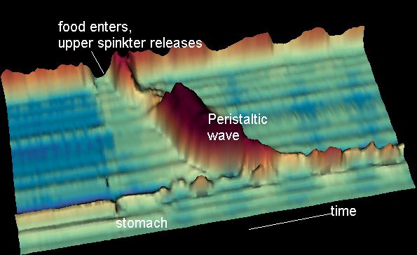
Peristalsis ( , ) is a type of intestinal motility, characterized by symmetry in biology#Radial symmetry, radially symmetrical contraction and relaxation of muscles that propagate in a wave down a tube, in an wikt:anterograde, anterograde direction. Peristalsis is progression of coordinated contraction of involuntary circular muscles, which is preceded by a simultaneous contraction of the longitudinal muscle and relaxation of the circular muscle in the lining of the gut. In much of a digestive tract, such as the human gastrointestinal tract, smooth muscle tissue contracts in sequence to produce a peristaltic wave, which propels a ball of food (called a bolus (digestion), bolus before being transformed into chyme in the stomach) along the tract. The peristaltic movement comprises relaxation of circular smooth muscles, then their contraction behind the chewed material to keep it from moving backward, then longitudinal contraction to push it forward. Earthworms use a similar mec ... [...More Info...] [...Related Items...] OR: [Wikipedia] [Google] [Baidu] |
Intercalated Disc
Intercalated discs or lines of Eberth are microscopic identifying features of cardiac muscle. Cardiac muscle consists of individual heart muscle cells (cardiomyocytes) connected by intercalated discs to work as a single functional Syncytium#Cardiac muscle, syncytium. By contrast, skeletal muscle consists of multinucleated muscle fibers and exhibits no intercalated discs. Intercalated discs support synchronized contraction of cardiac tissue in a wave-like pattern so that the heart can work like a pump. They occur at the Z line of the sarcomere and can be visualized easily when observing a longitudinal section of the tissue. Structure Intercalated discs are complex structures that connect adjacent cardiac muscle cells. The three types of cell junction recognised as making up an intercalated disc are desmosomes, Fascia adherens, fascia adherens junctions, and gap junctions. * Fascia adherens are anchoring sites for actin, and connect to the closest sarcomere. * Desmosomes prevent se ... [...More Info...] [...Related Items...] OR: [Wikipedia] [Google] [Baidu] |
Cell Nucleus
The cell nucleus (; : nuclei) is a membrane-bound organelle found in eukaryote, eukaryotic cell (biology), cells. Eukaryotic cells usually have a single nucleus, but a few cell types, such as mammalian red blood cells, have #Anucleated_cells, no nuclei, and a few others including osteoclasts have Multinucleate, many. The main structures making up the nucleus are the nuclear envelope, a double membrane that encloses the entire organelle and isolates its contents from the cellular cytoplasm; and the nuclear matrix, a network within the nucleus that adds mechanical support. The cell nucleus contains nearly all of the cell's genome. Nuclear DNA is often organized into multiple chromosomes – long strands of DNA dotted with various proteins, such as histones, that protect and organize the DNA. The genes within these chromosomes are Nuclear organization, structured in such a way to promote cell function. The nucleus maintains the integrity of genes and controls the activities of the ... [...More Info...] [...Related Items...] OR: [Wikipedia] [Google] [Baidu] |


