|
Muscular System Of The Horse
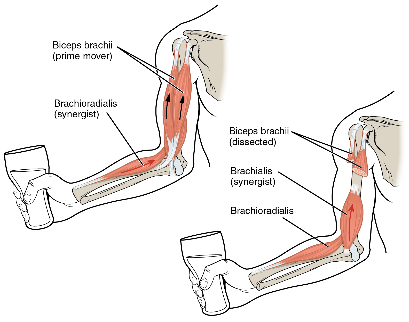
Types of muscle As in all vertebrates, horses have three types of muscle: * Skeletal muscle: this type of muscle contributes to movement and posture, and is consciously controlled (voluntary muscle). While some muscles attach solely to skin or cartilage (muscles of facial expression, etc.), contraction of skeletal muscles more commonly leads to the muscle pulling a tendon, which in turn pulls a bone, resulting in either flexion or extension of a joint. Skeletal muscles are usually arranged in antagonistic pairs in opposition to each other, with one flexing the joint (a flexor muscle) and the other extending it (extensor muscle). * Smooth muscle: this type of muscle is controlled by the autonomic nervous system (making it an involuntary muscle type). Smooth muscle is involved in digestion and other organs such as the eye * Cardiac: involuntary muscle that causes the heart to beat. This type of muscle has high numbers of mitochondria, allowing it to be fatigue-resistant. Build ... [...More Info...] [...Related Items...] OR: [Wikipedia] [Google] [Baidu] |
Anatomical Terms Of Muscle
Anatomical terminology is used to uniquely describe aspects of skeletal muscle, cardiac muscle, and smooth muscle such as their actions, structure, size, and location. Types There are three types of muscle tissue in the body: skeletal, smooth, and cardiac. Skeletal muscle Skeletal muscle, or "voluntary muscle", is a striated muscle tissue that primarily joins to bone with tendons. Skeletal muscle enables movement of bones, and maintains posture. The widest part of a muscle that pulls on the tendons is known as the belly. Muscle slip A muscle slip is a slip of muscle that can either be an anatomical variant, or a branching of a muscle as in rib connections of the serratus anterior muscle. Smooth muscle Smooth muscle is involuntary and found in parts of the body where it conveys action without conscious intent. The majority of this type of muscle tissue is found in the digestive and urinary systems where it acts by propelling forward food, chyme, and feces in the former and u ... [...More Info...] [...Related Items...] OR: [Wikipedia] [Google] [Baidu] |
Dorsum (biology)
Standard anatomical terms of location are used to describe unambiguously the anatomy of humans and other animals. The terms, typically derived from Latin or Greek roots, describe something in its standard anatomical position. This position provides a definition of what is at the front ("anterior"), behind ("posterior") and so on. As part of defining and describing terms, the body is described through the use of anatomical planes and axes. The meaning of terms that are used can change depending on whether a vertebrate is a biped or a quadruped, due to the difference in the neuraxis, or if an invertebrate is a non-bilaterian. A non-bilaterian has no anterior or posterior surface for example but can still have a descriptor used such as proximal or distal in relation to a body part that is nearest to, or furthest from its middle. International organisations have determined vocabularies that are often used as standards for subdisciplines of anatomy. For example, '' Terminolog ... [...More Info...] [...Related Items...] OR: [Wikipedia] [Google] [Baidu] |
Equine Exertional Rhabdomyolysis
Equine exertional rhabdomyolysis (ER) is a syndrome that affects the skeletal muscles within a horse. This syndrome causes the muscle to break down which is generally associated with exercise and diet regime. Depending on the severity, there are various types of ER, including sporadic (i.e., Tying-Up, Monday Morning Sickness/Disease, Azoturia) and chronic (i.e., Polysaccharide Storage Myopathy (PSSM) and Recurrent Exertional Rhabdomyolysis (RER)). Types of equine exertional rhabdomyolysis (ER) Equine Exertional Rhabdomyolysis (ER) is a general term used to define both sporadic - (infrequent) and chronic - (repeated) manifestations of the condition. The severity of the condition defines what type of ER a horse has. Sporadic equine exertional rhabdomyolysis (ER) The types of equine ER that are considered sporadic include tying-up, also commonly referred to as Monday morning sickness and/or Monday morning disease, and azoturia also known as black water disease, set fast, and/or pa ... [...More Info...] [...Related Items...] OR: [Wikipedia] [Google] [Baidu] |
Compartment Syndrome
Compartment syndrome is a serious medical condition in which increased pressure within a Fascial compartment, body compartment compromises blood flow and tissue function, potentially leading to permanent damage if not promptly treated. There are two types: Acute (medicine), acute and Chronic condition, chronic. Acute compartment syndrome can lead to a loss of the affected limb due to tissue death. Symptoms of acute compartment syndrome (ACS) include severe pain, decreased blood flow, decreased movement, numbness, and a pale limb. It is most often due to Injury, physical trauma, like a bone fracture (up to 75% of cases) or a crush injury. It can also occur after Reperfusion injury, blood flow returns following a period of poor circulation. Diagnosis is Clinical diagnosis, clinical, based on symptoms, not a specific test. However, it may be supported by measuring the pressure inside the Fascial compartment, compartment. It is classically described by pain out of proportion to the in ... [...More Info...] [...Related Items...] OR: [Wikipedia] [Google] [Baidu] |
Bowed Tendon
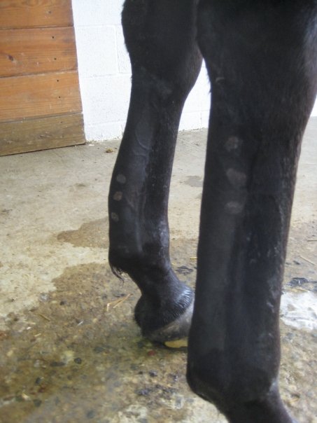
Tendinitis/tendonitis is inflammation of a tendon, often involving torn collagen fibers. A bowed tendon is a horseman's term for a tendon after a horse has sustained an injury that causes swelling in one or more tendons creating a "bowed" appearance. Description of tendinitis in horses Tendinitis usually involves disruption of the tendon fibers. It is most commonly seen in the superficial digital flexor tendon (SDFT) in a front leg—the tendon that runs down the back of the leg, closest to the surface. Tendinitis creating a "bow" is uncommon in the deep digital flexor tendon (DDFT) of a front leg, but is not uncommon in the pastern and foot regions. Tendinitis of the SDFT or DDFT in the hind leg is less common. When the SDFT is damaged significantly, there is a thickening of the tendon, giving it a bowed appearance when the leg is viewed from the side. Bows usually occur in the middle of the tendon region, although they may also be seen in the upper third, right below the kne ... [...More Info...] [...Related Items...] OR: [Wikipedia] [Google] [Baidu] |
Trochanter
A trochanter is a tubercle of the femur near its joint with the hip bone. In humans and most mammals, the trochanters serve as important muscle attachment sites. Humans have two, sometimes three, trochanters. Etymology The anatomical term ''trochanter'' (the bony protrusions on the femur) derives from the Greek τροχαντήρ (''trochantḗr''). This Greek word itself is generally broken down into: * τροχάζω (''trokházō''), meaning “to run quickly,” “to gallop,” or “to move rapidly.” * -τήρ (''-tḗr''), a suffix in Greek that often signifies an agent or instrument (“one who oes something�� or “that which oes something��). While the exact origin of the anatomical term trochanter is uncertain, multiple possible connections could be suggested. One possibility is that the term was derived directly from the Greek roots without influence from the maritime meaning, with the name referencing the trochanter’s role in enabling swift movement throu ... [...More Info...] [...Related Items...] OR: [Wikipedia] [Google] [Baidu] |
Aponeurosis
An aponeurosis (; : aponeuroses) is a flattened tendon by which muscle attaches to bone or fascia. Aponeuroses exhibit an ordered arrangement of collagen fibres, thus attaining high tensile strength in a particular direction while being vulnerable to tensional or shear forces in other directions. They have a shiny, whitish-silvery color, are histologically similar to tendons, and are very sparingly supplied with blood vessels and nerves. When dissected, aponeuroses are papery and peel off by sections. The primary regions with thick aponeuroses are in the ventral abdominal region, the dorsal lumbar region, the ventriculus in birds, and the palmar (palms) and plantar (soles) regions. Anatomy Anterior abdominal aponeuroses The anterior abdominal aponeuroses are located just superficial to the rectus abdominis muscle. It has for its borders the external oblique, pectoralis muscles, and the latissimus dorsi. Posterior lumbar aponeuroses The posterior lumbar aponeuroses are sit ... [...More Info...] [...Related Items...] OR: [Wikipedia] [Google] [Baidu] |
Abduction (kinesiology)
Motion, the process of movement, is described using specific anatomical terminology, anatomical terms. Motion includes movement of Organ (anatomy), organs, joints, Limb (anatomy), limbs, and specific sections of the body. The terminology used describes this motion according to its direction relative to the anatomical position of the body parts involved. Anatomy, Anatomists and others use a unified set of terms to describe most of the movements, although other, more specialized terms are necessary for describing unique movements such as those of the hands, feet, and eyes. In general, motion is classified according to the anatomical plane it occurs in. ''Flexion'' and ''extension'' are examples of ''angular'' motions, in which two axes of a joint are brought closer together or moved further apart. ''Rotational'' motion may occur at other joints, for example the shoulder, and are described as ''internal'' or ''external''. Other terms, such as ''elevation'' and ''depression'', descri ... [...More Info...] [...Related Items...] OR: [Wikipedia] [Google] [Baidu] |
Rear (horse)
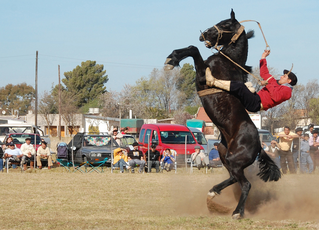
Rearing occurs when a horse or other equidae, equine "stands up" on its hind legs with the forelegs off the ground. Rearing may be linked to fright, aggression, excitement, disobedience, non experienced rider, or pain. It is not uncommon to see stallions rearing in the wild when they fight, while striking at their opponent with their front legs. Mares are generally more likely to kick when acting in aggression, but may rear if they need to strike at a threat in front of them. When a horse rears around people, in most cases, it is considered a dangerous habit for riding horses, as not only can a rider fall off from a considerable height, but also because it is possible for the animal to fall over backwards, which could cause injuries or death to both horse and rider. It is therefore strongly discouraged. A horse that has a habit of rearing generally requires extensive retraining by an experienced horse trainer, and if the habit cannot be corrected, they horse may be deemed too ... [...More Info...] [...Related Items...] OR: [Wikipedia] [Google] [Baidu] |
Olecranon
The olecranon (, ), is a large, thick, curved bony process on the proximal, posterior end of the ulna. It forms the protruding part of the elbow and is opposite to the cubital fossa or elbow pit (trochlear notch). The olecranon serves as a lever for the extensor muscles that straighten the elbow joint. Structure The olecranon is situated at the proximal end of the ulna, one of the two bones in the forearm. When the hand faces forward ( supination) the olecranon faces towards the back (posteriorly). It is bent forward at the summit so as to present a prominent lip which is received into the olecranon fossa of the humerus during extension of the forearm. Its base is contracted where it joins the body and the narrowest part of the upper end of the ulna. Its posterior surface, directed backward, is triangular, smooth, subcutaneous, and covered by a bursa. Its superior surface is of quadrilateral form, marked behind by a rough impression for the insertion of the triceps brachi ... [...More Info...] [...Related Items...] OR: [Wikipedia] [Google] [Baidu] |
Stay Apparatus
The stay apparatus is an arrangement of muscles, tendons, and ligaments that work together so that an animal can remain standing with virtually no muscular effort. It is best known as the mechanism by which horses can enter a light sleep while still standing up. The effect is that an animal can distribute its weight on three limbs while resting a fourth in a flexed, non-weight-bearing position. The animal can periodically shift its weight to rest a different leg, and thus all limbs are able to be individually rested, reducing overall wear and tear. The relatively slim legs of certain large mammals, such as horses and cows, would be subject to dangerous levels of fatigue if not for the stay apparatus. The lower part of the stay apparatus consists of the suspensory apparatus, which is the same in both front and hind legs, while the upper portion of the stay apparatus is different between the fore and hind limbs. In the front legs, the stay apparatus engages when the animal's musc ... [...More Info...] [...Related Items...] OR: [Wikipedia] [Google] [Baidu] |
Hyoid
The hyoid-bone (lingual-bone or tongue-bone) () is a horseshoe-shaped bone situated in the anterior midline of the neck between the chin and the thyroid-cartilage. At rest, it lies between the base of the mandible and the third cervical vertebra. Unlike other bones, the hyoid is only distantly articulated to other bones by muscles or ligaments. It is the only bone in the human body that is not connected to any other bones. The hyoid is anchored by muscles from the anterior, posterior and inferior directions, and aids in tongue movement and swallowing. The hyoid bone provides attachment to the muscles of the floor of the mouth and the tongue above, the larynx below, and the epiglottis and pharynx behind. Its name is derived . Structure The hyoid bone is classed as an irregular bone and consists of a central part called the body, and two pairs of horns, the greater and lesser horns. Body The body of the hyoid bone is the central part of the hyoid bone. *At the front ... [...More Info...] [...Related Items...] OR: [Wikipedia] [Google] [Baidu] |


