|
Munro's Microabscesses
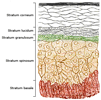
Munro's microabscess is an abscess (collection of neutrophils) in the stratum corneum of the epidermis due to the infiltration of neutrophils from papillary dermis into the epidermal stratum corneum. They are a cardinal sign of psoriasis where they are seen in the hyperkeratotic and parakeratotic areas of the stratum corneum. Munro microabscesses are not seen in seborrheic dermatitis, pityriasis rosea, lichen ruber planus nor dermatitis herpetiformis Dermatitis herpetiformis (DH) is a chronic autoimmune blistering skin condition, characterised by intensely itchy blisters filled with a watery fluid. DH is a cutaneous manifestation of coeliac disease, although the exact causal mechanism is not .... It is named for William John Munro (1863–1908).Johnson, A. 1983. The Man behind the Eponym. William John Munro (1863–1908). ''The American Journal of Dermatopathology'' 5(5): 477–478. References Psoriasis {{Cutaneous-condition-stub ... [...More Info...] [...Related Items...] OR: [Wikipedia] [Google] [Baidu] |
Abscess
An abscess is a collection of pus that has built up within the tissue of the body, usually caused by bacterial infection. Signs and symptoms of abscesses include redness, pain, warmth, and swelling. The swelling may feel fluid-filled when pressed. The area of redness often extends beyond the swelling. Carbuncles and boils are types of abscess that often involve hair follicles, with carbuncles being larger. A cyst is related to an abscess, but it contains a material other than pus, and a cyst has a clearly defined wall. Abscesses can also form internally on internal organs and after surgery. They are usually caused by a bacterial infection. Often many different types of bacteria are involved in a single infection. In many areas of the world, the most common bacteria present is ''methicillin-resistant Staphylococcus aureus''. Rarely, parasites can cause abscesses; this is more common in the developing world. Diagnosis of a skin abscess is usually made based on what it looks ... [...More Info...] [...Related Items...] OR: [Wikipedia] [Google] [Baidu] |
Neutrophil
Neutrophils are a type of phagocytic white blood cell and part of innate immunity. More specifically, they form the most abundant type of granulocytes and make up 40% to 70% of all white blood cells in humans. Their functions vary in different animals. They are also known as neutrocytes, heterophils or polymorphonuclear leukocytes. They are formed from stem cells in the bone marrow and differentiated into subpopulations of neutrophil-killers and neutrophil-cagers. They are short-lived (between 5 and 135 hours, see ) and highly mobile, as they can enter parts of tissue where other cells/molecules cannot. Neutrophils may be subdivided into segmented neutrophils and banded neutrophils (or bands). They form part of the polymorphonuclear cells family (PMNs) together with basophils and eosinophils. The name ''neutrophil'' derives from staining characteristics on hematoxylin and eosin ( H&E) histological or cytological preparations. Whereas basophilic white blood cells ... [...More Info...] [...Related Items...] OR: [Wikipedia] [Google] [Baidu] |
Stratum Corneum
The stratum corneum (Latin language, Latin for 'horny layer') is the outermost layer of the epidermis (skin), epidermis. Consisting of dead tissue, it protects underlying tissue from infection, dehydration, chemicals and mechanical stress. It is composed of 15–20 layers of flattened cells with no nuclei and cell organelles. Among its properties are mechanical shear, impact resistance, water flux and hydration regulation, microbial proliferation and invasion regulation, initiation of inflammation through cytokine activation and dendritic cell activity, and selective permeability to exclude toxins, irritants, and allergens. The cytoplasm of its cells shows filamentous keratin. These corneocytes are embedded in a lipid matrix composed of ceramides, cholesterol, and fatty acids. Desquamation is the process of cell shedding from the surface of the stratum corneum, balancing proliferating keratinocytes that form in the stratum basale. These cells migrate through the epidermis tow ... [...More Info...] [...Related Items...] OR: [Wikipedia] [Google] [Baidu] |
Epidermis (skin)
The epidermis is the outermost of the three layers that comprise the skin, the inner layers being the dermis and hypodermis. The epidermal layer provides a barrier to infection from environmental pathogens and regulates the amount of water released from the body into the atmosphere through transepidermal water loss. The epidermis is composed of multiple layers of flattened cells that overlie a base layer ( stratum basale) composed of columnar cells arranged perpendicularly. The layers of cells develop from stem cells in the basal layer. The thickness of the epidermis varies from 31.2μm for the penis to 596.6μm for the sole of the foot with most being roughly 90μm. Thickness does not vary between the sexes but becomes thinner with age. The human epidermis is an example of epithelium, particularly a stratified squamous epithelium. The word epidermis is derived through Latin , itself and . Something related to or part of the epidermis is termed epidermal. Structure ... [...More Info...] [...Related Items...] OR: [Wikipedia] [Google] [Baidu] |
Papillary Dermis
The dermis or corium is a layer of skin between the epidermis (with which it makes up the cutis) and subcutaneous tissues, that primarily consists of dense irregular connective tissue and cushions the body from stress and strain. It is divided into two layers, the superficial area adjacent to the epidermis called the papillary region and a deep thicker area known as the reticular dermis.James, William; Berger, Timothy; Elston, Dirk (2005). ''Andrews' Diseases of the Skin: Clinical Dermatology'' (10th ed.). Saunders. Pages 1, 11–12. . The dermis is tightly connected to the epidermis through a basement membrane. Structural components of the dermis are collagen, elastic fibers, and extrafibrillar matrix.Marks, James G; Miller, Jeffery (2006). ''Lookingbill and Marks' Principles of Dermatology'' (4th ed.). Elsevier Inc. Page 8–9. . It also contains mechanoreceptors that provide the sense of touch and thermoreceptors that provide the sense of heat. In addition, hair follicles, swe ... [...More Info...] [...Related Items...] OR: [Wikipedia] [Google] [Baidu] |
Medical Sign
Signs and symptoms are diagnostic indications of an illness, injury, or condition. Signs are objective and externally observable; symptoms are a person's reported subjective experiences. A sign for example may be a higher or lower temperature than normal, raised or lowered blood pressure or an abnormality showing on a medical scan. A symptom is something out of the ordinary that is experienced by an individual such as feeling feverish, a headache or other pains in the body, which occur as the body's immune system fights off an infection. Signs and symptoms Signs A medical sign is an objective observable indication of a disease, injury, or medical condition that may be detected during a physical examination. These signs may be visible, such as a rash or bruise, or otherwise detectable such as by using a stethoscope or taking blood pressure. Medical signs, along with symptoms, help in forming a diagnosis. Some examples of signs are nail clubbing of either the fingernails o ... [...More Info...] [...Related Items...] OR: [Wikipedia] [Google] [Baidu] |
Psoriasis
Psoriasis is a long-lasting, noncontagious autoimmune disease characterized by patches of abnormal skin. These areas are red, pink, or purple, dry, itchy, and scaly. Psoriasis varies in severity from small localized patches to complete body coverage. Injury to the skin can trigger psoriatic skin changes at that spot, which is known as the Koebner phenomenon. The five main types of psoriasis are plaque, guttate, inverse, pustular, and erythrodermic. Plaque psoriasis, also known as psoriasis vulgaris, makes up about 90% of cases. It typically presents as red patches with white scales on top. Areas of the body most commonly affected are the back of the forearms, shins, navel area, and scalp. Guttate psoriasis has drop-shaped lesions. Pustular psoriasis presents as small, noninfectious, pus-filled blisters. Inverse psoriasis forms red patches in skin folds. Erythrodermic psoriasis occurs when the rash becomes very widespread and can develop from any of the other types. ... [...More Info...] [...Related Items...] OR: [Wikipedia] [Google] [Baidu] |
Seborrheic Dermatitis
Seborrhoeic dermatitis (also spelled seborrheic dermatitis in American English) is a long-term skin disorder. Symptoms include flaky, scaly, greasy, and occasionally itchy and inflamed skin. Areas of the skin rich in sebum, oil-producing glands are often affected including the scalp, face, and chest. It can result in social or self-esteem problems. In babies, when the scalp is primarily involved, it is called cradle cap. Mild seborrhoeic dermatitis of the scalp may be described in lay terms as dandruff due to the dry, flaky character of the skin. However, as dandruff may refer to any dryness or scaling of the scalp, not all dandruff is seborrhoeic dermatitis. Seborrhoeic dermatitis is sometimes inaccurately referred to as seborrhoea. The cause is unclear but believed to involve a number of genetic and environmental factors. Risk factors for seborrhoeic dermatitis include immunocompromised, poor immune function, Parkinson's disease, and alcoholic pancreatitis. The condition ma ... [...More Info...] [...Related Items...] OR: [Wikipedia] [Google] [Baidu] |
Pityriasis Rosea
Pityriasis rosea is a type of skin rash. Classically, it begins with a single red and slightly scaly area known as a "herald patch". This is then followed, days to weeks later, by an eruption of many smaller scaly spots; pinkish with a red edge in people with light skin and greyish in darker skin. About 20% of cases show atypical deviations from this pattern. It usually lasts less than three months and goes away without treatment. Sometimes malaise or a fever may occur before the start of the rash or itchiness, but often there are few other symptoms. While the cause is not entirely clear, it is believed to be related to human herpesvirus 6 (HHV6) or human herpesvirus 7 (HHV7). It does not appear to be contagious disease, contagious. Certain medications may result in a similar rash. Diagnosis is based on the symptoms. Evidence for specific treatment is limited. About 1.3% of people are affected at some point in time. It most often occurs in those between the ages of 10 and 35. ... [...More Info...] [...Related Items...] OR: [Wikipedia] [Google] [Baidu] |
Lichen Ruber Planus
Lichen planus (LP) is a chronic inflammatory and autoimmune disease that affects the skin, nails, hair, and mucous membranes. It is not an actual lichen, but is named for its appearance. It is characterized by polygonal, flat-topped, violaceous papules and plaques with overlying, reticulated, fine white scale ( Wickham's striae), commonly affecting dorsal hands, flexural wrists and forearms, trunk, anterior lower legs and oral mucosa. The hue may be gray-brown in people with darker skin. Although there is a broad clinical range of LP manifestations, the skin and oral cavity remain as the major sites of involvement. The cause is unknown, but it is thought to be the result of an autoimmune process with an unknown initial trigger. There is no cure, but many different medications and procedures have been used in efforts to control the symptoms. The term lichenoid reaction (lichenoid eruption or lichenoid lesion) refers to a lesion of similar or identical histopathologic and clinical ... [...More Info...] [...Related Items...] OR: [Wikipedia] [Google] [Baidu] |
Dermatitis Herpetiformis
Dermatitis herpetiformis (DH) is a chronic autoimmune blistering skin condition, characterised by intensely itchy blisters filled with a watery fluid. DH is a cutaneous manifestation of coeliac disease, although the exact causal mechanism is not known. DH is neither related to nor caused by herpes virus; the name means that it is a skin inflammation having an appearance (Latin: '' -formis'') similar to herpes. The age of onset is usually about 15 to 40, but DH also may affect children and the elderly. Men are slightly more affected than women. Estimates of DH prevalence vary from 1 in 400 to 1 in 10,000. It is most common in patients of northern European and northern Indian ancestry, and is associated with the human leukocyte antigen (HLA) haplotype HLA-DQ2 or HLA-DQ8 along with coeliac disease and gluten sensitivity. Dermatitis herpetiformis was first described by Louis Adolphus Duhring in 1884. A connection between DH and coeliac disease was recognized in 1967. Signs and ... [...More Info...] [...Related Items...] OR: [Wikipedia] [Google] [Baidu] |


