|
Middle Cerebral Artery
The middle cerebral artery (MCA) is one of the three major paired cerebral artery, cerebral arteries that supply blood to the cerebrum. The MCA arises from the internal carotid artery and continues into the lateral sulcus where it then branches and projects to many parts of the lateral cerebral cortex. It also supplies blood to the anterior temporal lobes and the insular cortex, insular cortices. The left and right MCAs rise from trifurcations of the internal carotid artery, internal carotid arteries and thus are connected to the anterior cerebral artery, anterior cerebral arteries and the posterior communicating artery, posterior communicating arteries, which connect to the posterior cerebral artery, posterior cerebral arteries. The MCAs are not considered a part of the Circle of Willis. Structure The middle cerebral artery divides into four segments, named by the region they supply as opposed to order of branching as the latter can be somewhat variable: *M1: The ''sphenoidal' ... [...More Info...] [...Related Items...] OR: [Wikipedia] [Google] [Baidu] |
Circle Of Willis
The circle of Willis (also called Willis' circle, loop of Willis, cerebral arterial circle, and Willis polygon) is a circulatory anastomosis that supplies blood to the brain and surrounding structures in reptiles, birds and mammals, including humans. It is named after Thomas Willis (1621–1675), an English physician. Structure The circle of Willis is a part of the cerebral circulation and is composed of the following arteries: * Anterior cerebral artery (left and right) at their A1 segments * Anterior communicating artery * Internal carotid artery (left and right) at its distal tip (carotid terminus) * Posterior cerebral artery (left and right) at their P1 segments * Posterior communicating artery (left and right) The middle cerebral arteries, supplying the brain, are also considered part of the Circle of Willis Origin of arteries The left and right internal carotid arteries arise from the left and right common carotid arteries. The posterior communicating artery is given ... [...More Info...] [...Related Items...] OR: [Wikipedia] [Google] [Baidu] |
Wernicke's Area
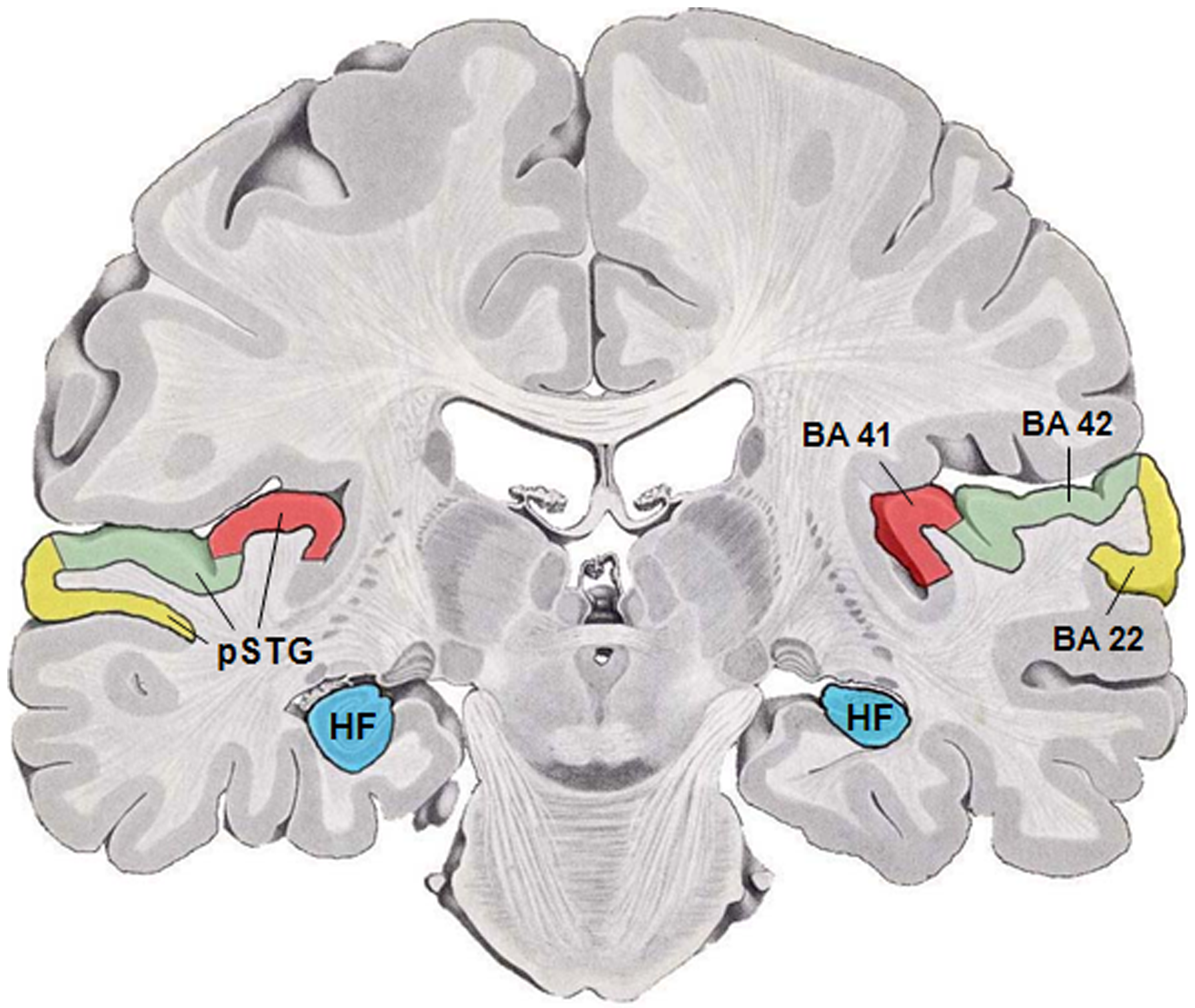
Wernicke's area (; ), also called Wernicke's speech area, is one of the two parts of the cerebral cortex that are linked to speech, the other being Broca's area. It is involved in the comprehension of written and spoken language, in contrast to Broca's area, which is primarily involved in the production of language. It is traditionally thought to reside in Brodmann area 22, located in the superior temporal gyrus in the dominant cerebral hemisphere, which is the left hemisphere in about 95% of right-handed individuals and 70% of left-handed individuals. Damage caused to Wernicke's area results in receptive, fluent aphasia. This means that the person with aphasia will be able to fluently connect words, but the phrases will lack meaning. This is unlike non-fluent aphasia, in which the person will use meaningful words, but in a non-fluent, telegraphic manner. Emerging research on the developmental trajectory of Wernicke's area highlights its evolving role in language acquisiti ... [...More Info...] [...Related Items...] OR: [Wikipedia] [Google] [Baidu] |
Broca's Area
Broca's area, or the Broca area (, also , ), is a region in the frontal lobe of the dominant Cerebral hemisphere, hemisphere, usually the left, of the Human brain, brain with functions linked to speech production. Language processing in the brain, Language processing has been linked to Broca's area since Pierre Paul Broca reported impairments in two patients. They had lost the ability to speak after injury to the posterior inferior frontal gyrus (pars triangularis) (BA45) of the brain. Since then, the approximate region he identified has become known as Broca's area, and the deficit in language production as Broca's aphasia, also called expressive aphasia. Broca's area is now typically defined in terms of the pars opercularis and pars triangularis of the inferior frontal gyrus, represented in Korbinian Brodmann, Brodmann's Brodmann area, cytoarchitectonic map as Brodmann area 44 and Brodmann area 45 of the dominant hemisphere. Functional magnetic resonance imaging (fMRI) has show ... [...More Info...] [...Related Items...] OR: [Wikipedia] [Google] [Baidu] |
Parietal Lobe
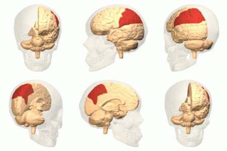
The parietal lobe is one of the four Lobes of the brain, major lobes of the cerebral cortex in the brain of mammals. The parietal lobe is positioned above the temporal lobe and behind the frontal lobe and central sulcus. The parietal lobe integrates sensory information among various sensory modality, modalities, including spatial sense and navigation (proprioception), the main sensory receptive area for the sense of touch in the somatosensory cortex which is just posterior to the central sulcus in the postcentral gyrus, and the two-streams hypothesis#Dorsal stream, dorsal stream of the visual system. The major sensory inputs from the skin (mechanoreceptor, touch, thermoreceptor, temperature, and nociceptor, pain receptors), relay through the thalamus to the parietal lobe. Several areas of the parietal lobe are important in language processing in the brain, language processing. The somatosensory cortex can be illustrated as a distorted figure – the cortical homunculus (Latin: "li ... [...More Info...] [...Related Items...] OR: [Wikipedia] [Google] [Baidu] |
Angular Gyrus
The angular gyrus is a region of the brain lying mainly in the posteroinferior region of the parietal lobe, occupying the posterior part of the inferior parietal lobule. It represents the Brodmann area 39. Its significance is in transferring visual information to Wernicke's area, in order to make meaning out of visually perceived words. It is also involved in a number of processes related to language, number processing and spatial cognition, memory retrieval, attention, and theory of mind. Anatomy Connections Left and right angular gyri are connected by the dorsal splenium and isthmus of the corpus callosum. Boundaries * Anteriorly by the supramarginal gyrus. * Superiorly by the intraparietal sulcus. * Posteriorly by the parieto-occipital sulcus. * Inferiorly the angular gyrus of the parietal lobe is continuous as the superior and middle temporal gyri. Also, the angular sulcus, which is capped by the angular gyrus, is continuous as the superior temporal sulcus inf ... [...More Info...] [...Related Items...] OR: [Wikipedia] [Google] [Baidu] |
Anastomosis
An anastomosis (, : anastomoses) is a connection or opening between two things (especially cavities or passages) that are normally diverging or branching, such as between blood vessels, leaf veins, or streams. Such a connection may be normal (such as the foramen ovale in a fetus' heart) or abnormal (such as the patent foramen ovale in an adult's heart); it may be acquired (such as an arteriovenous fistula) or innate (such as the arteriovenous shunt of a metarteriole); and it may be natural (such as the aforementioned examples) or artificial (such as a surgical anastomosis). The reestablishment of an anastomosis that had become blocked is called a reanastomosis. Anastomoses that are abnormal, whether congenital or acquired, are often called fistulas. The term is used in medicine, biology, mycology, geology, and geography. Etymology Anastomosis: medical or Modern Latin, from Greek ἀναστόμωσις, anastomosis, "outlet, opening", Greek ana- "up, on, upon", stoma "mouth" ... [...More Info...] [...Related Items...] OR: [Wikipedia] [Google] [Baidu] |
Pars Triangularis
The inferior frontal gyrus (IFG; also gyrus frontalis inferior) is the lowest positioned gyrus of the frontal gyri, of the frontal lobe, and is part of the prefrontal cortex. Its superior border is the inferior frontal sulcus (which divides it from the middle frontal gyrus), its inferior border is the lateral sulcus (which divides it from the superior temporal gyrus) and its posterior border is the inferior precentral sulcus. Above it is the middle frontal gyrus, behind it is the precentral gyrus. The inferior frontal gyrus contains Broca's area, which is involved in language processing and speech production. Structure The inferior frontal gyrus is highly convoluted and has three cytoarchitecturally diverse regions. The three subdivisions are an opercular part, a triangular part, and an orbital part. These divisions are marked by two rami arising from the lateral sulcus. The ascending ramus separates the opercular and triangular parts. The anterior (horizontal) ramus separate ... [...More Info...] [...Related Items...] OR: [Wikipedia] [Google] [Baidu] |
Operculum (brain)
In human brain anatomy, an operculum (Latin, meaning "little lid") (: opercula), may refer to the frontal, temporal, or parietal operculum, which together cover the insular cortex, insula as the opercula of insula. It can also refer to the occipital operculum, part of the occipital lobe. The insular lobe is a portion of the cerebral cortex that has invaginated to lie deep within the lateral sulcus. It sits like an island (the meaning of ''insular'') almost surrounded by the groove of the Circular sulcus of insula, circular sulcus and covered over and obscured by the insular opercula. A part of the parietal lobe, the frontoparietal operculum, covers the upper part of the insular lobe from the front to the back. The opercula lie on the precentral gyrus, precentral and postcentral gyrus, postcentral gyrus, gyri (on either side of the central sulcus). The part of the parietal operculum that forms the ceiling of the lateral sulcus functions as the secondary somatosensory cortex. De ... [...More Info...] [...Related Items...] OR: [Wikipedia] [Google] [Baidu] |
Insular Cortex
The insular cortex (also insula and insular lobe) is a portion of the cerebral cortex folded deep within the lateral sulcus (the fissure separating the temporal lobe from the parietal lobe, parietal and frontal lobes) within each brain hemisphere, hemisphere of the mammalian brain. The insulae are believed to be involved in consciousness and play a role in diverse functions usually linked to emotion or the regulation of the body's homeostasis. These functions include compassion, empathy, taste, perception, motor control, self-awareness, cognitive functioning, interpersonal relationships, and awareness of homeostatic emotions such as Hunger (physiology), hunger, pain and fatigue. In relation to these, it is involved in psychopathology. The insular cortex is divided by the central sulcus of the insula, into two parts: the anterior insula and the posterior insula in which more than a dozen field areas have been identified. The cortical area overlying the insula toward the lateral su ... [...More Info...] [...Related Items...] OR: [Wikipedia] [Google] [Baidu] |
Basal Ganglia
The basal ganglia (BG) or basal nuclei are a group of subcortical Nucleus (neuroanatomy), nuclei found in the brains of vertebrates. In humans and other primates, differences exist, primarily in the division of the globus pallidus into external and internal regions, and in the division of the striatum. Positioned at the base of the forebrain and the top of the midbrain, they have strong connections with the cerebral cortex, thalamus, brainstem and other brain areas. The basal ganglia are associated with a variety of functions, including regulating voluntary motor control, motor movements, procedural memory, procedural learning, habituation, habit formation, conditional learning, eye movements, cognition, and emotion. The main functional components of the basal ganglia include the striatum, consisting of both the dorsal striatum (caudate nucleus and putamen) and the ventral striatum (nucleus accumbens and olfactory tubercle), the globus pallidus, the ventral pallidum, the substa ... [...More Info...] [...Related Items...] OR: [Wikipedia] [Google] [Baidu] |


