|
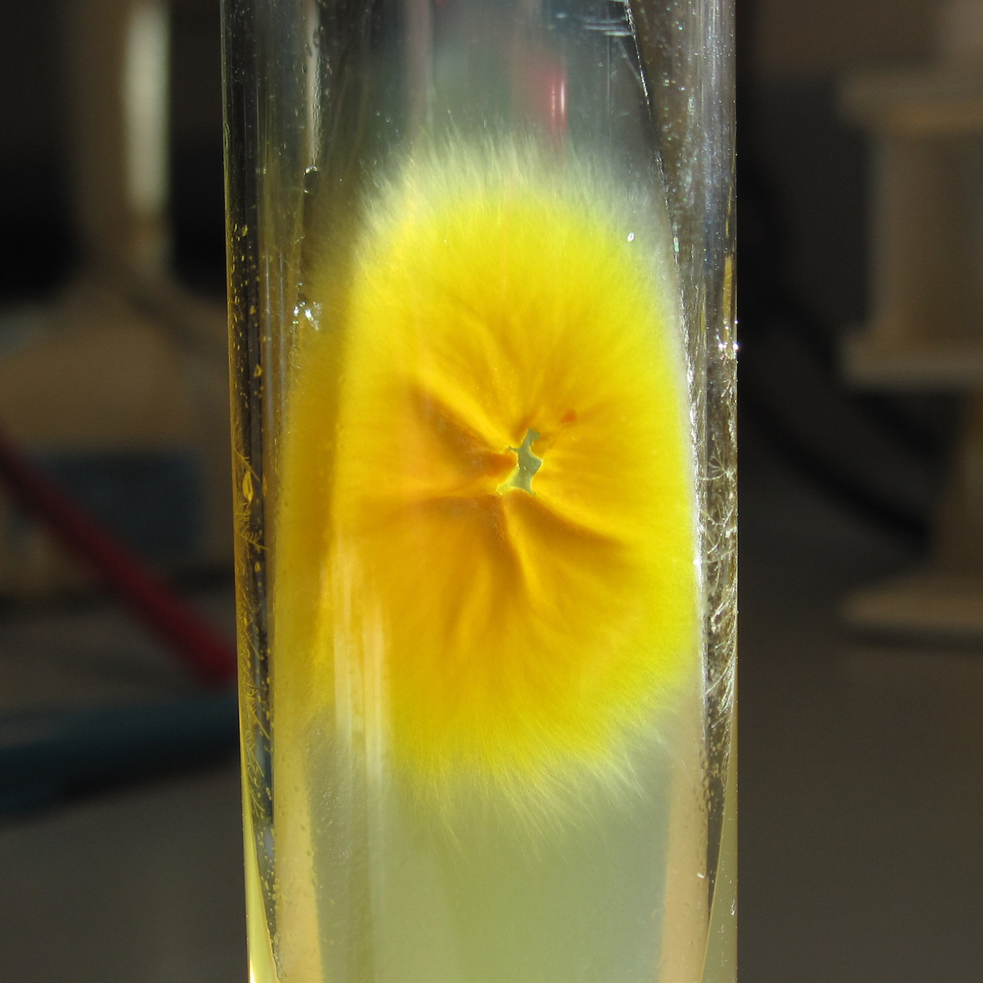
Microsporum Ripariae
''Microsporum'' is a genus of fungi that causes tinea capitis, tinea corporis, ringworm, and other dermatophytoses (fungal infections of the skin). ''Microsporum'' forms both macroconidia (large asexual reproductive structures) and microconidia (smaller asexual reproductive structures) on short conidiophores. Macroconidia are hyaline, multiseptate, variable in form, fusiform, spindle-shaped to obovate, 7–20 by 30–160 um in size, with thin or thick echinulate to verrucose cell walls. Their shape, size and cell wall features are important characteristics for species identification. Microconidia are hyaline, single-celled, pyriform to clavate, smooth-walled, 2.5–3.5 by 4–7 um in size and are not diagnostic for any one species. The separation of this genus from ''Trichophyton'' is essentially based on the roughness of the macroconidial cell wall, although in practice this may sometimes be difficult to observe. Seventeen species of ''Microsporum'' have been descri ... [...More Info...] [...Related Items...] OR: [Wikipedia] [Google] [Baidu] |
Microsporum Canis
''Microsporum canis'' is a pathogenic, asexual fungus in the phylum Ascomycota that infects the upper, dead layers of skin on domesticated cats, and occasionally dogs and humans. The species has a worldwide distribution. Taxonomy and evolution ''Microsporum canis'' reproduces by means of two conidial forms, large, spindle-shaped, multicelled macroconidia and small, single-celled microconidia. First records of ''M. canis'' date to 1902. Evolutionary studies have established that ''M. canis'', like the very closely related sibling species ''M. distortum'' and ''M. equinum'', is a genetic clone derived from the sexually reproducing species, ''Arthroderma otae''. Members of Ascomycota often possess conspicuous asexual and sexual forms that can coexist in time and space. ''Microsporum canis'' exemplifies a common situation in ascomycetous fungi in which, over time, one mating type strain has undergone habitat divergence from the other and established a self-sustaining reproductive ... [...More Info...] [...Related Items...] OR: [Wikipedia] [Google] [Baidu] |
Micrometre
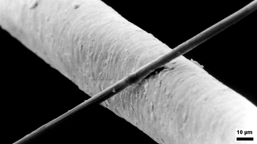
The micrometre (English in the Commonwealth of Nations, Commonwealth English as used by the International Bureau of Weights and Measures; SI symbol: μm) or micrometer (American English), also commonly known by the non-SI term micron, is a unit of length in the International System of Units (SI) equalling (SI standard prefix "micro-" = ); that is, one millionth of a metre (or one thousandth of a millimetre, , or about ). The nearest smaller common SI Unit, SI unit is the nanometre, equivalent to one thousandth of a micrometre, one millionth of a millimetre or one billionth of a metre (). The micrometre is a common unit of measurement for wavelengths of infrared radiation as well as sizes of biological cell (biology), cells and bacteria, and for grading wool by the diameter of the fibres. The width of a single human hair ranges from approximately 20 to . Examples Between 1 μm and 10 μm: * 1–10 μm – length of a typical bacterium * 3–8 μm – width of str ... [...More Info...] [...Related Items...] OR: [Wikipedia] [Google] [Baidu] |
Microsporum Gallinae
''Microsporum gallinae'' is a fungus of the genus ''Microsporum'' that causes dermatophytosis, commonly known as ringworm. Chickens represent the host population of ''Microsporum gallinae'' but its opportunistic nature allows it to enter other populations of fowl, mice, squirrels, cats, dogs and monkeys. Human cases of ''M. gallinae'' are rare, and usually mild, non-life-threatening superficial infections. Taxonomy and naming ''Microsporum gallinae'' was first identified in 1881 by Megnin from chicken favus, and named ''Epidermophyton gallinae''. It was later transferred from the Epidermophyton genus, and classified in the Trichophyton genus, as ''T. gallinae''. The identification of rough-walled macroconidia, a hallmark of the Microsporum genus, lead to the dermatophyte being classified as ''M. gallinae''. There is still debate about the phylogenetic placement of this dermatophyte, but the accepted name is ''Microsporum gallinae''. Analysis of its DNA sequences by PCR shows ' ... [...More Info...] [...Related Items...] OR: [Wikipedia] [Google] [Baidu] |
Microsporum Fulvum
''Microsporum fulvum'' is a wildly-distributed dermatophyte species in the Fungi Kingdom. It is known to be a close relative to other dermatophytes such as ''Trichophyton and'' ''Epidermophyton.'' The fungus is common within soil environments and grows well on keratinized material, such as hair, nails and dead skin. It is recognized as an opportunistic fungal pathogen capable of causing cutaneous mycoses in humans and animals. Originally, the fungus was thought to be ''Microsporum gypseum'' until enhanced genetic examination separated the two as distinct species in 1963. History and taxonomy ''Microsporum fulvum'' was first documented in 1909 as ''Microsporum gypseum'' by Weitzman et al. ( Argentina Medical Society)''.'' The fungus was thought to be the imperfect state of the anamorphic, asexually reproducing, ''M. gypseum.'' However, in Stockdale (1963) ''M. fulvum'' was considered and described as its own species, '' Nannizzia fulva'', the perfect state of the fungus. In the ... [...More Info...] [...Related Items...] OR: [Wikipedia] [Google] [Baidu] |
Microsporum Ferrugineum
''Microsporum'' is a genus of fungi that causes tinea capitis, tinea corporis, ringworm, and other dermatophytoses (fungal infections of the skin). ''Microsporum'' forms both macroconidia (large asexual reproductive structures) and microconidia (smaller asexual reproductive structures) on short conidiophores. Macroconidia are hyaline, multiseptate, variable in form, fusiform, spindle-shaped to obovate, 7–20 by 30–160 um in size, with thin or thick echinulate to verrucose cell walls. Their shape, size and cell wall features are important characteristics for species identification. Microconidia are hyaline, single-celled, pyriform to clavate, smooth-walled, 2.5–3.5 by 4–7 um in size and are not diagnostic for any one species. The separation of this genus from ''Trichophyton'' is essentially based on the roughness of the macroconidial cell wall, although in practice this may sometimes be difficult to observe. Seventeen species of ''Microsporum'' have been descri ... [...More Info...] [...Related Items...] OR: [Wikipedia] [Google] [Baidu] |
Trichophyton
''Trichophyton'' is a genus Genus (; : genera ) is a taxonomic rank above species and below family (taxonomy), family as used in the biological classification of extant taxon, living and fossil organisms as well as Virus classification#ICTV classification, viruses. In bino ... of fungus, which includes the parasitic varieties that cause tinea, including athlete's foot, ringworm, jock itch, and similar infections of the nail, beard, skin and scalp. Trichophyton fungi are molds characterized by the development of both smooth-walled macro- and microconidia. Macroconidia are mostly borne laterally directly on the hyphae or on short pedicels, and are thin- or thick-walled, clavate to fusiform, and range from 4 to 8 by 8 to 50 μm in size. Macroconidia are few or absent in many species. Microconidia are spherical, pyriform to clavate or of irregular shape, and range from 2 to 3 by 2 to 4 μm in size. Species and their habitat preference According to current classificati ... [...More Info...] [...Related Items...] OR: [Wikipedia] [Google] [Baidu] |

