|
Meningeal Branches Of Spinal Nerve
The meningeal branches of the spinal nerves (also known as recurrent meningeal nerves, sinuvertebral nerves, or recurrent nerves of Luschka) are a number of small nerves that branch from the segmental spinal nerve near the origin of the anterior and posterior rami, but before the rami communicans; rami communicantes are branches which communicate between the spinal nerves and the sympathetic trunk. They then re-enter the intervertebral foramen, and innervate the facet joints, the anulus fibrosus of the intervertebral disc An intervertebral disc (British English), also spelled intervertebral disk (American English), lies between adjacent vertebrae in the vertebral column. Each disc forms a fibrocartilaginous joint (a symphysis), to allow slight movement of the ver ..., and the ligaments and periosteum of the spinal canal, carrying pain sensation. The nucleus pulposus of the intervertebral disk has no pain innervation. References * Drake RL, Vogl W, Mitchell AWM. ''Gray's ... [...More Info...] [...Related Items...] OR: [Wikipedia] [Google] [Baidu] |
Anulus Fibrosus Disci Intervertebralis
An intervertebral disc (British English), also spelled intervertebral disk (American English), lies between adjacent vertebrae in the vertebral column. Each disc forms a fibrocartilaginous joint (a symphysis), to allow slight movement of the vertebrae, to act as a ligament to hold the vertebrae together, and to function as a shock absorber for the spine. Structure Intervertebral discs consist of an outer fibrous ring, the ''anulus (or annulus) fibrosus disci intervertebralis'', which surrounds an inner gel-like center, the ''nucleus pulposus''. The ''anulus fibrosus'' consists of several layers (laminae) of fibrocartilage made up of both type I and type II collagen. Type I is concentrated toward the edge of the ring, where it provides greater strength. The stiff laminae can withstand compressive forces. The fibrous intervertebral disc contains the ''nucleus pulposus'' and this helps to distribute pressure evenly across the disc. This prevents the development of stress conc ... [...More Info...] [...Related Items...] OR: [Wikipedia] [Google] [Baidu] |
Spinal Nerve
A spinal nerve is a mixed nerve, which carries Motor neuron, motor, Sensory neuron, sensory, and Autonomic nervous system, autonomic signals between the spinal cord and the body. In the human body there are 31 pairs of spinal nerves, one on each side of the vertebral column. These are grouped into the corresponding cervical vertebrae, cervical, thoracic vertebrae, thoracic, lumbar vertebrae, lumbar, sacral vertebrae, sacral and coccygeal vertebrae, coccygeal regions of the spine. There are eight pairs of cervical nerves, twelve pairs of thoracic nerves, five pairs of lumbar nerves, five pairs of sacral nerves, and one pair of coccygeal nerves. The spinal nerves are part of the peripheral nervous system. Structure Each spinal nerve is a mixed nerve, formed from the combination of nerve root axon, fibers from its Dorsal root of spinal nerve, dorsal and Ventral root of spinal nerve, ventral roots. The dorsal root is the afferent nerve fiber, afferent sensory root and carries sen ... [...More Info...] [...Related Items...] OR: [Wikipedia] [Google] [Baidu] |
Rami Communicans
Ramus communicans (: rami communicantes) is the Latin term used for a nerve which connects two other nerves, and can be translated as "communicating branch". Structure When used without further definition, it almost always refers to a communicating branch between a spinal nerve and the sympathetic trunk. More specifically, it usually refers to one of the following : * Gray ramus communicans * White ramus communicans The grey and white rami communicantes are responsible for conveying autonomic signals, specifically for the sympathetic nervous system. Their difference in colouration is caused by differences in myelination of the nerve fibres contained within, i.e. there are more myelinated than unmyelinated fibres in the white rami communicantes while the converse is true for the grey rami communicantes. Gray ramus communicans The grey rami communicantes exist at every level of the spinal cord and are responsible for carrying postganglionic nerve fibres from the paravertebral ... [...More Info...] [...Related Items...] OR: [Wikipedia] [Google] [Baidu] |
Intervertebral Foramen
The intervertebral foramen (also neural foramen) (often abbreviated as IV foramen or IVF) is an opening between (the intervertebral notches of) two pedicles (one above and one below) of adjacent vertebra in the articulated spine. Each intervertebral foramen gives passage to a spinal nerve and spinal blood vessels, and lodges a posterior (dorsal) root ganglion. Cervical, thoracic, and lumbar vertebrae all have intervertebral foramina. Anatomy Structure In the thoracic region and lumbar region, each vertebral foramen is additionally bounded anteriorly by (the inferior portion of) the body of vertebra (particularly in the thoracic region) and adjacent intervertebral disc (particularly in the lumbar region). In the cervical region, a small part of the body of vertebra inferior to the intervertebral disc also forms the anterior boundary of the IVF (due to the fact that the junction of the pedicle with the body of vertebra is situated somewhat more inferiorly on the body). ... [...More Info...] [...Related Items...] OR: [Wikipedia] [Google] [Baidu] |
Zygapophysial Joint
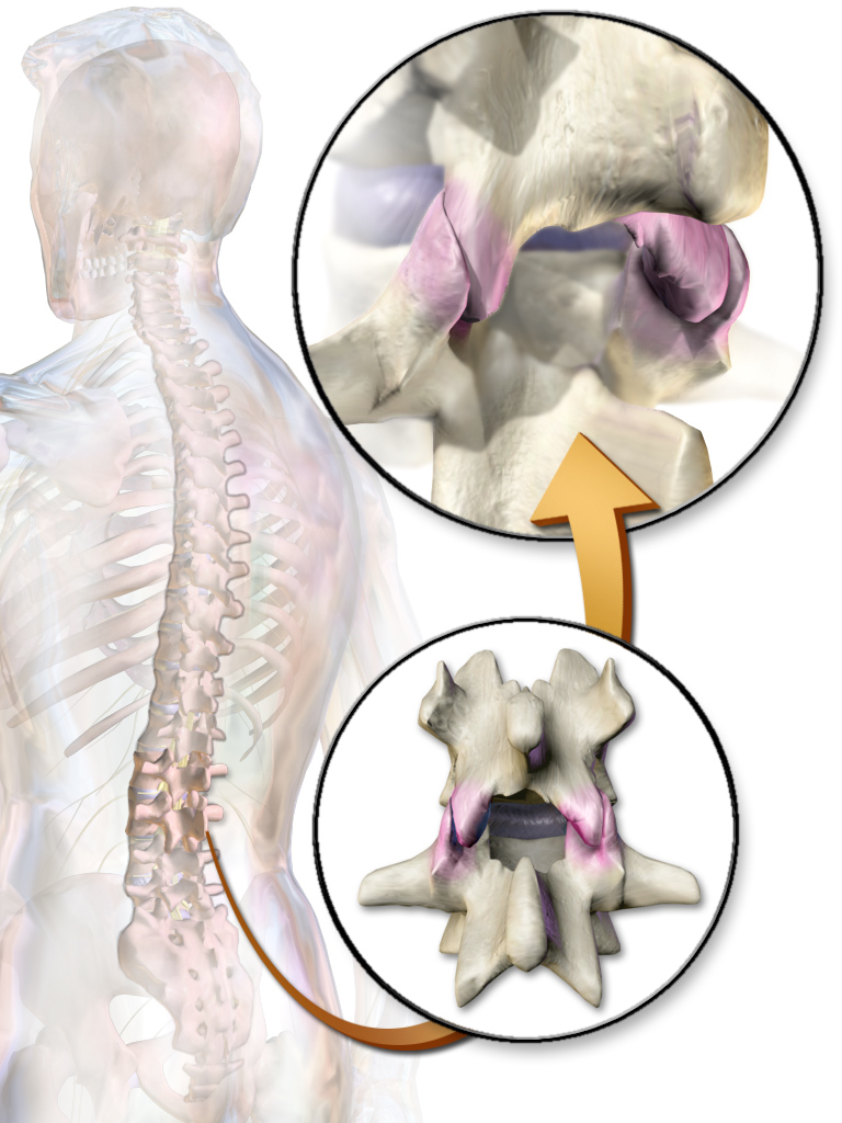
The facet joints (also zygapophysial joints, zygapophyseal, apophyseal, or Z-joints) are a set of synovial, plane joints between the articular processes of two adjacent vertebrae. There are two facet joints in each spinal motion segment and each facet joint is innervated by the recurrent meningeal nerves. Innervation Innervation to the facet joints vary between segments of the spinal, but they are generally innervated by medial branch nerves that come off the dorsal rami. It is thought that these nerves are for primary sensory input, though there is some evidence that they have some motor input local musculature. Within the cervical spine, most joints are innervated by the medial branch nerve (a branch of the dorsal rami) from the same levels. In other words, the facet joint between C4 and C5 vertebral segments is innervated by the C4 and C5 medial branch nerves. However, there are two exceptions: # The facet joint between C2 and C3 is innervated by the third occipital n ... [...More Info...] [...Related Items...] OR: [Wikipedia] [Google] [Baidu] |
Intervertebral Disk
An intervertebral disc (British English), also spelled intervertebral disk (American English), lies between adjacent vertebrae in the vertebral column. Each disc forms a fibrocartilaginous joint (a symphysis), to allow slight movement of the vertebrae, to act as a ligament to hold the vertebrae together, and to function as a shock absorber for the spine. Structure Intervertebral discs consist of an outer fibrous ring, the ''anulus (or annulus) fibrosus disci intervertebralis'', which surrounds an inner gel-like center, the ''nucleus pulposus''. The ''anulus fibrosus'' consists of several layers (laminae) of fibrocartilage made up of both type I and type II collagen. Type I is concentrated toward the edge of the ring, where it provides greater strength. The stiff laminae can withstand compressive forces. The fibrous intervertebral disc contains the ''nucleus pulposus'' and this helps to distribute pressure evenly across the disc. This prevents the development of stress concen ... [...More Info...] [...Related Items...] OR: [Wikipedia] [Google] [Baidu] |
Spinal Canal
In human anatomy, the spinal canal, vertebral canal or spinal cavity is an elongated body cavity enclosed within the dorsal bony arches of the vertebral column, which contains the spinal cord, spinal roots and dorsal root ganglia. It is a process of the dorsal body cavity formed by alignment of the vertebral foramina. Under the vertebral arches, the spinal canal is also covered anteriorly by the posterior longitudinal ligament and posteriorly by the ligamentum flavum. The potential space between these ligaments and the dura mater covering the spinal cord is known as the epidural space. Spinal nerves exit the spinal canal via the intervertebral foramina under the corresponding vertebral pedicles. In humans, the spinal cord gets outgrown by the vertebral column during development into adulthood, and the lower section of the spinal canal is occupied by the filum terminale and a bundle of spinal nerves known as the cauda equina instead of the actual spinal cord, which fi ... [...More Info...] [...Related Items...] OR: [Wikipedia] [Google] [Baidu] |
Nucleus Pulposus
An intervertebral disc (British English), also spelled intervertebral disk (American English), lies between adjacent vertebrae in the vertebral column. Each disc forms a fibrocartilaginous joint (a symphysis), to allow slight movement of the vertebrae, to act as a ligament to hold the vertebrae together, and to function as a shock absorber for the spine. Structure Intervertebral discs consist of an outer fibrous ring, the ''anulus (or annulus) fibrosus disci intervertebralis'', which surrounds an inner gel-like center, the ''nucleus pulposus''. The ''anulus fibrosus'' consists of several layers (laminae) of fibrocartilage made up of both type I and type II collagen. Type I is concentrated toward the edge of the ring, where it provides greater strength. The stiff laminae can withstand compressive forces. The fibrous intervertebral disc contains the ''nucleus pulposus'' and this helps to distribute pressure evenly across the disc. This prevents the development of stress concen ... [...More Info...] [...Related Items...] OR: [Wikipedia] [Google] [Baidu] |
Spinal Nerves
A spinal nerve is a mixed nerve, which carries motor, sensory, and autonomic signals between the spinal cord and the body. In the human body there are 31 pairs of spinal nerves, one on each side of the vertebral column. These are grouped into the corresponding cervical, thoracic, lumbar, sacral and coccygeal regions of the spine. There are eight pairs of cervical nerves, twelve pairs of thoracic nerves, five pairs of lumbar nerves, five pairs of sacral nerves, and one pair of coccygeal nerves. The spinal nerves are part of the peripheral nervous system. Structure Each spinal nerve is a mixed nerve, formed from the combination of nerve root fibers from its dorsal and ventral roots. The dorsal root is the afferent sensory root and carries sensory information to the brain. The ventral root is the efferent motor root and carries motor information from the brain. The spinal nerve emerges from the spinal column through an opening (intervertebral foramen) between adjacen ... [...More Info...] [...Related Items...] OR: [Wikipedia] [Google] [Baidu] |


