|
Mandible
In jawed vertebrates, the mandible (from the Latin ''mandibula'', 'for chewing'), lower jaw, or jawbone is a bone that makes up the lowerand typically more mobilecomponent of the mouth (the upper jaw being known as the maxilla). The jawbone is the skull's only movable, posable bone, sharing Temporomandibular joint, joints with the cranium's temporal bones. The mandible hosts the lower Human tooth, teeth (their depth delineated by the alveolar process). Many muscles attach to the bone, which also hosts nerves (some connecting to the teeth) and blood vessels. Amongst other functions, the jawbone is essential for chewing food. Owing to the Neolithic Revolution, Neolithic advent of agriculture (), human jaws evolved to be Human jaw shrinkage, smaller. Although it is the strongest bone of the facial skeleton, the mandible tends to deform in old age; it is also subject to Mandibular fracture, fracturing. Surgery allows for the removal of jawbone fragments (or its entirety) as well a ... [...More Info...] [...Related Items...] OR: [Wikipedia] [Google] [Baidu] |
Mandibular Fracture
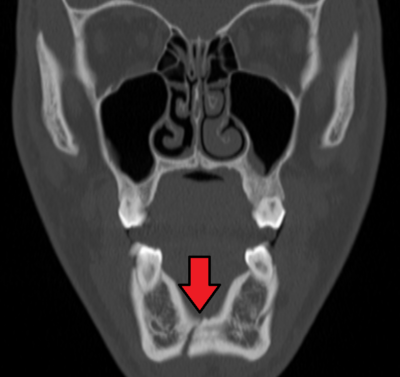
Mandibular fracture, also known as fracture of the jaw, is a break through the mandibular bone. In about 60% of cases the break occurs in two places. It may result in a decreased ability to fully open the mouth. Often the teeth will not feel properly aligned or there may be bleeding of the gums. Mandibular fractures occur most commonly among males in their 30s. Mandibular fractures are typically the result of trauma. This can include a fall onto the chin or a hit from the side. Rarely they may be due to osteonecrosis or tumors in the bone. The most common area of fracture is at the condyle (36%), body (21%), angle (20%) and symphysis (14%). Rarely the fracture may occur at the ramus (3%) or coronoid process (2%). While a diagnosis can occasionally be made with plain X-ray, modern CT scans are more accurate. Immediate surgery is not necessarily required. Occasionally people may go home and follow up for surgery in the next few days. A number of surgical techniques may b ... [...More Info...] [...Related Items...] OR: [Wikipedia] [Google] [Baidu] |
Temporomandibular Joint
In anatomy, the temporomandibular joints (TMJ) are the two joints connecting the jawbone to the skull. It is a bilateral Synovial joint, synovial articulation between the temporal bone of the skull above and the condylar process of mandible below; it is from these bones that its name is derived. The joints are unique in their bilateral function, being connected via the mandible. Structure The main components are the joint capsule, articular disc, mandibular condyles, articular surface of the temporal bone, temporomandibular ligament, stylomandibular ligament, sphenomandibular ligament, and lateral pterygoid muscle. Capsule The articular capsule (capsular ligament) is a thin, loose envelope, attached above to the circumference of the mandibular fossa and the articular tubercle immediately in front; below, to the neck of the condyle of the mandible. Its loose attachment to the neck of the mandible allows for free movement. Articular disc The unique feature of the temporomand ... [...More Info...] [...Related Items...] OR: [Wikipedia] [Google] [Baidu] |
Angle Of The Mandible
__NOTOC__ The angle of the mandible (a.k.a. gonial angle, Masseteric Tuberosity, and Masseteric Insertion) is located at the posterior border at the junction of the lower border of the ramus of the mandible. The angle of the mandible, which may be either inverted or everted, is marked by rough, oblique ridges on each side, for the attachment of the masseter laterally, and the pterygoideus internus ( medial pterygoid muscle) medially; the stylomandibular ligament is attached to the angle between these muscles. The forensic term for the midpoint of the mandibular angle is the gonion. The gonion is a cephalometric landmark located at the lowest, posterior, and lateral point on the angle. This site is at the apex of the maximum curvature of the mandible, where the ascending ramus becomes the body of the mandible. The mandibular angle has been named as a forensic tool for gender determination, but some studies have called into question whether there is any significant sex differ ... [...More Info...] [...Related Items...] OR: [Wikipedia] [Google] [Baidu] |
Pharyngeal Arch
The pharyngeal arches, also known as visceral arches'','' are transient structures seen in the Animal embryonic development, embryonic development of humans and other vertebrates, that are recognisable precursors for many structures. In fish, the arches support the Fish gill, gills and are known as the branchial arches, or gill arches. In the human embryo, the arches are first seen during the fourth week of human embryonic development, development. They appear as a series of outpouchings of mesoderm on both sides of the developing pharynx. The vasculature of the pharyngeal arches are the aortic arches that arise from the aortic sac. Structure In humans and other vertebrates, the pharyngeal arches are derived from all three germ layers (the primary layers of cells that form during embryonic development). Neural crest cells enter these arches where they contribute to features of the skull and facial skeleton such as bone and cartilage. However, the existence of pharyngeal structu ... [...More Info...] [...Related Items...] OR: [Wikipedia] [Google] [Baidu] |
Skull
The skull, or cranium, is typically a bony enclosure around the brain of a vertebrate. In some fish, and amphibians, the skull is of cartilage. The skull is at the head end of the vertebrate. In the human, the skull comprises two prominent parts: the neurocranium and the facial skeleton, which evolved from the first pharyngeal arch. The skull forms the frontmost portion of the axial skeleton and is a product of cephalization and vesicular enlargement of the brain, with several special senses structures such as the eyes, ears, nose, tongue and, in fish, specialized tactile organs such as barbels near the mouth. The skull is composed of three types of bone: cranial bones, facial bones and ossicles, which is made up of a number of fused flat and irregular bones. The cranial bones are joined at firm fibrous junctions called sutures and contains many foramina, fossae, processes, and sinuses. In zoology, the openings in the skull are called fenestrae, the most ... [...More Info...] [...Related Items...] OR: [Wikipedia] [Google] [Baidu] |
Human Skull
The skull, or cranium, is typically a bony enclosure around the brain of a vertebrate. In some fish, and amphibians, the skull is of cartilage. The skull is at the head end of the vertebrate. In the human, the skull comprises two prominent parts: the neurocranium and the facial skeleton, which evolved from the first pharyngeal arch. The skull forms the frontmost portion of the axial skeleton and is a product of cephalization and vesicular enlargement of the brain, with several special senses structures such as the eyes, ears, nose, tongue and, in fish, specialized tactile organs such as barbels near the mouth. The skull is composed of three types of bone: cranial bones, facial bones and ossicles, which is made up of a number of fused flat and irregular bones. The cranial bones are joined at firm fibrous junctions called sutures and contains many foramina, fossae, processes, and sinuses. In zoology, the openings in the skull are called fenestrae, the ... [...More Info...] [...Related Items...] OR: [Wikipedia] [Google] [Baidu] |
Alveolar Process
The alveolar process () is the portion of bone containing the tooth sockets on the jaw bones (in humans, the maxilla and the mandible). The alveolar process is covered by gums within the mouth, terminating roughly along the line of the mandibular canal. Partially comprising compact bone, it is penetrated by many small openings for blood vessels and connective fibres. The bone is of clinical, phonetic and forensic significance. Terminology The term ''alveolar'' () ('hollow') refers to the cavities of the tooth sockets, known as dental alveoli. The alveolar process is also called the ''alveolar bone'' or ''alveolar ridge''. In phonetics, the term refers more specifically to the ridges on the inside of the mouth which can be felt with the tongue, either on roof of the mouth between the upper teeth and the hard palate or on the bottom of the mouth behind the lower teeth. The curved portion of the process is referred to as the alveolar arch. The alveolar bone proper, also ca ... [...More Info...] [...Related Items...] OR: [Wikipedia] [Google] [Baidu] |
Maxilla
In vertebrates, the maxilla (: maxillae ) is the upper fixed (not fixed in Neopterygii) bone of the jaw formed from the fusion of two maxillary bones. In humans, the upper jaw includes the hard palate in the front of the mouth. The two maxillary bones are fused at the intermaxillary suture, forming the anterior nasal spine. This is similar to the mandible (lower jaw), which is also a fusion of two mandibular bones at the mandibular symphysis. The mandible is the movable part of the jaw. Anatomy Structure The maxilla is a paired bone - the two maxillae unite with each other at the intermaxillary suture. The maxilla consists of: * The body of the maxilla: pyramid-shaped; has an orbital, a nasal, an infratemporal, and a facial surface; contains the maxillary sinus. * Four processes: ** the zygomatic process ** the frontal process ** the alveolar process ** the palatine process It has three surfaces: * the anterior, posterior, medial Features of the maxilla include: * t ... [...More Info...] [...Related Items...] OR: [Wikipedia] [Google] [Baidu] |
Facial Skeleton
The facial skeleton comprises the ''facial bones'' that may attach to build a portion of the skull. The remainder of the skull is the neurocranium. In human anatomy and development, the facial skeleton is sometimes called the ''membranous viscerocranium'', which comprises the mandible and dermatocranial elements that are not part of the braincase. Structure In the human skull, the facial skeleton consists of fourteen bones in the face: * Inferior turbinal (2) * Lacrimal bones (2) * Mandible * Maxilla (2) * Nasal bones (2) * Palatine bones (2) * Vomer * Zygomatic bones (2) Variations Elements of the ''cartilaginous viscerocranium'' (i.e., splanchnocranial elements), such as the hyoid bone, are sometimes considered part of the facial skeleton. The ethmoid bone (or a part of it) and also the sphenoid bone are sometimes included, but otherwise considered part of the neurocranium. Because the maxillary bones are fused, they are often collectively listed as only one bone. ... [...More Info...] [...Related Items...] OR: [Wikipedia] [Google] [Baidu] |
Mouth
A mouth also referred to as the oral is the body orifice through which many animals ingest food and animal communication#Auditory, vocalize. The body cavity immediately behind the mouth opening, known as the oral cavity (or in Latin), is also the first part of the alimentary canal, which leads to the pharynx and the gullet. In tetrapod vertebrates, the mouth is bounded on the outside by the lips and cheeks — thus the oral cavity is also known as the buccal cavity (from Latin ', meaning "cheek") — and contains the tongue on the inside. Except for some groups like birds and lissamphibians, vertebrates usually have teeth in their mouths, although some fish species have pharyngeal teeth instead of oral teeth. Most bilaterian phylum, phyla, including arthropods, molluscs and chordates, have a two-opening gut tube with a mouth at one end and an anus at the other. Which end forms first in ontogeny is a criterion used to classify bilaterian animals into protostomes and deuterostomes ... [...More Info...] [...Related Items...] OR: [Wikipedia] [Google] [Baidu] |
Middle Ear
The middle ear is the portion of the ear medial to the eardrum, and distal to the oval window of the cochlea (of the inner ear). The mammalian middle ear contains three ossicles (malleus, incus, and stapes), which transfer the vibrations of the eardrum into waves in the fluid and membranes of the inner ear. The hollow space of the middle ear is also known as the tympanic cavity and is surrounded by the tympanic part of the temporal bone. The auditory tube (also known as the Eustachian tube or the pharyngotympanic tube) joins the tympanic cavity with the nasal cavity ( nasopharynx), allowing pressure to equalize between the middle ear and throat. The primary function of the middle ear is to efficiently transfer acoustic energy from compression waves in air to fluid–membrane waves within the cochlea. Structure Ossicles The middle ear contains three tiny bones known as the ossicles: '' malleus'', ''incus'', and '' stapes''. The ossicles were given their Latin names f ... [...More Info...] [...Related Items...] OR: [Wikipedia] [Google] [Baidu] |


