|
List Of Neurological Research Methods
There are numerous types of research methods used when conducting neurological research, all with the purpose of trying to view the activity that occurs within the brain during a certain activity or behavior. The disciplines within which these methods are used is quite broad, ranging from psychology to neuroscience to biomedical engineering to sociology. The following is a list of neuroimaging methods: *Electroencephalography, Electroencephalography (EEG) :*Quantitative electroencephalography, Quantitative electroencephalography (QEEG) :*Stereoelectroencephalography, Stereoelectroencephalography (SEEG) *Functional magnetic resonance imaging, Functional magnetic resonance imaging (fMRI) *Magnetoencephalography, Magnetoencephalography (MEG) *Near-infrared spectroscopy, Near-infrared spectroscopy (NIRS) *Positron emission tomography, Positron emission tomography (PET) *Single-unit recording *Transcranial direct current stimulation, Transcranial direct-current stimulation (TDCS) *Transc ... [...More Info...] [...Related Items...] OR: [Wikipedia] [Google] [Baidu] |
Psychology
Psychology is the scientific study of mind and behavior. Psychology includes the study of conscious and unconscious phenomena, including feelings and thoughts. It is an academic discipline of immense scope, crossing the boundaries between the natural and social sciences. Psychologists seek an understanding of the emergent properties of brains, linking the discipline to neuroscience. As social scientists, psychologists aim to understand the behavior of individuals and groups.Fernald LD (2008)''Psychology: Six perspectives'' (pp.12–15). Thousand Oaks, CA: Sage Publications.Hockenbury & Hockenbury. Psychology. Worth Publishers, 2010. Ψ (''psi''), the first letter of the Greek word ''psyche'' from which the term psychology is derived (see below), is commonly associated with the science. A professional practitioner or researcher involved in the discipline is called a psychologist. Some psychologists can also be classified as behavioral or cognitive scientists. Some ps ... [...More Info...] [...Related Items...] OR: [Wikipedia] [Google] [Baidu] |
Near-infrared Spectroscopy
Near-infrared spectroscopy (NIRS) is a spectroscopic method that uses the near-infrared region of the electromagnetic spectrum (from 780 nm to 2500 nm). Typical applications include medical and physiological diagnostics and research including blood sugar, pulse oximetry, functional neuroimaging, sports medicine, elite sports training, ergonomics, rehabilitation, neonatal research, brain computer interface, urology (bladder contraction), and neurology (neurovascular coupling). There are also applications in other areas as well such as pharmaceutical, food and agrochemical quality control, atmospheric chemistry, combustion research and astronomy. Theory Near-infrared spectroscopy is based on molecular overtone and combination vibrations. Such transitions are forbidden by the selection rules of quantum mechanics. As a result, the molar absorptivity in the near-IR region is typically quite small. (NIR absorption bands are typically 10–100 times weaker than the ... [...More Info...] [...Related Items...] OR: [Wikipedia] [Google] [Baidu] |
Functional Neuroimaging
Functional neuroimaging is the use of neuroimaging technology to measure an aspect of brain function, often with a view to understanding the relationship between activity in certain brain areas and specific mental functions. It is primarily used as a research tool in cognitive neuroscience, cognitive psychology, neuropsychology, and social neuroscience. Overview Common methods of functional neuroimaging include * Positron emission tomography (PET) * Functional magnetic resonance imaging (fMRI) * Electroencephalography (EEG) * Magnetoencephalography (MEG) * Functional near-infrared spectroscopy (fNIRS) * Single-photon emission computed tomography (SPECT) * Functional ultrasound imaging (fUS) PET, fMRI, fNIRS and fUS can measure localized changes in cerebral blood flow related to neural activity. These changes are referred to as ''activations''. Regions of the brain which are activated when a subject performs a particular task may play a role in the neural computations which ... [...More Info...] [...Related Items...] OR: [Wikipedia] [Google] [Baidu] |
Transcranial Magnetic Stimulation
Transcranial magnetic stimulation (TMS) is a noninvasive form of brain stimulation in which a changing magnetic field is used to induce an electric current at a specific area of the brain through electromagnetic induction. An electric pulse generator, or stimulator, is connected to a magnetic coil connected to the scalp. The stimulator generates a changing electric current within the coil which creates a varying magnetic field, inducing a current within a region in the brain itself.NICE. January 201Transcranial magnetic stimulation for treating and preventing migraine/ref>Michael Craig Miller for Harvard Health Publications. July 26, 201Magnetic stimulation: a new approach to treating depression?/ref> TMS has shown diagnostic and therapeutic potential in the central nervous system with a wide variety of disease states in neurology and mental health, with research still evolving. Adverse effects of TMS appear rare and include fainting and seizure. Other potential issues inclu ... [...More Info...] [...Related Items...] OR: [Wikipedia] [Google] [Baidu] |
Transcranial Direct Current Stimulation
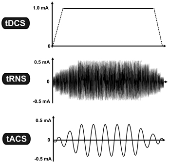
Transcranial direct current stimulation (tDCS) is a form of neuromodulation that uses constant, low direct current delivered via electrodes on the head. It was originally developed to help patients with brain injuries or neuropsychiatric conditions such as major depressive disorder. It can be contrasted with cranial electrotherapy stimulation, which generally uses alternating current the same way, as well as transcranial magnetic stimulation. Research shows increasing evidence for tDCS as a treatment for depression. There is mixed evidence about whether tDCS is useful for cognitive enhancement in healthy people. There is no strong evidence that tDCS is useful for memory deficits in Parkinson's disease and Alzheimer's disease, non-neuropathic pain, nor for improving arm or leg functioning and muscle strength in people recovering from a stroke. There is emerging supportive evidence for tDCS in the management of schizophreniaespecially for negative symptoms. Efficacy Depression ... [...More Info...] [...Related Items...] OR: [Wikipedia] [Google] [Baidu] |
Single-unit Recording
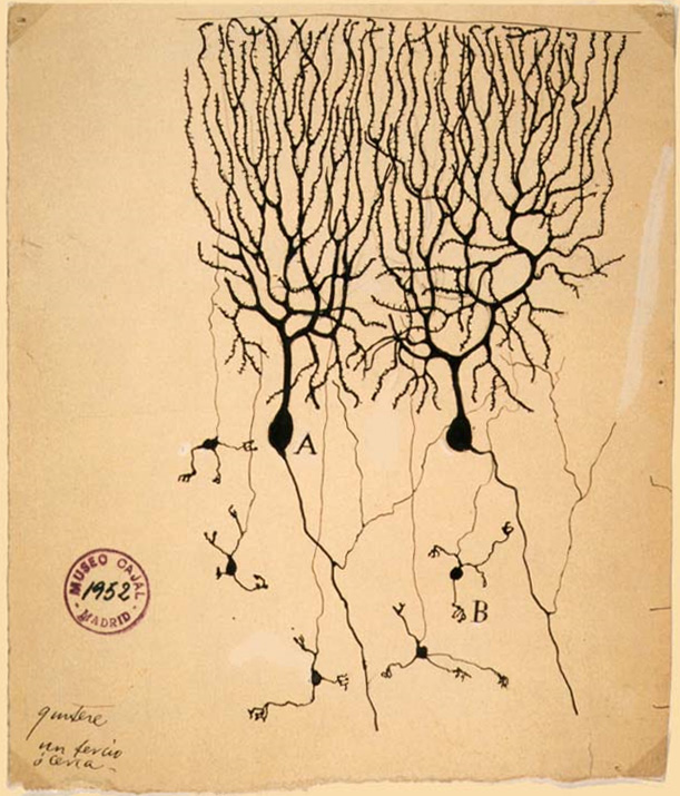
In neuroscience, single-unit recordings provide a method of measuring the electro-physiological responses of a single neuron using a microelectrode system. When a neuron generates an action potential, the signal propagates down the neuron as a current which flows in and out of the cell through excitable membrane regions in the soma and axon. A microelectrode is inserted into the brain, where it can record the rate of change in voltage with respect to time. These microelectrodes must be fine-tipped, high-impedance conductors; they are primarily glass micro-pipettes, metal microelectrodes made of platinum, tungsten, iridium or even iridium oxide. Microelectrodes can be carefully placed close to the cell membrane, allowing the ability to record extracellularly. Single-unit recordings are widely used in cognitive science, where it permits the analysis of human cognition and cortical mapping. This information can then be applied to brain–machine interface (BMI) technologies for ... [...More Info...] [...Related Items...] OR: [Wikipedia] [Google] [Baidu] |
Positron Emission Tomography
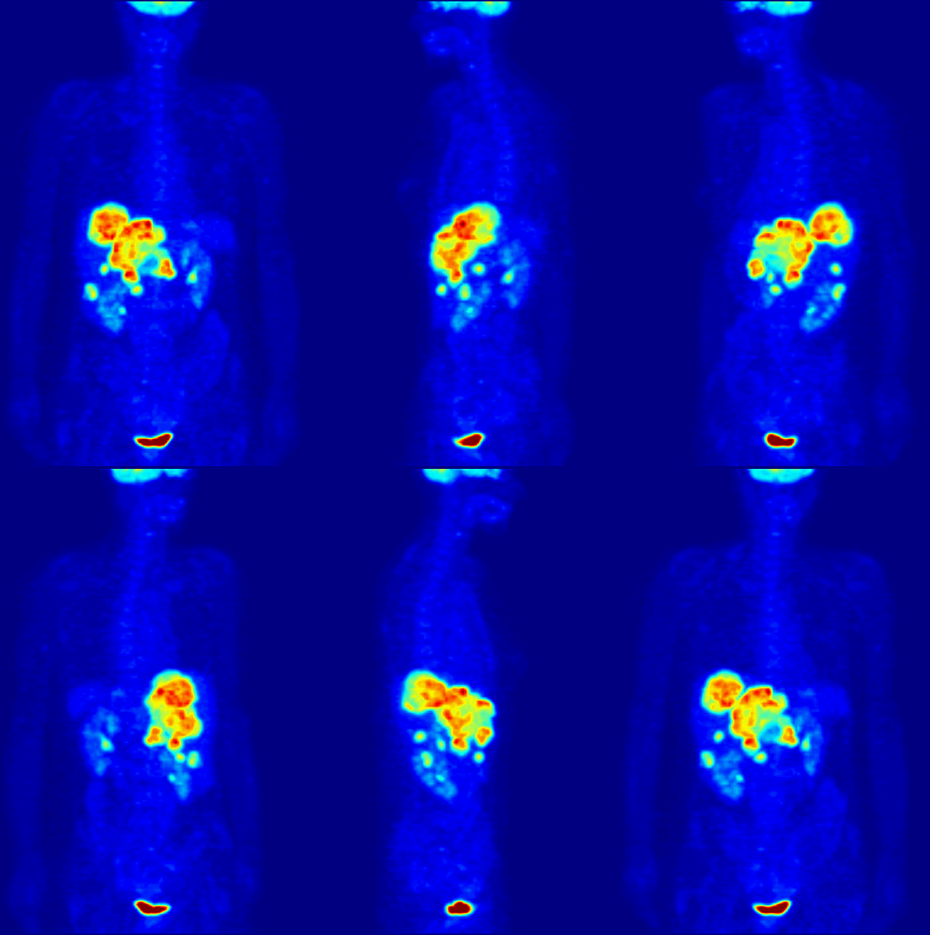
Positron emission tomography (PET) is a functional imaging technique that uses radioactive substances known as radiotracers to visualize and measure changes in metabolic processes, and in other physiological activities including blood flow, regional chemical composition, and absorption. Different tracers are used for various imaging purposes, depending on the target process within the body. For example: * Fluorodeoxyglucose ( 18F">sup>18FDG or FDG) is commonly used to detect cancer; * 18Fodium fluoride">sup>18Fodium fluoride (Na18F) is widely used for detecting bone formation; * Oxygen-15 (15O) is sometimes used to measure blood flow. PET is a common imaging technique, a medical scintillography technique used in nuclear medicine. A radiopharmaceutical – a radioisotope attached to a drug – is injected into the body as a radioactive tracer, tracer. When the radiopharmaceutical undergoes beta plus decay, a positron is emitted, and when the positron interacts with an or ... [...More Info...] [...Related Items...] OR: [Wikipedia] [Google] [Baidu] |
Magnetoencephalography
Magnetoencephalography (MEG) is a functional neuroimaging technique for mapping brain activity by recording magnetic fields produced by electrical currents occurring naturally in the brain, using very sensitive magnetometers. Arrays of SQUIDs (superconducting quantum interference devices) are currently the most common magnetometer, while the SERF (spin exchange relaxation-free) magnetometer is being investigated for future machines. Applications of MEG include basic research into perceptual and cognitive brain processes, localizing regions affected by pathology before surgical removal, determining the function of various parts of the brain, and neurofeedback. This can be applied in a clinical setting to find locations of abnormalities as well as in an experimental setting to simply measure brain activity. History MEG signals were first measured by University of Illinois physicist David Cohen in 1968, before the availability of the SQUID, using a copper induction coil as the ... [...More Info...] [...Related Items...] OR: [Wikipedia] [Google] [Baidu] |
Neuroscience
Neuroscience is the science, scientific study of the nervous system (the brain, spinal cord, and peripheral nervous system), its functions and disorders. It is a Multidisciplinary approach, multidisciplinary science that combines physiology, anatomy, molecular biology, developmental biology, cytology, psychology, physics, computer science, chemistry, medicine, statistics, and Mathematical Modeling, mathematical modeling to understand the fundamental and emergent properties of neurons, glia and neural circuits. The understanding of the biological basis of learning, memory, behavior, perception, and consciousness has been described by Eric Kandel as the "epic challenge" of the Biology, biological sciences. The scope of neuroscience has broadened over time to include different approaches used to study the nervous system at different scales. The techniques used by neuroscientists have expanded enormously, from molecular biology, molecular and cell biology, cellular studies of indivi ... [...More Info...] [...Related Items...] OR: [Wikipedia] [Google] [Baidu] |
Functional Magnetic Resonance Imaging
Functional magnetic resonance imaging or functional MRI (fMRI) measures brain activity by detecting changes associated with blood flow. This technique relies on the fact that cerebral blood flow and neuronal activation are coupled. When an area of the brain is in use, blood flow to that region also increases. The primary form of fMRI uses the blood-oxygen-level dependent (BOLD) contrast, discovered by Seiji Ogawa in 1990. This is a type of specialized brain and body scan used to map neural activity in the brain or spinal cord of humans or other animals by imaging the change in blood flow ( hemodynamic response) related to energy use by brain cells. Since the early 1990s, fMRI has come to dominate brain mapping research because it does not involve the use of injections, surgery, the ingestion of substances, or exposure to ionizing radiation. This measure is frequently corrupted by noise from various sources; hence, statistical procedures are used to extract the underlying sig ... [...More Info...] [...Related Items...] OR: [Wikipedia] [Google] [Baidu] |
Stereoelectroencephalography
Stereoelectroencephalography (SEEG) is the practice of recording electroencephalographic signals via depth electrodes (electrodes surgically implanted into the brain tissue). It may be used in patients with epilepsy not responding to medical treatment, and who are potential candidates to receive brain surgery in order to control seizures. This technique was introduced in the diagnostic work up of patients with epilepsy by the group of the S. Anne Hospital, Paris, France, in the second half of the 20th century. Intracerebral electrodes are placed within the desired brain areas to record the electrical activity during epileptic seizures, thus contributing to define with accuracy the boundaries of the "epileptogenic zone", i.e. the area of brain generating the seizures which should be eventually surgically resected to achieve freedom from epileptic seizures. Potential risks of the procedure, accounting for less than 1% of cases, include brain hemorrhage and infection, which can lead t ... [...More Info...] [...Related Items...] OR: [Wikipedia] [Google] [Baidu] |


