|
List Of MeSH Codes (A14)
The following is a partial list of the "A" codes for Medical Subject Headings (MeSH), as defined by the United States National Library of Medicine (NLM). This list continues the information at List of MeSH codes (A13). Codes following these are found at List of MeSH codes (A15). For other MeSH codes, see List of MeSH codes. The source for this content is the set o2006 MeSH Treesfrom the NLM. – stomatognathic system – cheek – facial muscles – jaw * – alveolar process * – tooth socket * – dental arch * – Human mandible, mandible * – chin * – mandibular condyle * – maxilla * – palate * – palate, hard – masticatory muscles * – masseter muscle * – pterygoid muscles (other), pterygoid muscles * – temporal muscle – mouth * – dentition * – dentition, mixed * – dentition, permanent * – dentition, primary * – Diastema (dentistry), diastema * – periodontium * – alveolar process * – tooth socket * � ... [...More Info...] [...Related Items...] OR: [Wikipedia] [Google] [Baidu] |
Medical Subject Headings
Medical Subject Headings (MeSH) is a comprehensive controlled vocabulary for the purpose of indexing journal articles and books in the life sciences. It serves as a thesaurus that facilitates searching. Created and updated by the United States National Library of Medicine (NLM), it is used by the MEDLINE/ PubMed article database and by NLM's catalog of book holdings. MeSH is also used by ClinicalTrials.gov registry to classify which diseases are studied by trials registered in ClinicalTrials. MeSH was introduced in the 1960s, with the NLM's own index catalogue and the subject headings of the Quarterly Cumulative Index Medicus (1940 edition) as precursors. The yearly printed version of MeSH was discontinued in 2007; MeSH is now available only online. It can be browsed and downloaded free of charge through PubMed. Originally in English, MeSH has been translated into numerous other languages and allows retrieval of documents from different origins. Structure MeSH vocabulary is ... [...More Info...] [...Related Items...] OR: [Wikipedia] [Google] [Baidu] |
Masticatory Muscles
There are four classical muscles of mastication. During mastication, three muscles of mastication (''musculi masticatorii'') are responsible for adduction of the jaw, and one (the lateral pterygoid) helps to abduct it. All four move the jaw laterally. Other muscles, usually associated with the hyoid, such as the mylohyoid muscle, are responsible for opening the jaw in addition to the lateral pterygoid. Structure The muscles are: * The masseter (composed of the superficial and deep head) * The temporalis (the sphenomandibularis is considered a part of the temporalis by some sources, and a distinct muscle by others) * The medial pterygoid * The lateral pterygoid In humans, the mandible, or lower jaw, is connected to the temporal bone of the skull via the temporomandibular joint. This is an extremely complex joint which permits movement in all planes. The muscles of mastication originate on the skull and insert into the mandible, thereby allowing for jaw movements during ... [...More Info...] [...Related Items...] OR: [Wikipedia] [Google] [Baidu] |
Tooth
A tooth ( : teeth) is a hard, calcified structure found in the jaws (or mouths) of many vertebrates and used to break down food. Some animals, particularly carnivores and omnivores, also use teeth to help with capturing or wounding prey, tearing food, for defensive purposes, to intimidate other animals often including their own, or to carry prey or their young. The roots of teeth are covered by gums. Teeth are not made of bone, but rather of multiple tissues of varying density and hardness that originate from the embryonic germ layer, the ectoderm. The general structure of teeth is similar across the vertebrates, although there is considerable variation in their form and position. The teeth of mammals have deep roots, and this pattern is also found in some fish, and in crocodilians. In most teleost fish, however, the teeth are attached to the outer surface of the bone, while in lizards they are attached to the inner surface of the jaw by one side. In cartilaginous fi ... [...More Info...] [...Related Items...] OR: [Wikipedia] [Google] [Baidu] |
Periodontal Ligament
The periodontal ligament, commonly abbreviated as the PDL, is a group of specialized connective tissue fibers that essentially attach a tooth to the alveolar bone within which it sits. It inserts into root cementum one side and onto alveolar bone on the other. Structure The PDL consists of principal fibres, loose connective tissue, blast and clast cells, oxytalan fibres and Cell Rest of Malassez. Alveolodental ligament The main principal fiber group is the alveolodental ligament, which consists of five fiber subgroups: alveolar crest, horizontal, oblique, apical, and interradicular on multirooted teeth. Principal fibers other than the alveolodental ligament are the transseptal fibers. All these fibers help the tooth withstand the naturally substantial compressive forces that occur during chewing and remain embedded in the bone. The ends of the principal fibers that are within either cementum or alveolar bone proper are considered Sharpey fibers. * Alveolar crest fibers ( ... [...More Info...] [...Related Items...] OR: [Wikipedia] [Google] [Baidu] |
Gingiva
The gums or gingiva (plural: ''gingivae'') consist of the mucosal tissue that lies over the mandible and maxilla inside the mouth. Gum health and disease can have an effect on general health. Structure The gums are part of the soft tissue lining of the mouth. They surround the teeth and provide a seal around them. Unlike the soft tissue linings of the lips and cheeks, most of the gums are tightly bound to the underlying bone which helps resist the friction of food passing over them. Thus when healthy, it presents an effective barrier to the barrage of periodontal insults to deeper tissue. Healthy gums are usually coral pink in light skinned people, and may be naturally darker with melanin pigmentation. Changes in color, particularly increased redness, together with swelling and an increased tendency to bleed, suggest an inflammation that is possibly due to the accumulation of bacterial plaque. Overall, the clinical appearance of the tissue reflects the underlying histology ... [...More Info...] [...Related Items...] OR: [Wikipedia] [Google] [Baidu] |
Dental Cementum
Cementum is a specialized calcified substance covering the root of a tooth. The cementum is the part of the periodontium that attaches the teeth to the alveolar bone by anchoring the periodontal ligament.Illustrated Dental Embryology, Histology, and Anatomy, Bath-Balogh and Fehrenbach, Elsevier, 2011, page 170. Structure The cells of cementum are the entrapped cementoblasts, the cementocytes. Each cementocyte lies in its lacuna, similar to the pattern noted in bone. These lacunae also have canaliculi or canals. Unlike those in bone, however, these canals in cementum do not contain nerves, nor do they radiate outward. Instead, the canals are oriented toward the periodontal ligament and contain cementocytic processes that exist to diffuse nutrients from the ligament because it is vascularized. After the apposition of cementum in layers, the cementoblasts that do not become entrapped in cementum line up along the cemental surface along the length of the outer covering of the perio ... [...More Info...] [...Related Items...] OR: [Wikipedia] [Google] [Baidu] |
Periodontium
The periodontium is the specialized tissues that both surround and support the teeth, maintaining them in the maxillary and mandibular bones. The word comes from the Greek terms περί ''peri''-, meaning "around" and -''odont'', meaning "tooth". Literally taken, it means that which is "around the tooth". Periodontics is the dental specialty that relates specifically to the care and maintenance of these tissues. It provides the support necessary to maintain teeth in function. It consists of four principal components, namely: * Gingiva * Periodontal ligament The periodontal ligament, commonly abbreviated as the PDL, is a group of specialized connective tissue fibers that essentially attach a tooth to the alveolar bone within which it sits. It inserts into root cementum one side and onto alveolar ... (PDL) * Cementum * Alveolar bone proper Each of these components is distinct in location, architecture, and biochemical properties, which adapt during the life of the struc ... [...More Info...] [...Related Items...] OR: [Wikipedia] [Google] [Baidu] |
Diastema (dentistry)
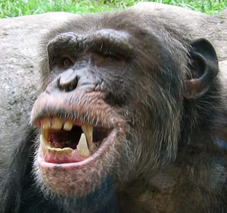
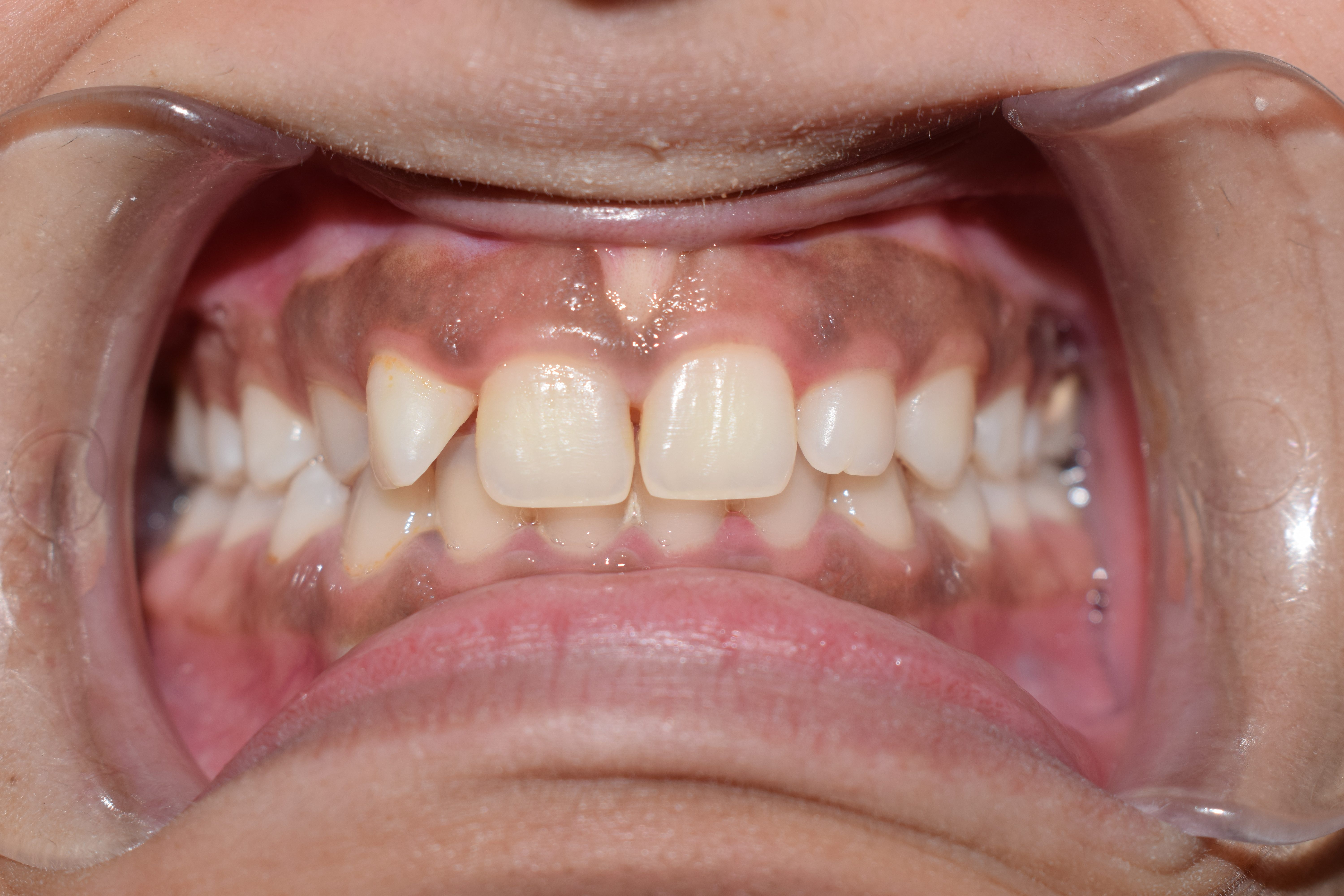
A diastema (plural diastemata, from Greek διάστημα, space) is a space or gap between two teeth. Many species of mammals have diastemata as a normal feature, most commonly between the incisors and molars. More colloquially, the condition may be referred to as gap teeth or tooth gap. In humans, the term is most commonly applied to an open space between the upper incisors (front teeth). It happens when there is an unequal relationship between the size of the teeth and the jaw. Diastemata are common for children and can exist in adult teeth as well. In humans Causes 1. Oversized Labial Frenulum: Diastema is sometimes caused or exacerbated by the action of a labial frenulum (the tissue connecting the lip to the gum), causing high mucosal attachment and less attached keratinized tissue. This is more prone to recession or by tongue thrusting, which can push the teeth apart. 2. Periodontal Disease: Periodontal disease, also known as gum disease, can result in bone los ... [...More Info...] [...Related Items...] OR: [Wikipedia] [Google] [Baidu] |
Dentition, Primary
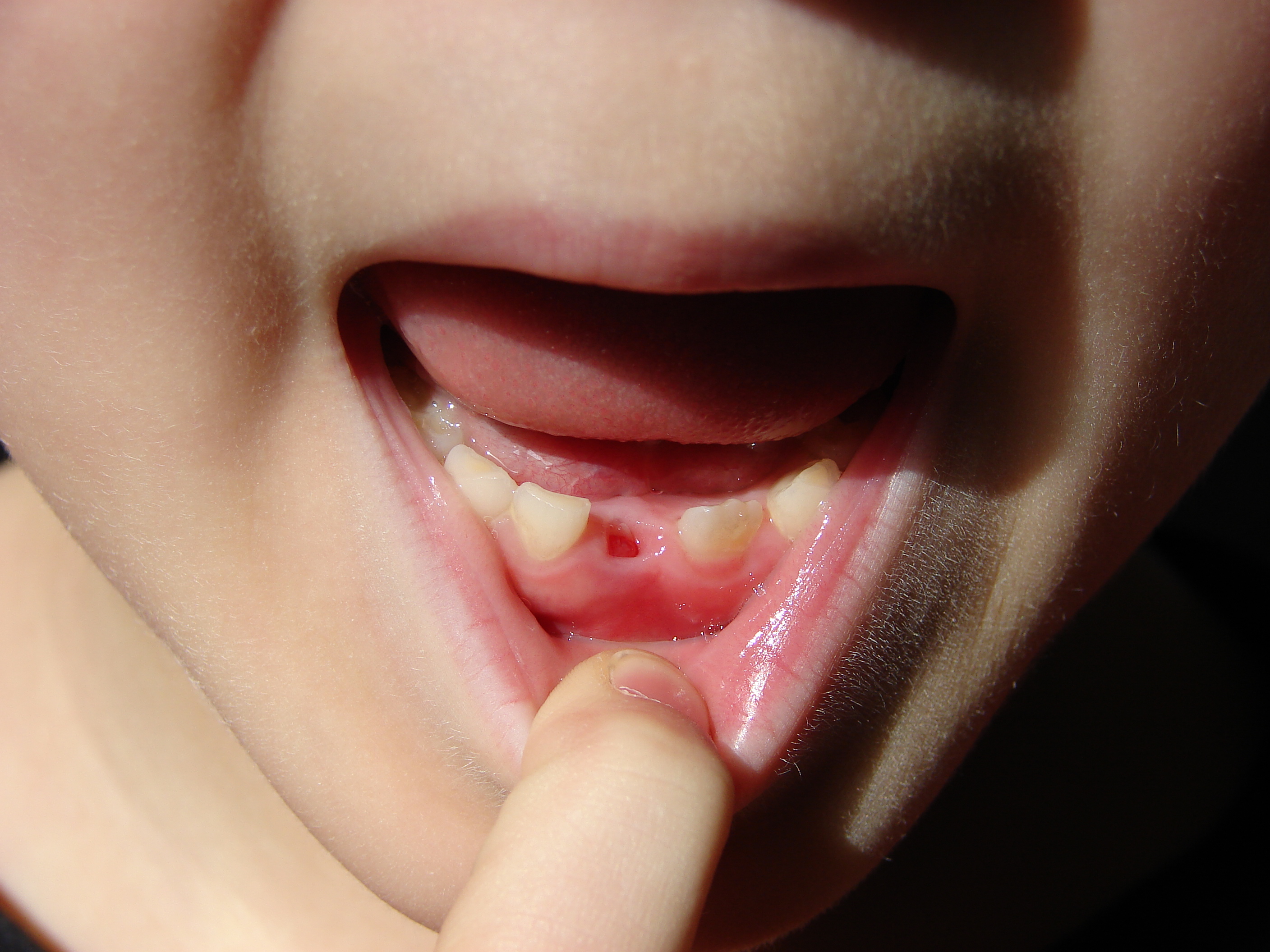
Deciduous teeth or primary teeth, also informally known as baby teeth, milk teeth, or temporary teeth,Illustrated Dental Embryology, Histology, and Anatomy, Bath-Balogh and Fehrenbach, Elsevier, 2011, page 255 are the first set of teeth in the growth and development of humans and other diphyodonts, which include most mammals but not elephants, kangaroos, or manatees which are polyphyodonts. Deciduous teeth develop during the embryonic stage of development and erupt (break through the gums and become visible in the mouth) during infancy. They are usually lost and replaced by permanent teeth, but in the absence of their permanent replacements, they can remain functional for many years into adulthood. Development Formation Primary teeth start to form during the embryonic phase of human life. The development of primary teeth starts at the sixth week of tooth development as the dental lamina. This process starts at the midline and then spreads back into the posterior region. By ... [...More Info...] [...Related Items...] OR: [Wikipedia] [Google] [Baidu] |
Dentition, Permanent
Permanent teeth or adult teeth are the second set of teeth formed in diphyodont mammals. In humans and old world simians, there are thirty-two permanent teeth, consisting of six maxillary and six mandibular molars, four maxillary and four mandibular premolars, two maxillary and two mandibular canines, four maxillary and four mandibular incisors. Timeline The first permanent tooth usually appears in the mouth at around six years of age, and the mouth will then be in a transition time with both primary (or deciduous dentition) teeth and permanent teeth during the mixed dentition period until the last primary tooth is lost or shed. The first of the permanent teeth to erupt are the permanent first molars, right behind the last 'milk' molars of the primary dentition. These first permanent molars are important for the correct development of a permanent dentition. Up to thirteen years of age, 28 of the 32 permanent teeth will appear. The full permanent dentition is completed much later ... [...More Info...] [...Related Items...] OR: [Wikipedia] [Google] [Baidu] |
Dentition
Dentition pertains to the development of teeth and their arrangement in the mouth. In particular, it is the characteristic arrangement, kind, and number of teeth in a given species at a given age. That is, the number, type, and morpho-physiology (that is, the relationship between the shape and form of the tooth in question and its inferred function) of the teeth of an animal. Animals whose teeth are all of the same type, such as most non-mammalian vertebrates, are said to have '' homodont'' dentition, whereas those whose teeth differ morphologically are said to have ''heterodont'' dentition. The dentition of animals with two successions of teeth (deciduous, permanent) is referred to as ''diphyodont'', while the dentition of animals with only one set of teeth throughout life is ''monophyodont''. The dentition of animals in which the teeth are continuously discarded and replaced throughout life is termed ''polyphyodont''. The dentition of animals in which the teeth are set in soc ... [...More Info...] [...Related Items...] OR: [Wikipedia] [Google] [Baidu] |
Mouth
In animal anatomy, the mouth, also known as the oral cavity, or in Latin cavum oris, is the opening through which many animals take in food and issue vocal sounds. It is also the cavity lying at the upper end of the alimentary canal, bounded on the outside by the lips and inside by the pharynx. In tetrapods, it contains the tongue and, except for some like birds, teeth. This cavity is also known as the buccal cavity, from the Latin ''bucca'' ("cheek"). Some animal phylum, phyla, including arthropods, molluscs and chordates, have a complete digestive system, with a mouth at one end and an anus at the other. Which end forms first in ontogeny is a criterion used to classify bilaterian animals into protostomes and deuterostomes. Development In the first Multicellular organism, multicellular animals, there was probably no mouth or gut and food particles were engulfed by the cells on the exterior surface by a process known as endocytosis. The particles became enclosed in vacuoles in ... [...More Info...] [...Related Items...] OR: [Wikipedia] [Google] [Baidu] |


