|
List Of MeSH Codes (A13)
The following is a partial list of the "A" codes for Medical Subject Headings (MeSH), as defined by the United States National Library of Medicine (NLM). This list continues the information at List of MeSH codes (A12). Codes following these are found at List of MeSH codes (A14). For other MeSH codes, see List of MeSH codes. The source for this content is the set o2006 MeSH Treesfrom the NLM. – animal structures – air sacs – anal sacs – beak – bursa of fabricius – cloaca – comb and wattles – corpora allata – crop, avian – egg shell – electric organ – embryo, nonmammalian * – chick embryo * – chorioallantoic membrane – fat body – feathers – forelimb * – carpus, animal * – wing – ganglia, invertebrate – gills – harderian gland – hemolymph – hepatopancreas – hindlimb * – stifle * – tarsus, animal – hoof and claw – horns * – antlers – interrenal gland – malpig ... [...More Info...] [...Related Items...] OR: [Wikipedia] [Google] [Baidu] |
Medical Subject Headings
Medical Subject Headings (MeSH) is a comprehensive controlled vocabulary for the purpose of indexing Academic journal, journal articles and books in the Life science, life sciences. It serves as a thesaurus of index terms that facilitates searching. Created and updated by the United States National Library of Medicine (NLM), it is used by the MEDLINE/PubMed article database and by NLM's catalog of book holdings. MeSH is also used by ClinicalTrials.gov registry to classify which diseases are studied by trials registered in ClinicalTrials. MeSH was introduced in the 1960s, with the NLM's own index catalogue and the subject headings of the Quarterly Cumulative Index Medicus (1940 edition) as precursors. The yearly printed version of MeSH was discontinued in 2007; MeSH is now available only online. It can be browsed and downloaded free of charge through PubMed. Originally in English, MeSH has been translated into numerous other languages and allows retrieval of documents from differ ... [...More Info...] [...Related Items...] OR: [Wikipedia] [Google] [Baidu] |
Chorioallantoic Membrane
The chorioallantoic membrane (CAM), also known as the chorioallantois, is a highly vascularized membrane found in the eggs of certain amniotes like birds and reptiles. It is formed by the fusion of the mesodermal layers of two extra-embryonic membranes – the chorion and the allantois. It is the avian homologue of the mammalian placenta. It is the outermost extra-embryonic membrane which lines the non-vascular egg shell membrane. Structure The chorioallantoic membrane is composed of three layers. The first is the chorionic epithelium that is the external layer present immediately below the shell membrane. It consist of epithelial cells that arise from chorionic ectoderm. The second is the intermediate mesodermal layer that consists of mesenchymal tissue formed by the fusion of the mesodermal layer of the chorion and the mesodermal layer of the allantois. This layer is highly vascularized and rich in stromal components. The third is the allantoic epithelium that consists of ... [...More Info...] [...Related Items...] OR: [Wikipedia] [Google] [Baidu] |
Mushroom Bodies
The mushroom bodies or ''corpora pedunculata'' are a pair of structures in the Supraesophageal ganglion, brain of arthropods, including insects and Crustacean, crustaceans, and some annelids (notably the ragworm ''Platynereis dumerilii''). They are known to play a role in olfactory memory, olfactory learning and memory. In most insects, the mushroom bodies and the lateral horn of insect brain, lateral horn are the two higher brain regions that receive olfactory information from the antennal lobe via projection neurons. They were first identified and described by French biologist Félix Dujardin in 1850. Structure Mushroom bodies are usually described as neuropils, i.e., as dense networks of neuronal processes (dendrite and axon terminals) and glia. They get their name from their roughly hemispherical ''calyx'', a protuberance that is joined to the rest of the brain by a central nerve tract or ''peduncle''. Most of our current knowledge of mushroom bodies comes from studies of a f ... [...More Info...] [...Related Items...] OR: [Wikipedia] [Google] [Baidu] |
Malpighian Tubules
The Malpighian tubule system is a type of excretory and osmoregulation, osmoregulatory system found in some insects, myriapods, arachnids and tardigrades. It has also been described in some crustacean species, and is likely the same organ as the posterior caeca which has been described in crustaceans. The system consists of branching tubules extending from the alimentary canal that absorbs solutes, water, and wastes from the surrounding hemolymph. The wastes then are released from the organism in the form of solid nitrogenous compounds and calcium oxalate. The system is named after Marcello Malpighi, a seventeenth-century anatomist. Structure Malpighian tubules are slender tubes normally found in the posterior regions of arthropod alimentary canals. Each tubule consists of a single layer of cells that is closed off at the distal end with the proximal end joining the alimentary canal at the junction between the midgut and hindgut. Most tubules are normally highly convoluted. The ... [...More Info...] [...Related Items...] OR: [Wikipedia] [Google] [Baidu] |
Antlers
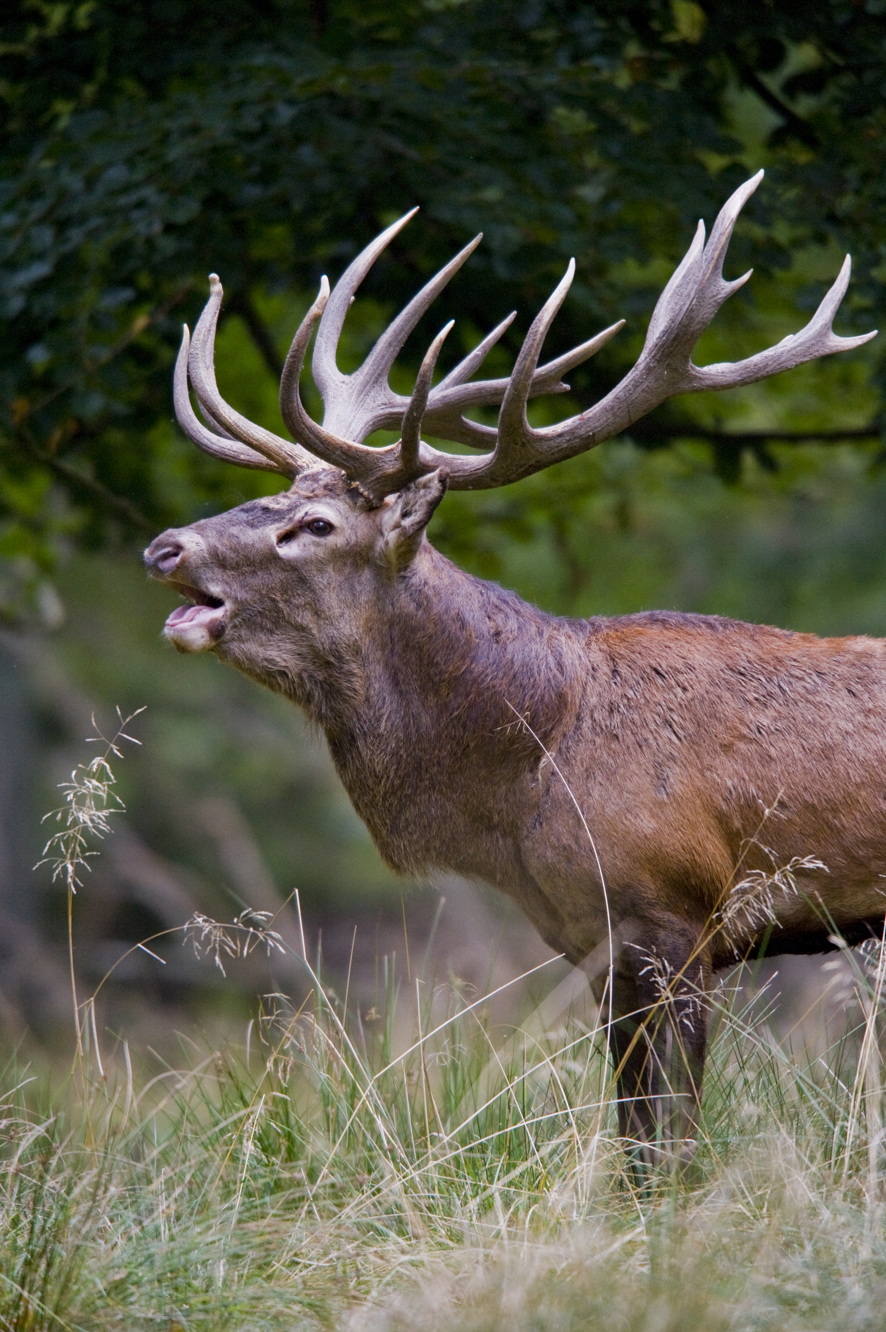
Antlers are extensions of an animal's skull found in members of the Cervidae (deer) Family (biology), family. Antlers are a single structure composed of bone, cartilage, fibrous tissue, skin, nerves, and blood vessels. They are generally found only on males, with the exception of Reindeer, reindeer/caribou. Antlers are Moulting, shed and regrown each year and function primarily as objects of sexual attraction and as Weapon (biology), weapons. Etymology Antler comes from the Old French ''antoillier ''(see present French : "Andouiller", from'' ant-, ''meaning before,'' oeil, ''meaning eye and'' -ier'', a suffix indicating an action or state of being) possibly from some form of an unattested Latin word ''*anteocularis'', "before the eye" (and applied to the word for "branch" or "horn (anatomy), horn"). Structure and development Antlers are unique to cervids. The ancestors of deer had tusks (long upper canine tooth, canine teeth). In most species, antlers appear to replace t ... [...More Info...] [...Related Items...] OR: [Wikipedia] [Google] [Baidu] |
Horn (anatomy)
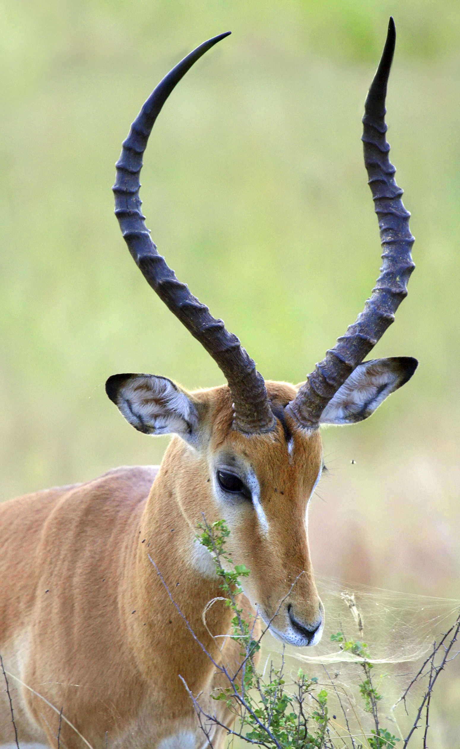
A horn is a permanent pointed projection on the head of various animals that consists of a covering of keratin and other proteins surrounding a core of live bone. Horns are distinct from antlers, which are not permanent. In mammals, true horns are found mainly among the ruminant artiodactyls, in the families Antilocapridae ( pronghorn) and Bovidae ( cattle, goats, antelope etc.). Cattle horns arise from subcutaneous connective tissue (under the scalp) and later fuse to the underlying frontal bone. One pair of horns is usual; however, two or more pairs occur in a few wild species and in some domesticated breeds of sheep. Polycerate (multi-horned) sheep breeds include the Hebridean, Icelandic, Jacob, Manx Loaghtan, and the Navajo-Churro. Horns usually have a curved or spiral shape, often with ridges or fluting. In many species, only males have horns. Horns start to grow soon after birth and continue to grow throughout the life of the animal (except in pronghorns, whi ... [...More Info...] [...Related Items...] OR: [Wikipedia] [Google] [Baidu] |
Stifle
The stifle joint (often simply stifle) is a complex joint in the hind limbs of quadruped mammals such as the sheep, horse or dog. It is the equivalent of the human knee and is often the largest synovial joint in the animal's body. The stifle joint joins three bones: the femur, patella, and tibia. The joint consists of three smaller ones: the femoropatellar joint, medial joint, and lateral femorotibial joint. The stifle joint consists of the femorotibial articulation ( femoral and tibial condyles), femoropatellar articulation (femoral trochlea and the patella), and the proximal articulation. The joint is stabilized by paired collateral ligaments which act to prevent abduction/adduction at the joint, as well as paired cruciate ligaments. The cranial cruciate ligament and the caudal cruciate ligament restrict cranial and caudal translation (respectively) of the tibia on the femur. The cranial cruciate also resists over-extension and inward rotation, and is the most commonly d ... [...More Info...] [...Related Items...] OR: [Wikipedia] [Google] [Baidu] |
Hindlimb
A hindlimb or back limb is one of the paired articulated appendages ( limbs) attached on the caudal ( posterior) end of a terrestrial tetrapod vertebrate's torso.http://www.merriam-webster.com/medical/hind%20limb, Merriam Webster Dictionary-Hindlimb With reference to quadrupeds, the term hindleg or back leg is often used instead. In bipedal animals with an upright posture (e.g. humans and some primates), the term lower limb is often used. Location It is located on the limb of an animal. Hindlimbs are present in a large number of quadrupeds. Though it is a posterior limb, it can cause lameness in some animals. The way of walking through hindlimbs are called bipedalism. Benefits of hindlimbs Hindlimbs are helpful in many ways, some examples are: Frogs Frogs can easily adapt at the surroundings using hindlimbs. The main reason is it can jump high to easily escape to its predator and also to catch prey. It can perform some tricks using the hindlimbs such as the somersault a ... [...More Info...] [...Related Items...] OR: [Wikipedia] [Google] [Baidu] |
Hepatopancreas
The hepatopancreas, digestive gland or midgut gland is an organ of the digestive tract of arthropods and molluscs. It provides the functions which in mammals are provided separately by the liver and pancreas, including the production of digestive enzymes, and absorption of digested food. Arthropods Arthropods, especially detritivores in the Order Isopoda, Suborder Oniscidea (woodlice), have been shown to be able to store heavy metals in their hepatopancreas. This could lead to bioaccumulation through the food chain and implications for food web destruction, if the accumulation gets high enough in polluted areas; for example, high metal concentrations are seen in spiders of the genus '' Dysdera'' which feed on woodlice, including their hepatopancreas, the major metal storage organ of isopods in polluted sites. Molluscs The hepatopancreas is a centre for lipid metabolism and for storage of lipids in gastropods Gastropods (), commonly known as slugs and snails, belong to a ... [...More Info...] [...Related Items...] OR: [Wikipedia] [Google] [Baidu] |
Hemolymph
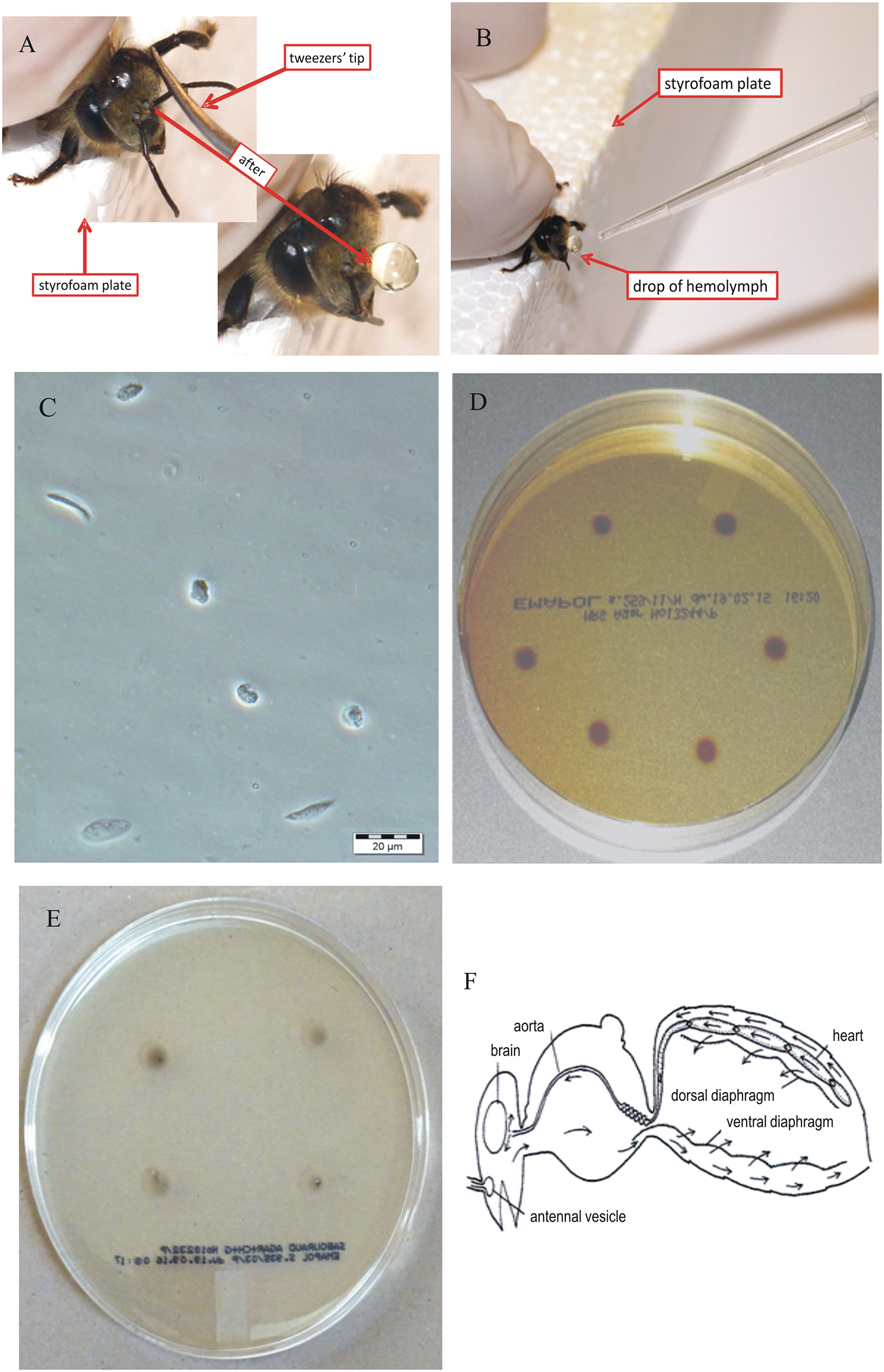
Hemolymph, or haemolymph, is a fluid, similar to the blood in invertebrates, that circulates in the inside of the arthropod's body, remaining in direct contact with the animal's tissues. It is composed of a fluid plasma in which hemolymph cells called hemocytes are dispersed. In addition to hemocytes, the plasma also contains many chemicals. It is the major tissue type of the open circulatory system characteristic of arthropods (for example, arachnids, crustaceans and insects). In addition, some non-arthropods such as mollusks possess a hemolymphatic circulatory system. Oxygen-transport systems were long thought unnecessary in insects, but ancestral and functional hemocyanin has been found in the hemolymph. Insect "blood" generally does not carry hemoglobin, although hemoglobin may be present in the tracheal system instead and play some role in respiration. Method of transport In the grasshopper, the closed portion of the system consists of tubular hearts and an ao ... [...More Info...] [...Related Items...] OR: [Wikipedia] [Google] [Baidu] |
Harderian Gland
The Harderian gland is a gland found within the eye's orbit that occurs in tetrapods (reptiles, amphibians, birds and mammals) that possess a nictitating membrane. The gland can be compound tubular or compound tubuloalveolar, and the fluid it secretes ( mucous, serous or lipid) varies between different groups of animals. In some animals, it acts as an accessory to the lacrimal gland, secreting fluid that eases movement of the nictitating membrane. Research has proposed that the gland has several other functions, including that of a photoprotective organ, a location of immune response, a source of thermoregulatory lipids, a source of pheromones, and a site of osmoregulation. In mammals, the gland secretes an oily substance used to preen the fur. The presence or absence of this gland is one of the cues used by palaeontologists to determine when fur evolved in the ancestors of mammals. The Harderian gland was first described in 1694 by Swiss anatomist Anatomy () is th ... [...More Info...] [...Related Items...] OR: [Wikipedia] [Google] [Baidu] |
Gills
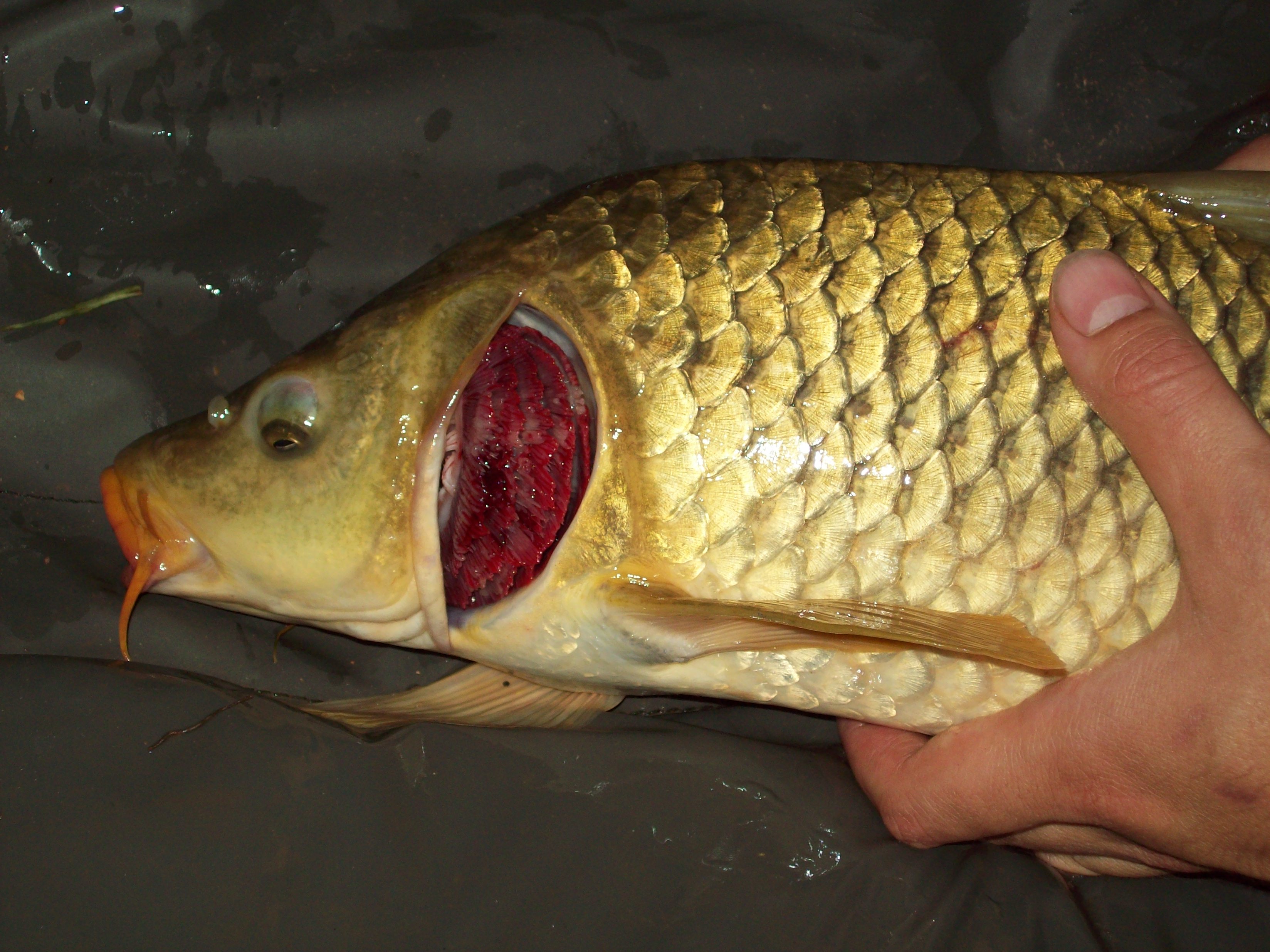
A gill () is a respiratory organ that many aquatic organisms use to extract dissolved oxygen from water and to excrete carbon dioxide. The gills of some species, such as hermit crabs, have adapted to allow respiration on land provided they are kept moist. The microscopic structure of a gill presents a large surface area to the external environment. Branchia (: branchiae) is the zoologists' name for gills (from Ancient Greek ). With the exception of some aquatic insects, the filaments and lamellae (folds) contain blood or coelomic fluid, from which gases are exchanged through the thin walls. The blood carries oxygen to other parts of the body. Carbon dioxide passes from the blood through the thin gill tissue into the water. Gills or gill-like organs, located in different parts of the body, are found in various groups of aquatic animals, including molluscs, crustaceans, insects, fish, and amphibians. Semiterrestrial marine animals such as crabs and mudskippers have gill chamber ... [...More Info...] [...Related Items...] OR: [Wikipedia] [Google] [Baidu] |


