|
Levator Labii Superioris
The levator labii superioris (: ''levatores labii superioris'', also called quadratus labii superioris, : ''quadrati labii superioris'') is a muscle of the human body used in facial expression. It is a broad sheet, the origin of which extends from the side of the nose to the zygomatic bone. Structure Its medial fibers form the ''angular head'' (also known as the levator labii superioris alaeque nasi muscle) which arises by a pointed extremity from the upper part of the frontal process of the maxilla and passing obliquely downward and lateralward divides into two slips. One of these is inserted into the greater alar cartilage and skin of the nose; the other is prolonged into the lateral part of the upper lip, blending with the infraorbital head and with the orbicularis oris. The intermediate portion or ''infraorbital head'' arises from the lower margin of the orbit immediately above the infraorbital foramen, some of its fibers being attached to the maxilla, others to the zygo ... [...More Info...] [...Related Items...] OR: [Wikipedia] [Google] [Baidu] |
Medial Infra-orbital Margin
The infraorbital margin is the lower margin of the eye socket. Structure It consists of the zygomatic bone and the maxilla, on which it separates the anterior Standard anatomical terms of location are used to describe unambiguously the anatomy of humans and other animals. The terms, typically derived from Latin or Greek roots, describe something in its standard anatomical position. This position pro ... and the orbital surface of the body of the maxilla. Function It is an attachment for the levator labii superioris muscle. Bones of the head and neck {{musculoskeletal-stub ... [...More Info...] [...Related Items...] OR: [Wikipedia] [Google] [Baidu] |
Maxilla
In vertebrates, the maxilla (: maxillae ) is the upper fixed (not fixed in Neopterygii) bone of the jaw formed from the fusion of two maxillary bones. In humans, the upper jaw includes the hard palate in the front of the mouth. The two maxillary bones are fused at the intermaxillary suture, forming the anterior nasal spine. This is similar to the mandible (lower jaw), which is also a fusion of two mandibular bones at the mandibular symphysis. The mandible is the movable part of the jaw. Anatomy Structure The maxilla is a paired bone - the two maxillae unite with each other at the intermaxillary suture. The maxilla consists of: * The body of the maxilla: pyramid-shaped; has an orbital, a nasal, an infratemporal, and a facial surface; contains the maxillary sinus. * Four processes: ** the zygomatic process ** the frontal process ** the alveolar process ** the palatine process It has three surfaces: * the anterior, posterior, medial Features of the maxilla include: * t ... [...More Info...] [...Related Items...] OR: [Wikipedia] [Google] [Baidu] |
Otorhinolaryngology
Otorhinolaryngology ( , abbreviated ORL and also known as otolaryngology, otolaryngology–head and neck surgery (ORL–H&N or OHNS), or ear, nose, and throat (ENT)) is a surgical subspecialty within medicine that deals with the surgical and medical management of conditions of the head and neck. Doctors who specialize in this area are called otorhinolaryngologists, otolaryngologists, head and neck surgeons, or ENT surgeons or physicians. Patients seek treatment from an otorhinolaryngologist for diseases of the ear, Human nose, nose, throat, base of skull, base of the skull, head, and neck. These commonly include functional diseases that affect the senses and activities of eating, drinking, speaking, breathing, swallowing, and hearing. In addition, ENT surgery encompasses the surgical management of cancers and benign tumors and reconstruction of the head and neck as well as plastic surgery of the face, scalp, and neck. Etymology The term is a combination of Neo-Latin classic ... [...More Info...] [...Related Items...] OR: [Wikipedia] [Google] [Baidu] |
Muscles Of The Head And Neck
Muscle is a soft tissue, one of the four basic types of animal tissue. There are three types of muscle tissue in vertebrates: skeletal muscle, cardiac muscle, and smooth muscle. Muscle tissue gives skeletal muscles the ability to contract. Muscle tissue contains special contractile proteins called actin and myosin which interact to cause movement. Among many other muscle proteins, present are two regulatory proteins, troponin and tropomyosin. Muscle is formed during embryonic development, in a process known as myogenesis. Skeletal muscle tissue is striated consisting of elongated, multinucleate muscle cells called muscle fibers, and is responsible for movements of the body. Other tissues in skeletal muscle include tendons and perimysium. Smooth and cardiac muscle contract involuntarily, without conscious intervention. These muscle types may be activated both through the interaction of the central nervous system as well as by innervation from peripheral plexus or endocri ... [...More Info...] [...Related Items...] OR: [Wikipedia] [Google] [Baidu] |
Levator Labii Superioris Alaeque Nasi
The levator labii superioris alaeque nasi muscle (occasionally shortened alaeque nasi muscle) is, translated from Latin, the "lifter of both the upper lip and of the wing of the nose". The muscle is attached to the upper frontal process of the maxilla and inserts into the skin of the lateral part of the nostril and upper lip. At 44 characters, its name is longer than that of any other muscle. Overview Historically known as Vidar's muscle, it dilates the nostril and elevates the upper lip, enabling one to snarl. ''Snore'' is used because it is the labial elevator closest to the nose. The levator labii superioris alaeque nasi is sometimes referred to as the "angular head" of the levator labii superioris muscle. See also * Frontalis muscle The frontalis muscle () is a muscle which covers parts of the forehead of the skull. Some sources consider the frontalis muscle to be a distinct muscle. However, Terminologia Anatomica currently classifies it as part of the occipitofrontalis mu ... [...More Info...] [...Related Items...] OR: [Wikipedia] [Google] [Baidu] |
Zygomaticus Minor Muscle
The zygomaticus minor muscle is a muscle of facial expression. It originates from the zygomatic bone, lateral to the rest of the levator labii superioris muscle, and inserts into the outer part of the upper lip. It draws the upper lip backward, upward, and outward and is used in smiling. It is innervated by the facial nerve (VII). Structure The zygomaticus minor muscle passes inferomedially from its origin to its insertion at an angle of approximately 30°. It has a mean width of around 0.5 cm. Origin It originates from the lateral aspect of just posterior to the zygomaticomaxillary suture. Insertion It inserts into the muscular tissue of the upper lip, blending distally with levator labii superioris muscle. Innervation The zygomaticus minor muscle receives motor innervation from the zygomatic branches and buccal branches of the facial nerve (CN VII). Relations The zygomaticus minor lies lateral to the rest of levator labii superioris muscle, and medial to its s ... [...More Info...] [...Related Items...] OR: [Wikipedia] [Google] [Baidu] |
Levator Anguli Oris
The levator anguli oris (caninus) is a facial muscle of the mouth arising from the canine fossa, immediately below the infraorbital foramen. It elevates angle of mouth medially. Its fibers are inserted into the angle of the mouth, intermingling with those of the zygomaticus, triangularis, and orbicularis oris. Specifically, the levator anguli oris is innervated by the buccal branches of the facial nerve The buccal branches of the facial nerve (infraorbital branches), are of larger size than the rest of the branches, pass horizontally forward to be distributed below the orbit and around the mouth. Branches The ''superficial branches'' run beneat .... Additional images File:Sobo 1909 264.png File:Sobo 1909 263.png, Seen from the inside. References External links PTCentral Muscles of the head and neck {{muscle-stub ... [...More Info...] [...Related Items...] OR: [Wikipedia] [Google] [Baidu] |
Orbit (anatomy)
In anatomy Anatomy () is the branch of morphology concerned with the study of the internal structure of organisms and their parts. Anatomy is a branch of natural science that deals with the structural organization of living things. It is an old scien ..., the orbit is the Body cavity, cavity or socket/hole of the skull in which the eye and Accessory visual structures, its appendages are situated. "Orbit" can refer to the bony socket, or it can also be used to imply the contents. In the adult human, the volume of the orbit is about , of which the eye occupies . The orbital contents comprise the eye, the Orbital fascia, orbital and retrobulbar fascia, extraocular muscles, cranial nerves optic nerve, II, oculomotor nerve, III, trochlear nerve, IV, trigeminal nerve, V, and abducens nerve, VI, blood vessels, fat, the lacrimal gland with its Lacrimal sac, sac and nasolacrimal duct, duct, the eyelids, Medial palpebral ligament, medial and Lateral palpebral raphe, lateral palpebr ... [...More Info...] [...Related Items...] OR: [Wikipedia] [Google] [Baidu] |
Orbicularis Oris
In human anatomy, the orbicularis oris muscle is a complex of muscles in the lips that encircles the mouth. It is not a true sphincter, as was once thought, as it is actually composed of four independent quadrants that interlace and give only an appearance of circularity.Saladin, "Anatomy & Physiology: The Unity of Form and Function". 5th edition. McGraw Hill. Page 330 It is also one of the muscles used in the playing of all brass instruments and some woodwind instrument Woodwind instruments are a family of musical instruments within the greater category of wind instruments. Common examples include flute, clarinet, oboe, bassoon, and saxophone. There are two main types of woodwind instruments: flutes and ...s. This muscle closes the mouth and puckers the lips when it contracts. Structure The orbicularis oris is not a simple sphincter muscle like the orbicularis oculi; it consists of numerous strata of muscular fibers surrounding the orifice of the mouth, but having ... [...More Info...] [...Related Items...] OR: [Wikipedia] [Google] [Baidu] |
Greater Alar Cartilage
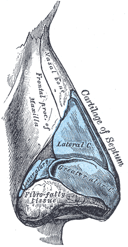
The major alar cartilage (greater alar cartilage) (lower lateral cartilage) is a thin, flexible plate, situated immediately below the lateral nasal cartilage, and bent upon itself in such a manner as to form the Nasal septum, medial wall and lateral wall of the nostril of its own side. The portion which forms the Nasal septum, medial wall (crus mediale) is loosely connected with the corresponding portion of the opposite cartilage, the two forming, together with the thickened integument and subjacent tissue, the nasal septum. The part which forms the lateral wall (crus laterale) is curved to correspond with the Human nose#External nose, ala of the nose; it is oval and flattened, narrow behind, where it is connected with the frontal process of the maxilla by a tough fibrous membrane, in which are found three or four small cartilaginous plates, the lesser alar cartilages (cartilagines alares minores; sesamoid cartilages). Above, it is connected by fibrous tissue to the lateral carti ... [...More Info...] [...Related Items...] OR: [Wikipedia] [Google] [Baidu] |
Frontal Process Of Maxilla
The frontal process of the maxilla is a strong plate, which projects upward, medialward, and backward from the maxilla, forming part of the lateral boundary of the nose. Its ''lateral surface'' is smooth, continuous with the anterior surface of the body, and gives attachment to the quadratus labii superioris, the orbicularis oculi, and the medial palpebral ligament. Its ''medial surface'' forms part of the lateral wall of the nasal cavity; at its upper part is a rough, uneven area, which articulates with the ethmoid, closing in the anterior ethmoidal cells; below this is an oblique ridge, the ethmoidal crest, the posterior end of which articulates with the middle nasal concha, while the anterior part is termed the agger nasi; the crest forms the upper limit of the atrium of the middle meatus. The ''upper border'' articulates with the frontal bone and the ''anterior'' with the nasal; the ''posterior border'' is thick, and hollowed into a groove, which is continuous below wit ... [...More Info...] [...Related Items...] OR: [Wikipedia] [Google] [Baidu] |


