|
Lateral Plate Mesoderm
The lateral plate mesoderm is the mesoderm that is found at the periphery of the embryo. It is to the side of the paraxial mesoderm, and further to the axial mesoderm. The lateral plate mesoderm is separated from the paraxial mesoderm by a narrow region of intermediate mesoderm. The mesoderm is the middle layer of the three germ layers, between the outer ectoderm and inner endoderm. During the third week of embryonic development the lateral plate mesoderm splits into two layers forming the intraembryonic coelom. The outer layer of lateral plate mesoderm adheres to the ectoderm to become the somatic or parietal layer known as the somatopleure. The inner layer adheres to the endoderm to become the splanchnic or visceral layer known as the splanchnopleure. Development The lateral plate mesoderm will split into two layers, the somatopleuric mesenchyme, and the splanchnopleuric mesenchyme. * The ''somatopleuric layer'' forms the future body wall. * The ''splanchnopleuric layer'' forms ... [...More Info...] [...Related Items...] OR: [Wikipedia] [Google] [Baidu] |
Axial Mesoderm
Axial mesoderm, or chordamesoderm, is the mesoderm in the embryo that lies along the central axis under the neural tube. * will give rise to notochord * starts as the notochordal process, whose formation finishes at day 20 in humans. * important not only in forming the notochord itself but also in inducing development of the overlying ectoderm into the neural tube * will eventually induce the formation of vertebral bodies. * ventral floor of the notochordal process fuses with endoderm. * The notochord will form the nucleus pulposus of intervertebral discs. There is some discussion as to whether these cells contributed from the notochord are replaced by others from the adjacent mesoderm. It gives rise to the notochordal process, which later becomes the notochord The notochord is an elastic, rod-like structure found in chordates. In vertebrates the notochord is an embryonic structure that disintegrates, as the vertebrae develop, to become the nucleus pulposus in the inter ... [...More Info...] [...Related Items...] OR: [Wikipedia] [Google] [Baidu] |
Endoderm
Endoderm is the innermost of the three primary germ layers in the very early embryo. The other two layers are the ectoderm (outside layer) and mesoderm (middle layer). Cells migrating inward along the archenteron form the inner layer of the gastrula, which develops into the endoderm. The endoderm consists at first of flattened cells, which subsequently become columnar. It forms the epithelial lining of multiple systems. In plant biology, endoderm corresponds to the innermost part of the cortex ( bark) in young shoots and young roots often consisting of a single cell layer. As the plant becomes older, more endoderm will lignify. Production The following chart shows the tissues produced by the endoderm. The embryonic endoderm develops into the interior linings of two tubes in the body, the digestive and respiratory tube. Liver and pancreas cells are believed to derive from a common precursor. In humans, the endoderm can differentiate into distinguishable organs after 5 w ... [...More Info...] [...Related Items...] OR: [Wikipedia] [Google] [Baidu] |
Septum Transversum
The septum transversum is a thick mass of cranial mesenchyme, formed in the embryo, that gives rise to parts of the thoracic diaphragm and the ventral mesentery of the foregut in the developed human being and other mammals. Origins The septum transversum originally arises as the most cranial part of the mesenchyme on day 22. During craniocaudal folding, it assumes a position cranial to the developing heart at the level of the cervical vertebrae. During subsequent weeks the dorsal end of the embryo grows much faster than its ventral counterpart resulting in an ''apparent descent'' of the ventrally located septum transversum. At week 8, it can be found at the level of the thoracic vertebrae. Nerve supply After successful craniocaudal folding the septum transversum picks up innervation from the adjacent ventral rami of spinal nerves C3, C4 and C5, thus forming the precursor of the phrenic nerve. During the descent of the septum, the phrenic nerve is carried along and assumes its d ... [...More Info...] [...Related Items...] OR: [Wikipedia] [Google] [Baidu] |
Peritoneal
The peritoneum is the serous membrane forming the lining of the abdominal cavity or coelom in amniotes and some invertebrates, such as annelids. It covers most of the intra-abdominal (or coelomic) organs, and is composed of a layer of mesothelium supported by a thin layer of connective tissue. This peritoneal lining of the cavity supports many of the abdominal organs and serves as a conduit for their blood vessels, lymphatic vessels, and nerves. The abdominal cavity (the space bounded by the vertebrae, abdominal muscles, diaphragm, and pelvic floor) is different from the intraperitoneal space (located within the abdominal cavity but wrapped in peritoneum). The structures within the intraperitoneal space are called "intraperitoneal" (e.g., the stomach and intestines), the structures in the abdominal cavity that are located behind the intraperitoneal space are called "retroperitoneal" (e.g., the kidneys), and those structures below the intraperitoneal space are called "subperit ... [...More Info...] [...Related Items...] OR: [Wikipedia] [Google] [Baidu] |
Pleural
The pleural cavity, or pleural space (or sometimes intrapleural space), is the potential space between the pulmonary pleurae, pleurae of the pleural sac that surrounds each lung. A small amount of serous fluid, serous pleural fluid is maintained in the pleural cavity to enable lubrication between the pleurae, membranes, and also to create a pressure gradient. The serous membrane that covers the surface of the lung is the visceral pleura and is separated from the outer membrane, the parietal pleura, by just the film of pleural fluid in the pleural cavity. The visceral pleura follows the fissures of the lung and the root of the lung structures. The parietal pleura is attached to the mediastinum, the upper surface of the thoracic diaphragm, diaphragm, and to the inside of the ribcage. Structure In humans, the left and right lungs are completely separated by the mediastinum, and there is no communication between their pleural cavities. Therefore, in cases of a unilateral pneumothor ... [...More Info...] [...Related Items...] OR: [Wikipedia] [Google] [Baidu] |
Pericardium
The pericardium (: pericardia), also called pericardial sac, is a double-walled sac containing the heart and the roots of the great vessels. It has two layers, an outer layer made of strong inelastic connective tissue (fibrous pericardium), and an inner layer made of serous membrane (serous pericardium). It encloses the pericardial cavity, which contains pericardial fluid, and defines the middle mediastinum. It separates the heart from interference of other structures, protects it against infection and blunt trauma, and lubricates the heart's movements. The English name originates from the Ancient Greek prefix ''peri-'' (περί) 'around' and the suffix ''-cardion'' (κάρδιον) 'heart'. Anatomy The pericardium is a tough fibroelastic sac which covers the heart from all sides except at the cardiac root (where the great vessels join the heart) and the bottom (where only the serous pericardium exists to cover the upper surface of the central tendon of diaphragm). ... [...More Info...] [...Related Items...] OR: [Wikipedia] [Google] [Baidu] |
Dorsal Mesentery
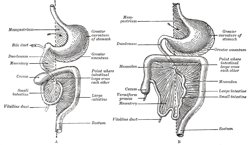
In human anatomy, the mesentery is an organ that attaches the intestines to the posterior abdominal wall, consisting of a double fold of the peritoneum. It helps (among other functions) in storing fat and allowing blood vessels, lymphatics, and nerves to supply the intestines. The (the part of the mesentery that attaches the colon to the abdominal wall) was formerly thought to be a fragmented structure, with all named parts—the ascending, transverse, descending, and sigmoid mesocolons, the mesoappendix, and the mesorectum—separately terminating their insertion into the posterior abdominal wall. However, in 2012, new microscopic and electron microscopic examinations showed the mesocolon to be a single structure derived from the duodenojejunal flexure and extending to the distal mesorectal layer. Thus the mesentery is an internal organ. Structure The mesentery of the small intestine arises from the root of the mesentery (or mesenteric root) and is the part connected ... [...More Info...] [...Related Items...] OR: [Wikipedia] [Google] [Baidu] |
Somatopleuric
In the anatomy of an embryo, the somatopleure is a structure created during embryogenesis when the lateral plate mesoderm splits into two layers. The outer (or somatic) layer becomes applied to the inner surface of the ectoderm, and with it (partially) forms the somatopleure. The combination of ectoderm and mesoderm, or somatopleure, forms the amnion, the chorion and the lateral body wall of the embryo. Limb formation, from the somatic mesoderm, is induced by hox genes and the expression of other molecules through an epithelial-mesenchyme transition. The embryonic somatopleure is then divided into 3 sections, the anterior limb bud formation, the posterior limb bud formation and the non limb forming wall. The bud forming sections grow in size. The somatic mesoderm under the ectoderm proliferates in mesenchyme form. In chicken, the extraembryonic tissues are separated into two layers: the splanchnopleure composed of the endoderm and splanchnic mesoderm, and the somatopleure com ... [...More Info...] [...Related Items...] OR: [Wikipedia] [Google] [Baidu] |
Splanchnopleuric
In the anatomy of an embryo, the splanchnopleuric mesenchyme is a structure created during embryogenesis when the lateral mesodermal germ layer splits into two layers. The inner (or splanchnic) layer adheres to the endoderm, and with it forms the splanchnopleure (mesoderm external to the coelom plus the endoderm). See also Post development the somato and splanchnopleuric junction lies at the duodeno-jejunal flexure. * somatopleure * mesenchyme Mesenchyme () is a type of loosely organized animal embryonic connective tissue of undifferentiated cells that give rise to most tissues, such as skin, blood, or bone. The interactions between mesenchyme and epithelium help to form nearly ever ... References External links * * Overview at Kennesaw State University Embryology {{developmental-biology-stub ... [...More Info...] [...Related Items...] OR: [Wikipedia] [Google] [Baidu] |
Bone Morphogenetic Protein
Bone morphogenetic proteins (BMPs) are a group of growth factors also known as cytokines and as metabologens. Professor Marshall Urist and Professor Hari Reddi discovered their ability to induce the formation of bone and cartilage, BMPs are now considered to constitute a group of pivotal morphogenetic signals, orchestrating tissue architecture throughout the body. The important functioning of BMP signals in physiology is emphasized by the multitude of roles for dysregulated BMP signalling in pathological processes. Cancerous disease often involves misregulation of the BMP signalling system. Absence of BMP signalling is, for instance, an important factor in the progression of colon cancer, and conversely, overactivation of BMP signalling following reflux-induced esophagitis provokes Barrett's esophagus and is thus instrumental in the development of esophageal adenocarcinoma. Recombinant human BMPs (rhBMPs) are used in orthopedic applications such as spinal fusions, nonunions, an ... [...More Info...] [...Related Items...] OR: [Wikipedia] [Google] [Baidu] |
Circulatory System
In vertebrates, the circulatory system is a system of organs that includes the heart, blood vessels, and blood which is circulated throughout the body. It includes the cardiovascular system, or vascular system, that consists of the heart and blood vessels (from Greek meaning ''heart'', and Latin meaning ''vessels''). The circulatory system has two divisions, a systemic circulation or circuit, and a pulmonary circulation or circuit. Some sources use the terms ''cardiovascular system'' and ''vascular system'' interchangeably with ''circulatory system''. The network of blood vessels are the great vessels of the heart including large elastic arteries, and large veins; other arteries, smaller arterioles, capillaries that join with venules (small veins), and other veins. The circulatory system is closed in vertebrates, which means that the blood never leaves the network of blood vessels. Many invertebrates such as arthropods have an open circulatory system with a he ... [...More Info...] [...Related Items...] OR: [Wikipedia] [Google] [Baidu] |
Splanchnopleuric Mesenchyme
In the anatomy of an embryo, the splanchnopleuric mesenchyme is a structure created during embryogenesis when the lateral mesodermal germ layer splits into two layers. The inner (or splanchnic) layer adheres to the endoderm, and with it forms the splanchnopleure (mesoderm external to the coelom plus the endoderm). See also Post development the somato and splanchnopleuric junction lies at the duodeno-jejunal flexure. * somatopleure * mesenchyme Mesenchyme () is a type of loosely organized animal embryonic connective tissue of undifferentiated cells that give rise to most tissues, such as skin, blood, or bone. The interactions between mesenchyme and epithelium help to form nearly ever ... References External links * * Overview at Kennesaw State University Embryology {{developmental-biology-stub ... [...More Info...] [...Related Items...] OR: [Wikipedia] [Google] [Baidu] |


