|
Jumper's Knee
Patellar tendinitis, also known as jumper's knee, is an overuse injury of the tendon that straightens the knee. Symptoms include pain in the front of the knee. Typically the pain and tenderness is at the lower part of the kneecap, though the upper part may also be affected. Generally there is no pain when the person is at rest. Complications may include patellar tendon rupture. Risk factors include being involved in athletics and being overweight. It is particularly common in athletes who are involved in jumping sports such as basketball and volleyball. Other risk factors include sex, age, occupation, and physical activity level. It is increasingly more likely to be developed with increasing age. The underlying mechanism involves small tears in the tendon connecting the kneecap with the shinbone. Diagnosis is generally based on symptoms and examination. Other conditions that can appear similar include infrapatellar bursitis, chondromalacia patella and patellofemoral syn ... [...More Info...] [...Related Items...] OR: [Wikipedia] [Google] [Baidu] |
Orthopedics
Orthopedic surgery or orthopedics (American and British English spelling differences, alternative spelling orthopaedics) is the branch of surgery concerned with conditions involving the musculoskeletal system. Orthopedic surgeons use both surgical and nonsurgical means to treat musculoskeletal Physical trauma, trauma, Spinal disease, spine diseases, Sports injury, sports injuries, degenerative diseases, infections, tumors and congenital disorders. Etymology Nicholas Andry coined the word in French as ', derived from the Ancient Greek words ("correct", "straight") and ("child"), and published ''Orthopedie'' (translated as ''Orthopædia: Or the Art of Correcting and Preventing Deformities in Children'') in 1741. The word was Assimilation (linguistics), assimilated into English as ''orthopædics''; the Typographic ligature, ligature ''æ'' was common in that era for ''ae'' in Greek- and Latin-based words. As the name implies, the discipline was initially developed with atte ... [...More Info...] [...Related Items...] OR: [Wikipedia] [Google] [Baidu] |
Patellar Tendon
The patellar tendon is the distal portion of the common tendon of the quadriceps femoris, which is continued from the patella to the tibial tuberosity. It is also sometimes called the patellar ligament as it forms a bone to bone connection when the patella is fully ossified. Structure The patellar tendon is a strong, flat ligament, which originates on the apex of the patella distally and adjoining margins of the patella and the rough depression on its posterior surface; below, it inserts on the tuberosity of the tibia; its superficial fibers are continuous over the front of the patella with those of the tendon of the quadriceps femoris. It is about 4.5 cm long in adults (range from 3 to 6 cm). The medial and lateral portions of the quadriceps tendon pass down on either side of the patella to be inserted into the upper extremity of the tibia on either side of the tuberosity; these portions merge into the capsule, as stated above, forming the medial and lateral patellar r ... [...More Info...] [...Related Items...] OR: [Wikipedia] [Google] [Baidu] |
Quadriceps
The quadriceps femoris muscle (, also called the quadriceps extensor, quadriceps or quads) is a large muscle group that includes the four prevailing muscles on the front of the thigh. It is the sole extensor muscle of the knee, forming a large fleshy mass which covers the front and sides of the femur. The name derives . Structure Parts The quadriceps femoris muscle is subdivided into four separate muscles (the 'heads'), with the first superficial to the other three over the femur (from the trochanters to the condyles): * The rectus femoris muscle occupies the middle of the thigh, covering most of the other three quadriceps muscles. It originates on the ilium. It is named for its straight course. * The vastus lateralis muscle is on the ''lateral side'' of the femur (i.e. on the outer side of the thigh). * The vastus medialis muscle is on the ''medial side'' of the femur (i.e. on the inner part thigh). * The vastus intermedius muscle lies between vastus lateralis and vas ... [...More Info...] [...Related Items...] OR: [Wikipedia] [Google] [Baidu] |
Muscle Contraction
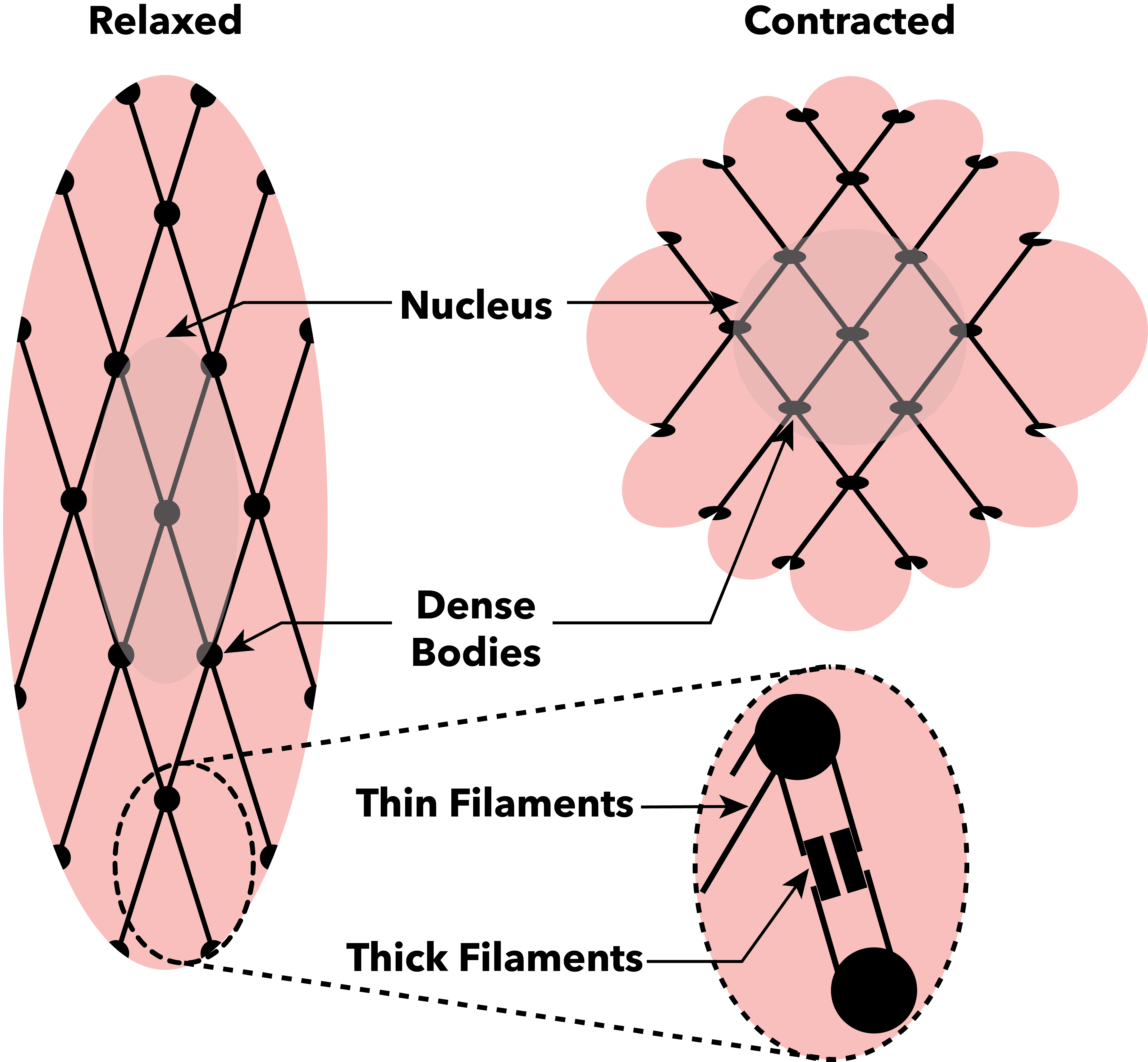
Muscle contraction is the activation of Tension (physics), tension-generating sites within muscle cells. In physiology, muscle contraction does not necessarily mean muscle shortening because muscle tension can be produced without changes in muscle length, such as when holding something heavy in the same position. The termination of muscle contraction is followed by muscle relaxation, which is a return of the muscle fibers to their low tension-generating state. For the contractions to happen, the muscle cells must rely on the change in action of two types of Myofilament, filaments: thin and thick filaments. The major constituent of thin filaments is a chain formed by helical coiling of two strands of actin, and thick filaments dominantly consist of chains of the Motor protein, motor-protein myosin. Together, these two filaments form myofibrils - the basic functional organelles in the skeletal muscle system. In vertebrates, Muscle cell#Muscle contraction in striated muscle, skele ... [...More Info...] [...Related Items...] OR: [Wikipedia] [Google] [Baidu] |
Rest, Ice, Compression, And Elevation
RICE is a mnemonic acronym for the four elements of a treatment regimen that was once recommended for soft tissue injuries: rest, ice, compression, and elevation. It was considered a first-aid treatment rather than a cure and aimed to control inflammation. It was thought that the reduction in pain and swelling that occurred as a result of decreased inflammation helped with healing. The protocol was often used to treat sprains, strains, cuts, bruises, and other similar injuries. Ice has been used for injuries since at least the 1960s, in a case where a 12-year-old boy needed to have a limb reattached. The limb was preserved before surgery by using ice. As news of the successful operation spread, the use of ice to treat acute injuries became common. The mnemonic was introduced by Dr. Gabe Mirkin in 1978. He withdrew his support of this regimen in 2014 after learning of the role of inflammation in the healing process. The implementation of RICE for soft tissue injuries as descr ... [...More Info...] [...Related Items...] OR: [Wikipedia] [Google] [Baidu] |
Magnetic Resonance Imaging
Magnetic resonance imaging (MRI) is a medical imaging technique used in radiology to generate pictures of the anatomy and the physiological processes inside the body. MRI scanners use strong magnetic fields, magnetic field gradients, and radio waves to form images of the organs in the body. MRI does not involve X-rays or the use of ionizing radiation, which distinguishes it from computed tomography (CT) and positron emission tomography (PET) scans. MRI is a medical application of nuclear magnetic resonance (NMR) which can also be used for imaging in other NMR applications, such as NMR spectroscopy. MRI is widely used in hospitals and clinics for medical diagnosis, staging and follow-up of disease. Compared to CT, MRI provides better contrast in images of soft tissues, e.g. in the brain or abdomen. However, it may be perceived as less comfortable by patients, due to the usually longer and louder measurements with the subject in a long, confining tube, although ... [...More Info...] [...Related Items...] OR: [Wikipedia] [Google] [Baidu] |
Ultrasound
Ultrasound is sound with frequency, frequencies greater than 20 Hertz, kilohertz. This frequency is the approximate upper audible hearing range, limit of human hearing in healthy young adults. The physical principles of acoustic waves apply to any frequency range, including ultrasound. Ultrasonic devices operate with frequencies from 20 kHz up to several gigahertz. Ultrasound is used in many different fields. Ultrasonic devices are used to detect objects and measure distances. Ultrasound imaging or sonography is often used in medicine. In the nondestructive testing of products and structures, ultrasound is used to detect invisible flaws. Industrially, ultrasound is used for cleaning, mixing, and accelerating chemical processes. Animals such as bats and porpoises use ultrasound for locating prey and obstacles. History Acoustics, the science of sound, starts as far back as Pythagoras in the 6th century BC, who wrote on the mathematical properties of String instrument ... [...More Info...] [...Related Items...] OR: [Wikipedia] [Google] [Baidu] |
Hamstrings
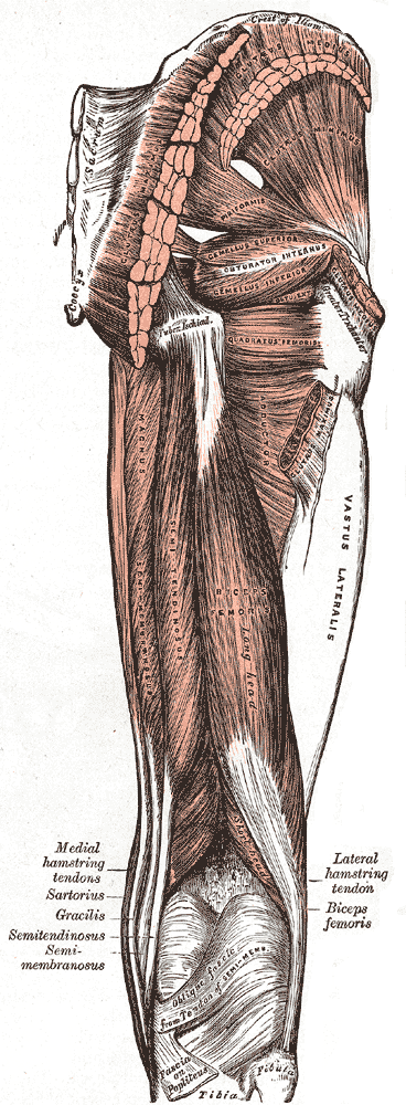
A hamstring () is any one of the three posterior thigh muscles in human anatomy between the hip and the knee: from anatomical_terms_of_location#Medial_and_lateral, medial to anatomical_terms_of_location#Medial_and_lateral, lateral, the semimembranosus, semitendinosus and biceps femoris. Etymology The word "ham" is derived from the Old English “ham” or “hom” meaning the hollow or bend of the knee, from a Germanic base where it meant "crooked". It gained the meaning of the leg of an animal around the 15th century. ''String'' refers to tendons, and thus the hamstrings' string-like tendons felt on either side of the back of the knee. Criteria The common criteria of any hamstring muscles are: # Muscles should originate from ischial tuberosity. # Muscles should be inserted over the knee joint, in the tibia or in the fibula. # Muscles will be innervated by the tibial nerve, tibial branch of the sciatic nerve. # Muscle will participate in flexion of the knee joint and extension ... [...More Info...] [...Related Items...] OR: [Wikipedia] [Google] [Baidu] |
Quadriceps Muscle
The quadriceps femoris muscle (, also called the quadriceps extensor, quadriceps or quads) is a large muscle group that includes the four prevailing muscles on the front of the thigh. It is the sole extensor muscle of the knee, forming a large fleshy mass which covers the front and sides of the femur. The name derives . Structure Parts The quadriceps femoris muscle is subdivided into four separate muscles (the 'heads'), with the first superficial to the other three over the femur (from the trochanters to the condyles): * The rectus femoris muscle occupies the middle of the thigh, covering most of the other three quadriceps muscles. It originates on the ilium. It is named for its straight course. * The vastus lateralis muscle is on the ''lateral side'' of the femur (i.e. on the outer side of the thigh). * The vastus medialis muscle is on the ''medial side'' of the femur (i.e. on the inner part thigh). * The vastus intermedius muscle lies between vastus lateralis and vastu ... [...More Info...] [...Related Items...] OR: [Wikipedia] [Google] [Baidu] |
Calf (anatomy)
The calf (: calves; Latin: ''sura'') is the back portion of the lower leg in human anatomy. The muscles within the calf correspond to the posterior compartment of the leg. The two largest muscles within this compartment are known together as the calf muscle and attach to the heel via the Achilles tendon. Several other, smaller muscles attach to the knee, the ankle, and via long tendons to the toes. Etymology From Middle English ''calf'', ''kalf'', from Old Norse ''kalfi'', possibly derived from the same Germanic root as English '' calf'' ("young cow"). Cognate with Icelandic ''kálfi'' ("calf of the leg"). ''Calf'' and ''calf of the leg'' are documented in use in Middle English circa AD 1350 and AD 1425 respectively. Historically, the ''absence of calf'', meaning a lower leg without a prominent calf muscle, was regarded by some authors as a sign of inferiority: ''it is well known that monkeys have no calves, and still less do they exist among the lower orders of mammals''. ... [...More Info...] [...Related Items...] OR: [Wikipedia] [Google] [Baidu] |
Gluteal Muscles
The gluteal muscles, often called glutes, are a group of three muscles which make up the gluteal region commonly known as the buttocks: the gluteus maximus muscle, gluteus maximus, gluteus medius muscle, gluteus medius and gluteus minimus muscle, gluteus minimus. The three muscles originate from the ilium bone, ilium and sacrum and insert on the femur. The functions of the muscles include Extension (kinesiology), extension, Abduction (kinesiology), abduction, external rotation, and internal rotation of the hip joint. Structure The gluteus maximus is the largest and most wiktionary:superficial, superficial of the three gluteal muscles. It makes up a large part of the shape and appearance of the hips. It is a narrow and thick fleshy mass of a quadrilateral shape, and forms the prominence of the buttocks. The gluteus medius is a broad, thick, radiating muscle, situated on the outer surface of the pelvis. It lies profound to the gluteus maximus and its posterior third is covered by ... [...More Info...] [...Related Items...] OR: [Wikipedia] [Google] [Baidu] |


