|
Ischium
The ischium (; : ischia) is a paired bone forming the lower and back part of the hip bone. Situated below the ilium (bone), ilium and behind the pubis (bone), pubis, it is one of three regions whose fusion creates the coxal bone. The superior portion of this region forms approximately one-third of the acetabulum. Structure The ischium is made up of three parts–the body, the superior ramus and the inferior ramus. The body contains a prominent ischial spine, spine, which serves as the origin for the superior gemellus muscle. The indentation inferior to the spine is the lesser sciatic notch. Continuing down the posterior side, the ischial tuberosity is a thick, rough-surfaced prominence below the lesser sciatic notch. This is the portion ...[...More Info...] [...Related Items...] OR: [Wikipedia] [Google] [Baidu] |
Hip Bone
The hip bone (os coxae, innominate bone, pelvic bone or coxal bone) is a large flat bone, constricted in the center and expanded above and below. In some vertebrates (including humans before puberty) it is composed of three parts: the Ilium (bone), ilium, ischium, and the Pubis (bone), pubis. The two hip bones join at the pubic symphysis and together with the sacrum and coccyx (the pelvic part of the vertebral column, spine) comprise the human skeleton, skeletal component of the pelvis – the pelvic girdle which surrounds the pelvic cavity. They are connected to the sacrum, which is part of the axial skeleton, at the sacroiliac joint. Each hip bone is connected to the corresponding femur (thigh bone) (forming the primary connection between the bones of the lower limb and the axial skeleton) through the large ball and socket joint of the hip joint, hip. Structure The hip bone is formed by three parts: the Ilium (bone), ilium, ischium, and Pubis (bone), pubis. At birth, these thre ... [...More Info...] [...Related Items...] OR: [Wikipedia] [Google] [Baidu] |
Pelvis
The pelvis (: pelves or pelvises) is the lower part of an Anatomy, anatomical Trunk (anatomy), trunk, between the human abdomen, abdomen and the thighs (sometimes also called pelvic region), together with its embedded skeleton (sometimes also called bony pelvis or pelvic skeleton). The pelvic region of the trunk includes the bony pelvis, the pelvic cavity (the space enclosed by the bony pelvis), the pelvic floor, below the pelvic cavity, and the perineum, below the pelvic floor. The pelvic skeleton is formed in the area of the back, by the sacrum and the coccyx and anteriorly and to the left and right sides, by a pair of hip bones. The two hip bones connect the spine with the lower limbs. They are attached to the sacrum posteriorly, connected to each other anteriorly, and joined with the two femurs at the hip joints. The gap enclosed by the bony pelvis, called the pelvic cavity, is the section of the body underneath the abdomen and mainly consists of the reproductive organs and ... [...More Info...] [...Related Items...] OR: [Wikipedia] [Google] [Baidu] |
Obturator Foramen
The obturator foramen is the large, Bilateral symmetry, bilaterally paired opening of the bony pelvis. It is formed by the pubis and ischium. It is mostly closed by the obturator membrane except for a small opening, the obturator canal, through which the obturator nerve and vessels pass. Structure The obturator foramen is situated inferior and somewhat anterior to the acetabulum. It is bounded by the pubis bone and the ischium: superiorly by the (grooved obturator surface) of the Superior rami of the pubes, superior ramus of pubis, inferiorly by the Ischium#Structure, ramus of ischium, and laterally by (the anterior edge of) the body of ischium (including by the margin of the acetabulum). The margin of the foramen is thin and uneven, and gives attachment to the obturator membrane. Superiorly, it presents a deep groove - the obturator groove - which passes obliquely inferomedially from the pelvis. The foramen is largely closed by the obturator membrane save for a small opening at ... [...More Info...] [...Related Items...] OR: [Wikipedia] [Google] [Baidu] |
Adductor Magnus Muscle
The adductor magnus is a large triangular muscle, situated on the medial side of the thigh. It consists of two parts. The portion which arises from the ischiopubic ramus (a small part of the inferior ramus of the pubis, and the inferior ramus of the ischium) is called the pubofemoral portion, adductor portion, or adductor minimus, and the portion arising from the tuberosity of the ischium is called the ischiocondylar portion, extensor portion, or "hamstring portion". Due to its common embryonic origin, innervation, and action the ischiocondylar portion (or hamstring portion) is often considered part of the hamstring group of muscles. The ischiocondylar portion of the adductor magnus is considered a muscle of the posterior compartment of the thigh while the pubofemoral portion of the adductor magnus is considered a muscle of the medial compartment. Structure Pubofemoral (adductor) portion Those fibers which arise from the ramus of the pubis are short, horizontal in directio ... [...More Info...] [...Related Items...] OR: [Wikipedia] [Google] [Baidu] |
Adductor Magnus
The adductor magnus is a large triangular muscle, situated on the medial side of the thigh. It consists of two parts. The portion which arises from the ischiopubic ramus (a small part of the inferior ramus of the pubis, and the inferior ramus of the ischium) is called the pubofemoral portion, adductor portion, or adductor minimus, and the portion arising from the tuberosity of the ischium is called the ischiocondylar portion, extensor portion, or "hamstring portion". Due to its common embryonic origin, innervation, and action the ischiocondylar portion (or hamstring portion) is often considered part of the hamstring group of muscles. The ischiocondylar portion of the adductor magnus is considered a muscle of the posterior compartment of the thigh while the pubofemoral portion of the adductor magnus is considered a muscle of the medial compartment. Structure Pubofemoral (adductor) portion Those fibers which arise from the ramus of the pubis are short, horizontal in direc ... [...More Info...] [...Related Items...] OR: [Wikipedia] [Google] [Baidu] |
Superficial Transverse Perineal Muscle
The transverse perineal muscles (transversus perinei) are the superficial and the deep transverse perineal muscles. Superficial transverse perineal The superficial transverse perineal muscle (transversus superficialis perinei or Lloyd-Beanie muscle) is a narrow muscular slip, which passes more or less transversely across the perineal space in front of the anus. It arises by tendinous fibers from the inner and forepart of the ischial tuberosity and, running medially, is inserted into the central tendinous point of the perineum (perineal body), joining in this situation with the muscle of the opposite side, with the external anal sphincter muscle behind, and with the bulbospongiosus muscle in front. In some cases, the fibers of the deeper layer of the external anal sphincter cross over in front of the anus and are continued into thi ... [...More Info...] [...Related Items...] OR: [Wikipedia] [Google] [Baidu] |
Internal Obturator Muscle
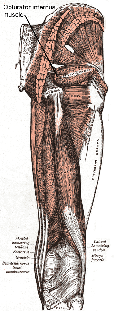
The internal obturator muscle or obturator internus muscle originates on the medial surface of the obturator membrane, the ischium near the membrane, and the rim of the pubis. It exits the pelvic cavity through the lesser sciatic foramen. The internal obturator is situated partly within the lesser pelvis, and partly at the back of the hip-joint. It functions to help laterally rotate femur with hip extension and abduct femur with hip flexion, as well as to steady the femoral head in the acetabulum. Structure Origin The internal obturator muscle arises from the inner surface of the antero-lateral wall of the pelvis. It surrounds the obturator foramen. It is attached to the inferior pubic ramus and ischium, and at the side to the inner surface of the hip bone below and behind the pelvic brim. It reaches from the upper part of the greater sciatic foramen above and behind to the obturator foramen below and in front. It also arises from the pelvic surface of the obturator mem ... [...More Info...] [...Related Items...] OR: [Wikipedia] [Google] [Baidu] |
Ischial Tuberosity
The ischial tuberosity (or tuberosity of the ischium, tuber ischiadicum), also known colloquially as the sit bones or sitz bones, or as a pair the sitting bones, is a large posterior bony protuberance on the superior ramus of the ischium. It marks the lateral boundary of the pelvic outlet. When sitting, the weight is frequently placed upon the ischial tuberosity. The gluteus maximus provides cover in the upright posture, but leaves it free in the seated position.Platzer (2004), p 236 The distance between a cyclist's ischial tuberosities is one of the factors in the choice of a bicycle saddle. Divisions The tuberosity is divided into two portions: a lower, rough, somewhat triangular part, and an upper, smooth, quadrilateral portion. * The ''lower portion'' is subdivided by a prominent longitudinal ridge, passing from base to apex, into two parts: ** The outer gives attachment to the adductor magnus ** The inner to the sacrotuberous ligament * The ''upper portion'' is subdiv ... [...More Info...] [...Related Items...] OR: [Wikipedia] [Google] [Baidu] |
Tuberosity Of The Ischium
The ischial tuberosity (or tuberosity of the ischium, tuber ischiadicum), also known colloquially as the sit bones or sitz bones, or as a pair the sitting bones, is a large posterior (anatomy), posterior bone, bony protuberance on the superior ramus of the ischium, superior ramus of the ischium. It marks the lateral boundary of the pelvic outlet. When sitting, the weight is frequently placed upon the ischial tuberosity. The Gluteus maximus muscle, gluteus maximus provides cover in the upright posture, but leaves it free in the seated position.Platzer (2004), p 236 The distance between a cyclist's ischial tuberosities is one of the factors in the choice of a bicycle saddle. Divisions The tuberosity is divided into two portions: a lower, rough, somewhat triangular part, and an upper, smooth, quadrilateral portion. * The ''lower portion'' is subdivided by a prominent longitudinal ridge, passing from base to apex, into two parts: ** The outer gives attachment to the adductor magnus ... [...More Info...] [...Related Items...] OR: [Wikipedia] [Google] [Baidu] |
Ischial Spine
The ischial spine is part of the posterior border of the body of the ischium bone of the pelvis. It is a thin and pointed triangular eminence, more or less elongated in different subjects. Structure The pudendal nerve travels close to the ischial spine. Clinical significance The ischial spine can serve as a landmark in pudendal anesthesia, as the pudendal nerve The pudendal nerve is the main nerve of the perineum. It is a Mixed nerve, mixed (motor and sensory) nerve and also conveys Sympathetic nervous system, sympathetic Autonomic nervous system, autonomic fibers. It carries sensation from the exter ... lies close to the ischial spine. Additional images File:Sciatic notches.png, Right hip bone, external surface, showing the greater and lesser sciatic notches, separated by the ischial spine File:Gray319.png, Articulations of pelvis. Anterior view. File:Slide3ADA.JPG, Pelvis. Anterior view. File:Ischial spine - animation02-1.gif, Animation showing the ischial spine (highl ... [...More Info...] [...Related Items...] OR: [Wikipedia] [Google] [Baidu] |
Acetabulum
The acetabulum (; : acetabula), also called the cotyloid cavity, is a wikt:concave, concave surface of the pelvis. The femur head, head of the femur meets with the pelvis at the acetabulum, forming the Hip#Articulation, hip joint. Structure There are three bones of the ''os coxae'' (hip bone) that come together to form the ''acetabulum''. Contributing a little more than two-fifths of the structure is the ischium, which provides lower and side boundaries to the acetabulum. The Ilium (bone), ilium forms the upper boundary, providing a little less than two-fifths of the structure of the acetabulum. The rest is formed by the Pubis (bone), pubis, near the midline. It is bounded by a prominent uneven rim, thick and strong on top, which serves as the point of attachment for the acetabular labrum. The acetabular labrum reduces the size of the opening of the acetabulum and deepens the surface of the hip joint. At the lower part of the acetabulum is the acetabular notch, which is continuo ... [...More Info...] [...Related Items...] OR: [Wikipedia] [Google] [Baidu] |

