|
Iris Pigment Epithelium
The iris pigment epithelium (IPE) is a one cell thick layer of cuboidal cells lying behind the iris. The epithelial cells are highly pigmented due to the numerous large melanosomes which pack the cytoplasm of each cell. Towards the central axis, the IPE terminates at the pupillary margin. Peripherally, the IPE is continuous with the inner, non-pigmented layer of the ciliary epithelium. The iris dilator muscle is strictly attached to the anterior side of the iris pigmented epithelium and represents the anterior continuation of the pigmented ciliary epithelium. The ciliary epithelia represent the anterior continuation of the multilayered retina, whose retinal pigmented epithelium (RPE) corresponds to the pigmented ciliary epithelium, while the multilayered sensory retina fades into the non-pigmented ciliary epithelium. Despite their very different functions and histological appearances, these regions have a common origin from the two layers of the embryological optic cup. The mel ... [...More Info...] [...Related Items...] OR: [Wikipedia] [Google] [Baidu] |
Iris (anatomy)
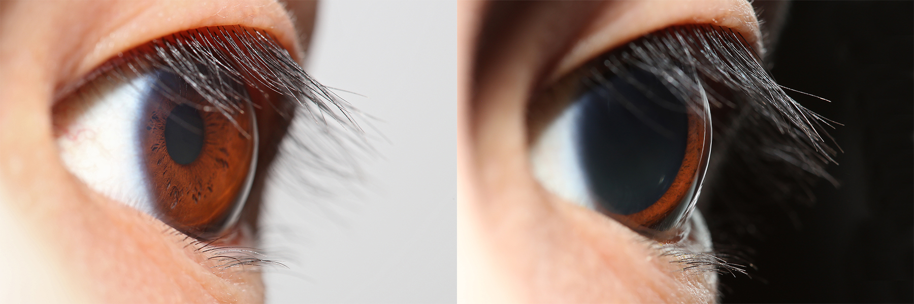
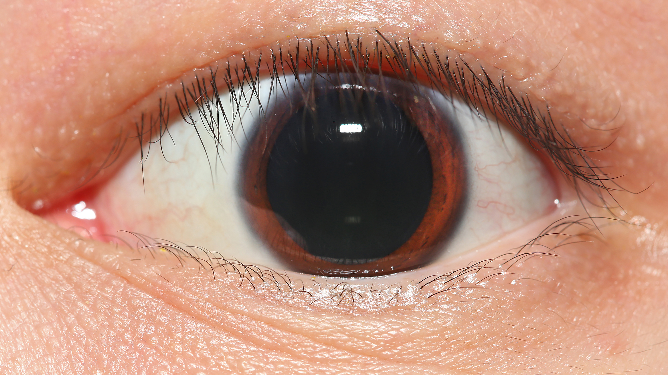
The iris (: irides or irises) is a thin, annular structure in the eye in most mammals and birds that is responsible for controlling the diameter and size of the pupil, and thus the amount of light reaching the retina. In optical terms, the pupil is the eye's aperture, while the iris is the diaphragm (optics), diaphragm. Eye color is defined by the iris. Etymology The word "iris" is derived from the Greek word for "rainbow", also Iris (mythology), its goddess plus messenger of the gods in the ''Iliad'', because of the many eye color, colours of this eye part. Structure The iris consists of two layers: the front pigmented Wikt:fibrovascular, fibrovascular layer known as a stroma of iris, stroma and, behind the stroma, pigmented epithelial cells. The stroma is connected to a sphincter muscle (sphincter pupillae), which contracts the pupil in a circular motion, and a set of dilator muscles (dilator pupillae), which pull the iris radially to enlarge the pupil, pulling it in folds. ... [...More Info...] [...Related Items...] OR: [Wikipedia] [Google] [Baidu] |
Melanosome
A melanosome is an organelle found in animal cells and is the site for synthesis, storage and transport of melanin, the most common light-absorbing pigment found in the animal kingdom. Melanosomes are responsible for color and photoprotection in animal cells and tissues. Melanosomes are synthesised in the skin in melanocyte cells, as well as the eye in choroidal melanocytes and retinal pigment epithelial (RPE) cells. In lower vertebrates, they are found in melanophores or chromatophores. Structure Melanosomes are relatively large organelles, measuring up to 500 nm in diameter. They are bound by a bilipid membrane and are, in general, rounded, sausage-like, or cigar-like in shape. The shape is constant for a given species and cell type. They have a characteristic ultrastructure on electron microscopy, which varies according to the maturity of the melanosome, and for research purposes a numeric staging system is sometimes used. Synthesis of melanin Melanosomes are dep ... [...More Info...] [...Related Items...] OR: [Wikipedia] [Google] [Baidu] |
Cytoplasm
The cytoplasm describes all the material within a eukaryotic or prokaryotic cell, enclosed by the cell membrane, including the organelles and excluding the nucleus in eukaryotic cells. The material inside the nucleus of a eukaryotic cell and contained within the nuclear membrane is termed the nucleoplasm. The main components of the cytoplasm are the cytosol (a gel-like substance), the cell's internal sub-structures, and various cytoplasmic inclusions. In eukaryotes the cytoplasm also includes the nucleus, and other membrane-bound organelles.The cytoplasm is about 80% water and is usually colorless. The submicroscopic ground cell substance, or cytoplasmic matrix, that remains after the exclusion of the cell organelles and particles is groundplasm. It is the hyaloplasm of light microscopy, a highly complex, polyphasic system in which all resolvable cytoplasmic elements are suspended, including the larger organelles such as the ribosomes, mitochondria, plant plasti ... [...More Info...] [...Related Items...] OR: [Wikipedia] [Google] [Baidu] |
Iris Dilator Muscle
The iris dilator muscle (pupil dilator muscle, pupillary dilator, radial muscle of iris, radiating fibers), is a smooth muscle of the eye, running radially in the iris and therefore fit as a dilator. The pupillary dilator consists of a spokelike arrangement of modified contractile cells called myoepithelial cells. These cells are stimulated by the sympathetic nervous system. When stimulated, the cells contract, widening the pupil and allowing more light to enter the eye. The ciliary muscle, pupillary sphincter muscle and pupillary dilator muscle sometimes are called intrinsic ocular muscles or intraocular muscles. Structure Innervation It is innervated by the sympathetic system, which acts by releasing noradrenaline, which acts on α1-receptors. Thus, when presented with a threatening stimulus that activates the fight-or-flight response, this innervation contracts the muscle and dilates the pupil, thus temporarily letting more light reach the retina. The dilator muscl ... [...More Info...] [...Related Items...] OR: [Wikipedia] [Google] [Baidu] |
Retina
The retina (; or retinas) is the innermost, photosensitivity, light-sensitive layer of tissue (biology), tissue of the eye of most vertebrates and some Mollusca, molluscs. The optics of the eye create a focus (optics), focused two-dimensional image of the visual world on the retina, which then processes that image within the retina and sends nerve impulses along the optic nerve to the visual cortex to create visual perception. The retina serves a function which is in many ways analogous to that of the photographic film, film or image sensor in a camera. The neural retina consists of several layers of neurons interconnected by Chemical synapse, synapses and is supported by an outer layer of pigmented epithelial cells. The primary light-sensing cells in the retina are the photoreceptor cells, which are of two types: rod cell, rods and cone cell, cones. Rods function mainly in dim light and provide monochromatic vision. Cones function in well-lit conditions and are responsible fo ... [...More Info...] [...Related Items...] OR: [Wikipedia] [Google] [Baidu] |
Optic Cup (embryological)
Optic cup may refer to: * Optic cup (anatomical), the white cup-like area in the center of the optic disc * Optic cup (embryology) During embryonic development of the eye, the outer wall of the bulb of the optic vesicles becomes thickened and invaginated, and the bulb is thus converted into a cup, the optic cup (or ophthalmic cup), consisting of two strata of cells. These tw ..., a structure in embryos that gives rise to the retina of the eye {{disambig ... [...More Info...] [...Related Items...] OR: [Wikipedia] [Google] [Baidu] |
Glaucoma
Glaucoma is a group of eye diseases that can lead to damage of the optic nerve. The optic nerve transmits visual information from the eye to the brain. Glaucoma may cause vision loss if left untreated. It has been called the "silent thief of sight" because the loss of vision usually occurs slowly over a long period of time. A major risk factor for glaucoma is increased pressure within the eye, known as Intraocular pressure, intraocular pressure (IOP). It is associated with old age, a family history of glaucoma, and certain medical conditions or the use of some medications. The word ''glaucoma'' comes from the Ancient Greek word (), meaning 'gleaming, blue-green, gray'. Of the different types of glaucoma, the most common are called open-angle glaucoma and closed-angle glaucoma. Inside the eye, a liquid called Aqueous humour, aqueous humor helps to maintain shape and provides nutrients. The aqueous humor normally drains through the trabecular meshwork. In open-angle glaucoma, ... [...More Info...] [...Related Items...] OR: [Wikipedia] [Google] [Baidu] |
Diabetes
Diabetes mellitus, commonly known as diabetes, is a group of common endocrine diseases characterized by sustained high blood sugar levels. Diabetes is due to either the pancreas not producing enough of the hormone insulin, or the cells of the body becoming unresponsive to insulin's effects. Classic symptoms include polydipsia (excessive thirst), polyuria (excessive urination), polyphagia (excessive hunger), weight loss, and blurred vision. If left untreated, the disease can lead to various health complications, including disorders of the cardiovascular system, eye, kidney, and nerves. Diabetes accounts for approximately 4.2 million deaths every year, with an estimated 1.5 million caused by either untreated or poorly treated diabetes. The major types of diabetes are type 1 and type 2. The most common treatment for type 1 is insulin replacement therapy (insulin injections), while anti-diabetic medications (such as metformin and semaglutide) and lifestyle modificatio ... [...More Info...] [...Related Items...] OR: [Wikipedia] [Google] [Baidu] |
Photophobia
Photophobia is a medical symptom of abnormal intolerance to visual perception of light. As a medical symptom, photophobia is not a morbid fear or phobia, but an experience of discomfort or pain to the eyes due to light exposure or by presence of actual physical sensitivity of the eyes, though the term is sometimes additionally applied to abnormal or irrational fear of light, such as heliophobia. The term ''photophobia'' comes . Causes Patients may develop photophobia as a result of several different medical conditions, related to the human eye, eye, the nervous system, genetic, or other causes. Photophobia may manifest itself in an increased response to light starting at any step in the visual system, such as: * Too much light entering the eye. Too much light can enter the eye if it is damaged, such as with corneal abrasion and retinal damage, or if its pupil is unable to normally constrict (seen with damage to the oculomotor nerve). * Due to albinism, the lack of pigment in th ... [...More Info...] [...Related Items...] OR: [Wikipedia] [Google] [Baidu] |


