|
Intraparenchymal Hemorrhage
Intraparenchymal hemorrhage is one form of intracerebral bleeding in which there is bleeding within brain parenchyma. The other form is intraventricular hemorrhage). Intraparenchymal hemorrhage accounts for approximately 8-13% of all strokes and results from a wide spectrum of disorders. It is more likely to result in death or major disability than ischemic stroke or subarachnoid hemorrhage, and therefore constitutes an immediate medical emergency. Intracerebral hemorrhages and accompanying edema may disrupt or compress adjacent brain tissue, leading to neurological dysfunction. Substantial displacement of brain parenchyma may cause elevation of intracranial pressure (ICP) and potentially fatal Herniation (brain), herniation syndromes. Signs and symptoms Clinical manifestations of intraparenchymal hemorrhage are determined by the size and location of hemorrhage, but may include the following: * Hypertension, fever, or cardiac arrhythmias * Nuchal rigidity * Subhyaloid retinal h ... [...More Info...] [...Related Items...] OR: [Wikipedia] [Google] [Baidu] |
Intracerebral Bleeding
Intracerebral hemorrhage (ICH), also known as hemorrhagic stroke, is a sudden bleeding into Intraparenchymal hemorrhage, the tissues of the brain (i.e. the parenchyma), into its Intraventricular hemorrhage, ventricles, or into both. An ICH is a type of bleeding within the skull and one kind of stroke (ischemic stroke being the other). Symptoms can vary dramatically depending on the severity (how much blood), acuity (over what timeframe), and location (anatomically) but can include headache, Hemiparesis, one-sided weakness, numbness, tingling, or Hemiplegia, paralysis, speech problems, vision or hearing problems, memory loss, attention problems, coordination problems, balance problems, dizziness or Presyncope, lightheadedness or vertigo, nausea/vomiting, seizures, decreased level of consciousness or Unconsciousness, total loss of consciousness, neck stiffness, and fever. Hemorrhagic stroke may occur on the background of alterations to the blood vessels in the brain, such as cer ... [...More Info...] [...Related Items...] OR: [Wikipedia] [Google] [Baidu] |
Anisocoria
Anisocoria is a condition characterized by an unequal size of the eyes' pupils. Affecting up to 20% of the population, anisocoria is often entirely harmless, but can be a sign of more serious medical problems. Causes Anisocoria is a common condition, defined by a diameter difference of 0.4 mm or more between the sizes of the pupils of the eyes. Anisocoria has various causes: * Physiological anisocoria: About 20% of the population has a slight difference in pupil size, which is known as physiological anisocoria. In this condition, the difference between pupils is usually less than 1 mm. * Horner's syndrome * Mechanical anisocoria: Occasionally, previous trauma, eye surgery, or inflammation ( uveitis, angle closure glaucoma) can lead to adhesions between the iris and the lens. * Adie tonic pupil: Tonic pupil is usually an isolated benign entity, presenting in young women. It may be associated with loss of deep tendon reflex (Adie's syndrome). Tonic pupil is charac ... [...More Info...] [...Related Items...] OR: [Wikipedia] [Google] [Baidu] |
Autonomic Instability
Dysautonomia, autonomic failure, or autonomic dysfunction is a condition in which the autonomic nervous system (ANS) does not work properly. This condition may affect the functioning of the heart, bladder, intestines, sweat glands, pupils, and blood vessels. Dysautonomia has many causes, not all of which may be classified as neuropathic. A number of conditions can feature dysautonomia, such as Parkinson's disease, multiple system atrophy, dementia with Lewy bodies, Ehlers–Danlos syndromes, autoimmune autonomic ganglionopathy and autonomic neuropathy, HIV/AIDS, mitochondrial cytopathy, pure autonomic failure, autism, and postural orthostatic tachycardia syndrome. Diagnosis is made by functional testing of the ANS, focusing on the affected organ system. Investigations may be performed to identify underlying disease processes that may have led to the development of symptoms or autonomic neuropathy. Symptomatic treatment is available for many symptoms associated with dy ... [...More Info...] [...Related Items...] OR: [Wikipedia] [Google] [Baidu] |
Tetraparesis
Tetraplegia, also known as quadriplegia, is defined as the dysfunction or loss of motor and/or sensory function in the cervical area of the spinal cord. A loss of motor function can present as either weakness or paralysis leading to partial or total loss of function in the arms, legs, trunk, and pelvis. ( Paraplegia is similar but affects the thoracic, lumbar, and sacral segments of the spinal cord and arm function is retained.) The paralysis may be flaccid or spastic. A loss of sensory function can present as an impairment or complete inability to sense light touch, pressure, heat, pinprick/pain, and proprioception. In these types of spinal cord injury, it is common to have a loss of both sensation and motor control. Signs and symptoms Although the most obvious symptom is impairment of the limbs, functioning is also impaired in the trunk and pelvic organs. This can lead to loss or impairment of controlling bowel and bladder, sexual function, digestion, breathing and ot ... [...More Info...] [...Related Items...] OR: [Wikipedia] [Google] [Baidu] |
Brain Stem
The brainstem (or brain stem) is the posterior stalk-like part of the brain that connects the cerebrum with the spinal cord. In the human brain the brainstem is composed of the midbrain, the pons, and the medulla oblongata. The midbrain is continuous with the thalamus of the diencephalon through the tentorial notch, and sometimes the diencephalon is included in the brainstem. The brainstem is very small, making up around only 2.6 percent of the brain's total weight. It has the critical roles of regulating heart and respiratory function, helping to control heart rate and breathing rate. It also provides the main motor and sensory nerve supply to the face and neck via the cranial nerves. Ten pairs of cranial nerves come from the brainstem. Other roles include the regulation of the central nervous system and the body's sleep cycle. It is also of prime importance in the conveyance of motor and sensory pathways from the rest of the brain to the body, and from the body back ... [...More Info...] [...Related Items...] OR: [Wikipedia] [Google] [Baidu] |
Caudate Nucleus
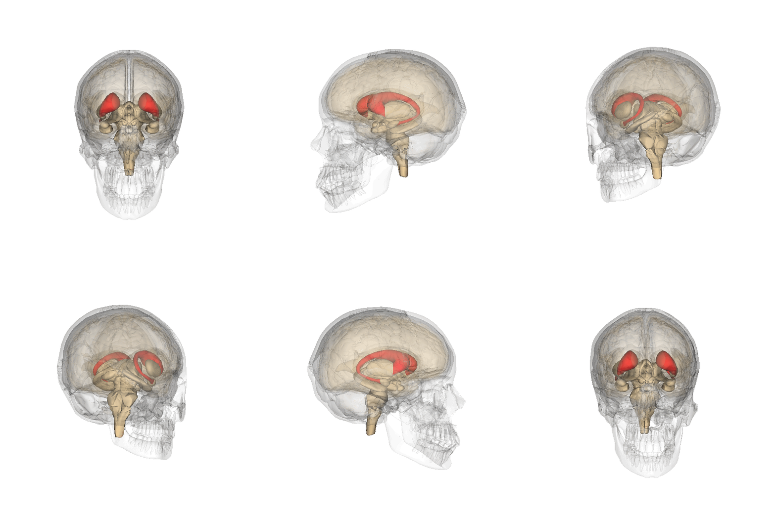
The caudate nucleus is one of the structures that make up the corpus striatum, which is part of the basal ganglia in the human brain. Although the caudate nucleus has long been associated with motor processes because of its relation to Parkinson's disease and Huntington's disease, it also plays important roles in nonmotor functions, such as procedural learning, associative learning, and inhibitory control of action. The caudate is also one of the brain structures that compose the reward system, and it functions as part of the cortico-basal ganglia-thalamo-cortical loop. Structure Along with the putamen, the caudate forms the dorsal striatum, which is considered a single functional structure; anatomically, it is separated by a large white-matter tract, the internal capsule, so it is sometimes also described as two structures—the medial dorsal striatum (the caudate) and the lateral dorsal striatum (the putamen). In this vein, the two are functionally distinct not bec ... [...More Info...] [...Related Items...] OR: [Wikipedia] [Google] [Baidu] |
Abulia
In neurology, abulia, or aboulia (from , meaning "will"),Bailly, A. (2000). Dictionnaire Grec Français, Éditions Hachette. refers to a lack of will or initiative and can be seen as a disorder of diminished motivation. Abulia falls in the middle of the spectrum of diminished motivation, with apathy being less extreme and akinetic mutism being more extreme than abulia.Marin, R. S., & Wilkosz, P. A. (2005)Disorders of diminished motivation. Journal of Head Trauma Rehabilitation, 20(4), 377-388. The condition was originally considered to be a disorder of the will,Berrios G.E. and Gili M. (1995) Abulia and impulsiveness revisited. ''Acta Psychiatrica Scandinavica'' 92: 161-167 and aboulic individuals are unable to act or make decisions independently; and their condition may range in severity from subtle to overwhelming. In the case of akinetic mutism, many patients describe that as soon as they "will" or attempt a movement, a "counter-will" or "resistance" rises up to meet them. ... [...More Info...] [...Related Items...] OR: [Wikipedia] [Google] [Baidu] |
Miosis
Miosis, or myosis (), is excessive constriction of the pupil. citing: Mosby's Medical Dictionary, 8th ed. The opposite condition, mydriasis, is the dilation of the pupil. Anisocoria is the condition of one pupil being more dilated than the other. Causes Age * Senile miosis (a reduction in the size of a person's pupil in old age)Diseases *[...More Info...] [...Related Items...] OR: [Wikipedia] [Google] [Baidu] |
Thalamus
The thalamus (: thalami; from Greek language, Greek Wikt:θάλαμος, θάλαμος, "chamber") is a large mass of gray matter on the lateral wall of the third ventricle forming the wikt:dorsal, dorsal part of the diencephalon (a division of the forebrain). Nerve fibers project out of the thalamus to the cerebral cortex in all directions, known as the thalamocortical radiations, allowing hub (network science), hub-like exchanges of information. It has several functions, such as the relaying of sensory neuron, sensory and motor neuron, motor signals to the cerebral cortex and the regulation of consciousness, sleep, and alertness. Anatomically, the thalami are paramedian symmetrical structures (left and right), within the vertebrate brain, situated between the cerebral cortex and the midbrain. It forms during embryonic development as the main product of the diencephalon, as first recognized by the Swiss embryologist and anatomist Wilhelm His Sr. in 1893. Anatomy The thalami ar ... [...More Info...] [...Related Items...] OR: [Wikipedia] [Google] [Baidu] |
Apraxia
Apraxia is a motor disorder caused by damage to the brain (specifically the posterior parietal cortex or corpus callosum), which causes difficulty with motor planning to perform tasks or movements. The nature of the damage determines the disorder's severity, and the absence of sensory loss or paralysis helps to explain the level of difficulty. Children may be born with apraxia; its cause is unknown, and symptoms are usually noticed in the early stages of development. Apraxia occurring later in life, known as ''acquired apraxia'', is typically caused by traumatic brain injury, stroke, dementia, Alzheimer's disease, brain tumor, or other neurodegenerative disorders. The multiple types of apraxia are categorized by the specific ability and/or body part affected. The term "apraxia" comes . Types The several types of apraxia include: * Apraxia of speech (AOS) is having difficulty planning and coordinating the movements necessary for speech (e.g. potato=totapo, topato).Heilma ... [...More Info...] [...Related Items...] OR: [Wikipedia] [Google] [Baidu] |
Aphasia
Aphasia, also known as dysphasia, is an impairment in a person's ability to comprehend or formulate language because of dysfunction in specific brain regions. The major causes are stroke and head trauma; prevalence is hard to determine, but aphasia due to stroke is estimated to be 0.1–0.4% in developed countries. Aphasia can also be the result of brain tumors, epilepsy, autoimmune neurological diseases, brain infections, or neurodegenerative diseases (such as dementias). To be diagnosed with aphasia, a person's language must be significantly impaired in one or more of the four aspects of communication. In the case of progressive aphasia, a noticeable decline in language abilities over a short period of time is required. The four aspects of communication include spoken language production, spoken language comprehension, written language production, and written language comprehension. Impairments in any of these aspects can impact functional communication. The difficulties o ... [...More Info...] [...Related Items...] OR: [Wikipedia] [Google] [Baidu] |
Homonymous Hemianopsia
Hemianopsia, or hemianopia, is a visual field loss on the left or right side of the vertical midline. It can affect one eye but usually affects both eyes. Homonymous hemianopsia (or homonymous hemianopia) is hemianopic visual field loss on the same side of both eyes. Homonymous hemianopsia occurs because the right half of the brain has visual pathways for the left hemifield of both eyes, and the left half of the brain has visual pathways for the right hemifield of both eyes. When one of these pathways is damaged, the corresponding visual field is lost. Signs and symptoms Paris as seen with right homonymous hemianopsia Mobility can be difficult for people with homonymous hemianopsia. "Patients frequently complain of bumping into obstacles on the side of the field loss, thereby bruising their arms and legs." People with homonymous hemianopsia often experience discomfort in crowds. "A patient with this condition may be unaware of what he or she cannot see and frequently bumps ... [...More Info...] [...Related Items...] OR: [Wikipedia] [Google] [Baidu] |


