|
Intraembryonic Coelom
In the development of the human embryo, the intraembryonic coelom (or somatic coelom) is a portion of the conceptus forming in the mesoderm during the third week of development. During the third week of development, the lateral plate mesoderm splits into a dorsal somatic mesoderm ( somatopleure) and a ventral splanchnic mesoderm ( splanchnopleure). The resulting cavity between the somatopleure and splanchnopleure is called the intraembryonic coelom. This space will give rise to the thoracic and abdominal cavities. The coelomic spaces in the lateral mesoderm and cardiogenic area are isolated. The isolated coelom The coelom (or celom) is the main body cavity in many animals and is positioned inside the body to surround and contain the digestive tract and other organs. In some animals, it is lined with mesothelium. In other animals, such as molluscs, i ... begins to organize into a horseshoe shape. The spaces soon join together and form a single horseshoe-shaped cavity: the int ... [...More Info...] [...Related Items...] OR: [Wikipedia] [Google] [Baidu] |
Lateral Plate Mesoderm
The lateral plate mesoderm is the mesoderm that is found at the periphery of the embryo. It is to the side of the paraxial mesoderm, and further to the axial mesoderm. The lateral plate mesoderm is separated from the paraxial mesoderm by a narrow region of intermediate mesoderm. The mesoderm is the middle layer of the three germ layers, between the outer ectoderm and inner endoderm. During the third week of embryonic development the lateral plate mesoderm splits into two layers forming the intraembryonic coelom. The outer layer of lateral plate mesoderm adheres to the ectoderm to become the somatic or parietal layer known as the somatopleure. The inner layer adheres to the endoderm to become the splanchnic or visceral layer known as the splanchnopleure. Development The lateral plate mesoderm will split into two layers, the somatopleuric mesenchyme, and the splanchnopleuric mesenchyme. * The ''somatopleuric layer'' forms the future body wall. * The ''splanchnopleuric layer'' forms ... [...More Info...] [...Related Items...] OR: [Wikipedia] [Google] [Baidu] |
Pericardial Cavity
The pericardium (: pericardia), also called pericardial sac, is a double-walled sac containing the heart and the roots of the great vessels. It has two layers, an outer layer made of strong inelastic connective tissue (fibrous pericardium), and an inner layer made of serous membrane (serous pericardium). It encloses the pericardial cavity, which contains pericardial fluid, and defines the middle mediastinum. It separates the heart from interference of other structures, protects it against infection and blunt trauma, and lubricates the heart's movements. The English name originates from the Ancient Greek prefix ''peri-'' (περί) 'around' and the suffix ''-cardion'' (κάρδιον) 'heart'. Anatomy The pericardium is a tough fibroelastic sac which covers the heart from all sides except at the cardiac root (where the great vessels join the heart) and the bottom (where only the serous pericardium exists to cover the upper surface of the central tendon of diaphragm). The ... [...More Info...] [...Related Items...] OR: [Wikipedia] [Google] [Baidu] |
Pleural Cavity
The pleural cavity, or pleural space (or sometimes intrapleural space), is the potential space between the pleurae of the pleural sac that surrounds each lung. A small amount of serous pleural fluid is maintained in the pleural cavity to enable lubrication between the membranes, and also to create a pressure gradient. The serous membrane that covers the surface of the lung is the visceral pleura and is separated from the outer membrane, the parietal pleura, by just the film of pleural fluid in the pleural cavity. The visceral pleura follows the fissures of the lung and the root of the lung structures. The parietal pleura is attached to the mediastinum, the upper surface of the diaphragm, and to the inside of the ribcage. Structure In humans, the left and right lungs are completely separated by the mediastinum, and there is no communication between their pleural cavities. Therefore, in cases of a unilateral pneumothorax, the contralateral lung will remain functioning n ... [...More Info...] [...Related Items...] OR: [Wikipedia] [Google] [Baidu] |
Peritoneal Cavity
The peritoneal cavity is a potential space located between the two layers of the peritoneum—the parietal peritoneum, the serous membrane that lines the abdominal wall, and visceral peritoneum, which surrounds the internal organs. While situated within the abdominal cavity, the term ''peritoneal cavity'' specifically refers to the potential space enclosed by these peritoneal membranes. The cavity contains a thin layer of lubricating serous fluid that enables the organs to move smoothly against each other, facilitating the movement and expansion of internal organs during digestion. The parietal and visceral peritonea are named according to their location and function. The peritoneal cavity, derived from the coelomic cavity in the embryo, is one of several body cavities, including the pleural cavities surrounding the lungs and the pericardial cavity around the heart. The peritoneal cavity is the largest serosal sac and fluid-filled cavity in the body, it secretes approximately ... [...More Info...] [...Related Items...] OR: [Wikipedia] [Google] [Baidu] |
Conceptus
A conceptus (from Latin Latin ( or ) is a classical language belonging to the Italic languages, Italic branch of the Indo-European languages. Latin was originally spoken by the Latins (Italic tribe), Latins in Latium (now known as Lazio), the lower Tiber area aroun ...: ''concipere'', to conceive) is an embryo and its appendages (adnexa), the associated membranes, placenta, and umbilical cord; the products of conception or, more broadly, "the product of conception at any point between fertilization and birth."'' The American Heritage Dictionary of the English Language'' (2011), 5th ed., p. 381. The conceptus includes all structures that develop from the zygote, both embryonic and extraembryonic. It includes the embryo as well as the embryonic part of the placenta and its associated membranes: amnion, chorion ( gestational sac), and yolk sac. References Embryology Human pregnancy {{Developmental-biology-stub ... [...More Info...] [...Related Items...] OR: [Wikipedia] [Google] [Baidu] |
Mesoderm
The mesoderm is the middle layer of the three germ layers that develops during gastrulation in the very early development of the embryo of most animals. The outer layer is the ectoderm, and the inner layer is the endoderm.Langman's Medical Embryology, 11th edition. 2010. The mesoderm forms mesenchyme, mesothelium and coelomocytes. Mesothelium lines coeloms. Mesoderm forms the muscles in a process known as myogenesis, septa (cross-wise partitions) and mesenteries (length-wise partitions); and forms part of the gonads (the rest being the gametes). Myogenesis is specifically a function of mesenchyme. The mesoderm differentiates from the rest of the embryo through intercellular signaling, after which the mesoderm is polarized by an organizing center. The position of the organizing center is in turn determined by the regions in which beta-catenin is protected from degradation by GSK-3. Beta-catenin acts as a co-factor that alters the activity of the transcription facto ... [...More Info...] [...Related Items...] OR: [Wikipedia] [Google] [Baidu] |
Lateral Plate Mesoderm
The lateral plate mesoderm is the mesoderm that is found at the periphery of the embryo. It is to the side of the paraxial mesoderm, and further to the axial mesoderm. The lateral plate mesoderm is separated from the paraxial mesoderm by a narrow region of intermediate mesoderm. The mesoderm is the middle layer of the three germ layers, between the outer ectoderm and inner endoderm. During the third week of embryonic development the lateral plate mesoderm splits into two layers forming the intraembryonic coelom. The outer layer of lateral plate mesoderm adheres to the ectoderm to become the somatic or parietal layer known as the somatopleure. The inner layer adheres to the endoderm to become the splanchnic or visceral layer known as the splanchnopleure. Development The lateral plate mesoderm will split into two layers, the somatopleuric mesenchyme, and the splanchnopleuric mesenchyme. * The ''somatopleuric layer'' forms the future body wall. * The ''splanchnopleuric layer'' forms ... [...More Info...] [...Related Items...] OR: [Wikipedia] [Google] [Baidu] |
Somatopleure
In the anatomy of an embryo, the somatopleure is a structure created during embryogenesis when the lateral plate mesoderm splits into two layers. The outer (or somatic) layer becomes applied to the inner surface of the ectoderm, and with it (partially) forms the somatopleure. The combination of ectoderm and mesoderm, or somatopleure, forms the amnion, the chorion and the lateral body wall of the embryo. Limb formation, from the somatic mesoderm, is induced by hox genes and the expression of other molecules through an epithelial-mesenchyme transition. The embryonic somatopleure is then divided into 3 sections, the anterior limb bud formation, the posterior limb bud formation and the non limb forming wall. The bud forming sections grow in size. The somatic mesoderm under the ectoderm proliferates in mesenchyme form. In chicken, the extraembryonic tissues are separated into two layers: the splanchnopleure composed of the endoderm and splanchnic mesoderm, and the somatopleure com ... [...More Info...] [...Related Items...] OR: [Wikipedia] [Google] [Baidu] |
Splanchnopleure
In the anatomy of an embryo, the splanchnopleuric mesenchyme is a structure created during embryogenesis when the lateral mesodermal germ layer splits into two layers. The inner (or splanchnic) layer adheres to the endoderm, and with it forms the splanchnopleure (mesoderm external to the coelom plus the endoderm). See also Post development the somato and splanchnopleuric junction lies at the duodeno-jejunal flexure. * somatopleure * mesenchyme References External links * * Overview at Kennesaw State University Kennesaw State University (KSU) is a public research university in the U.S. state of Georgia with two campuses in the Atlanta metropolitan area, one in the Kennesaw area and the other in Marietta on a combined of land. The school was founded ... Embryology {{developmental-biology-stub ... [...More Info...] [...Related Items...] OR: [Wikipedia] [Google] [Baidu] |
Coelom
The coelom (or celom) is the main body cavity in many animals and is positioned inside the body to surround and contain the digestive tract and other organs. In some animals, it is lined with mesothelium. In other animals, such as molluscs, it remains undifferentiated. In the past, and for practical purposes, coelom characteristics have been used to classify bilaterian animal phyla into informal groups. Etymology The term ''coelom'' derives from the Ancient Greek word () 'cavity'. Structure Development The coelom is the mesodermally lined cavity between the gut and the outer body wall. During the development of the embryo, coelom formation begins in the gastrulation stage. The developing digestive tube of an embryo forms as a blind pouch called the archenteron. In protostomes, the coelom forms by a process known as schizocoely. The archenteron initially forms, and the mesoderm splits into two layers: the first attaches to the body wall or ectoderm, forming ... [...More Info...] [...Related Items...] OR: [Wikipedia] [Google] [Baidu] |
Extraembryonic Coelom
The gestational sac is the large cavity of fluid surrounding the embryo. During early embryogenesis, it consists of the extraembryonic coelom, also called the chorionic cavity. The gestational sac is normally contained within the uterus. It is the only available structure that can be used to determine if an intrauterine pregnancy exists until the embryo can be identified. On obstetric ultrasound, the gestational sac is a dark ( anechoic) space surrounded by a white ( hyperechoic) rim. Structure The gestational sac is spherical in shape, and is usually located in the upper part (fundus) of the uterus. By approximately nine weeks of gestational age, due to folding of the trilaminar germ disc, the amniotic sac expands and occupy the majority of the volume of the gestational sac, eventually reducing the extraembryonic coelom (the gestational sac or the chorionic cavity) to a thin layer between the parietal somatopleuric and visceral splanchnopleuric layer of extraembryonic mesoderm. ... [...More Info...] [...Related Items...] OR: [Wikipedia] [Google] [Baidu] |
Cavitation (embryology)
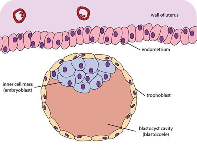
Cavitation is a process in early embryonic development that follows cleavage. Cavitation is the formation of the blastocoel, a fluid-filled cavity that defines the blastula, or in mammals the blastocyst. After fertilization, cell division of the zygote occurs which results in the formation of a solid ball of cells (blastomeres) called the morula. Further division of cells increases their number in the morula, and the morula differentiates them into two groups. The internal cells become the inner cell mass, and the outer cells become the trophoblast. Before cell differentiation takes place there are two transcription factors, Oct-4 and nanog that are uniformly expressed on all of the cells, but both of these transcription factors are turned off in the trophoblast once it has formed. The trophoblast cells form tight junction Tight junctions, also known as occluding junctions or ''zonulae occludentes'' (singular, ''zonula occludens''), are multiprotein Cell junction, junctiona ... [...More Info...] [...Related Items...] OR: [Wikipedia] [Google] [Baidu] |


