|
Infraorbital Canal
The infraorbital canal is a canal found at the base of the orbit that opens on to the maxilla. It is continuous with the infraorbital groove and opens onto the maxilla at the infraorbital foramen. The infraorbital nerve and infraorbital artery travel through the canal. Structure One of the canals of the orbital surface of the maxilla, the infraorbital canal, opens just below the margin of the orbit, the area of the skull containing the eye and related structures. It should not be confused with the infraorbital foramen, with which it is continuous. Function It transmits the infraorbital nerve as well as infraorbital artery, both of which enter this canal at the infraorbital groove and after coursing through the maxillary sinus exit via the infraorbital foramen In human anatomy, the infraorbital foramen is one of two small holes in the skull's upper jawbone ( maxillary bone), located below the eye socket and to the left and right of the nose. Both holes are used for blood vesse ... [...More Info...] [...Related Items...] OR: [Wikipedia] [Google] [Baidu] |
Infraorbital Groove
The infraorbital groove (or sulcus) is located in the middle of the posterior part of the orbital surface of the maxilla. Its function is to act as the passage of the infraorbital artery, the infraorbital vein, and the infraorbital nerve. Structure The infraorbital groove begins at the middle of the posterior border of the maxilla (with which it is continuous). This is near the upper edge of the infratemporal surface of the maxilla. It passes forward, and ends in a canal which subdivides into two branches. The infraorbital groove has an average length of 16.7 mm, with a small amount of variation between people. It is similar in men and women. Function The infraorbital groove creates space that allows for passage of the infraorbital artery, the infraorbital vein, and the infraorbital nerve. Clinical significance The infraorbital groove is an important Landmark, surgical landmark for Local anesthesia, local anaesthesia of the infraorbital nerve. See also * Infraorbital f ... [...More Info...] [...Related Items...] OR: [Wikipedia] [Google] [Baidu] |
Infraorbital Foramen
In human anatomy, the infraorbital foramen is one of two small holes in the skull's upper jawbone ( maxillary bone), located below the eye socket and to the left and right of the nose. Both holes are used for blood vessels and nerves. In anatomical terms, it is located below the infraorbital margin of the orbit. It transmits the infraorbital artery and vein, and the infraorbital nerve, a branch of the maxillary nerve. It is typically from the infraorbital margin. Structure Forming the exterior end of the infraorbital canal, the infraorbital foramen communicates with the infraorbital groove, the canal's opening on the interior side. The ramifications of the three principal branches of the trigeminal nerve—at the supraorbital, infraorbital, and mental foramen—are distributed on a vertical line (in anterior view) passing through the middle of the pupil. The infraorbital foramen is used as a pressure point to test the sensitivity of the infraorbital nerve. Palpation of th ... [...More Info...] [...Related Items...] OR: [Wikipedia] [Google] [Baidu] |
Canal (anatomy)
{{notability, date=April 2024 In anatomy, a canal (or ''canalis'' in Latin) is a tubular passage or channel which connects different regions of the body. Examples Cranial region * Alveolar canals * Carotid canal * Facial canal * Greater palatine canal * Incisive canals * Infraorbital canal * Mandibular canal * Optic canal * Palatovaginal canal * Pterygoid canal Abdominal region * Inguinal canal Pelvic region * Anal canal * Cervical canal * Pudendal canal Upper extremities * Suprascapular canal * Carpal canal * Ulnar canal * Radial canal Lower extremities * Adductor canal * Femoral canal * Obturator canal See also * Foramen In anatomy and osteology, a foramen (; : foramina, or foramens ; ) is an opening or enclosed gap within the dense connective tissue (bones and deep fasciae) of extant and extinct amniote animals, typically to allow passage of nerves, artery, ... References *''Dorland's Illustrated Medical Dictionary'', 27th ed. 1988 W.B. Saunders Co ... [...More Info...] [...Related Items...] OR: [Wikipedia] [Google] [Baidu] |
Orbit (anatomy)
In anatomy Anatomy () is the branch of morphology concerned with the study of the internal structure of organisms and their parts. Anatomy is a branch of natural science that deals with the structural organization of living things. It is an old scien ..., the orbit is the Body cavity, cavity or socket/hole of the skull in which the eye and Accessory visual structures, its appendages are situated. "Orbit" can refer to the bony socket, or it can also be used to imply the contents. In the adult human, the volume of the orbit is about , of which the eye occupies . The orbital contents comprise the eye, the Orbital fascia, orbital and retrobulbar fascia, extraocular muscles, cranial nerves optic nerve, II, oculomotor nerve, III, trochlear nerve, IV, trigeminal nerve, V, and abducens nerve, VI, blood vessels, fat, the lacrimal gland with its Lacrimal sac, sac and nasolacrimal duct, duct, the eyelids, Medial palpebral ligament, medial and Lateral palpebral raphe, lateral palpebr ... [...More Info...] [...Related Items...] OR: [Wikipedia] [Google] [Baidu] |
Maxilla
In vertebrates, the maxilla (: maxillae ) is the upper fixed (not fixed in Neopterygii) bone of the jaw formed from the fusion of two maxillary bones. In humans, the upper jaw includes the hard palate in the front of the mouth. The two maxillary bones are fused at the intermaxillary suture, forming the anterior nasal spine. This is similar to the mandible (lower jaw), which is also a fusion of two mandibular bones at the mandibular symphysis. The mandible is the movable part of the jaw. Anatomy Structure The maxilla is a paired bone - the two maxillae unite with each other at the intermaxillary suture. The maxilla consists of: * The body of the maxilla: pyramid-shaped; has an orbital, a nasal, an infratemporal, and a facial surface; contains the maxillary sinus. * Four processes: ** the zygomatic process ** the frontal process ** the alveolar process ** the palatine process It has three surfaces: * the anterior, posterior, medial Features of the maxilla include: * t ... [...More Info...] [...Related Items...] OR: [Wikipedia] [Google] [Baidu] |
Infraorbital Nerve
The infraorbital nerve is a branch of the maxillary nerve (itself a branch of the trigeminal nerve (CN V)). It arises in the pterygopalatine fossa. It passes through the inferior orbital fissure to enter the orbit. It travels through the orbit, then enters and traverses the infraorbital canal, exiting the canal at the infraorbital foramen to reach the face. It provides sensory innervation to the skin and mucous membranes around the middle of the face. Structure Origin The infraorbital nerve is a branch of the maxillary nerve (CN V2), itself a branch of the trigeminal nerve (CN V); it may be considered as the terminal branch of the maxillary nerve. It arises from the maxillary nerve in the pterygopalatine fossa. Course It travels through the inferior orbital fissure to enter the orbit. It runs anteriorly along the floor of the orbit in the infraorbital groove to the infraorbital canal of the maxilla. Within the infraorbital canal it has three branches, the posterior ... [...More Info...] [...Related Items...] OR: [Wikipedia] [Google] [Baidu] |
Infraorbital Artery
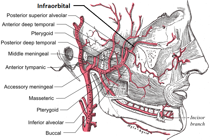
The infraorbital artery is a small artery in the head that arises from the maxillary artery and passes through the inferior orbital fissure to enter the orbit, then passes forward along the floor of the orbit, finally exiting the orbit through the infraorbital foramen to reach the face. Anatomy Origin The infraorbital artery arises from the maxillary artery; it often arises in conjunction with the posterior superior alveolar artery. It may be considered a continuation of the third part of the maxillary artery and continues the direction of the maxillary artery. Course It passes anterior-ward to enter the orbit through the inferior orbital fissure. In the orbit, it courses along the floor of the orbit with the infraorbital nerve first along the infraorbital groove and then the infraorbital canal. It exits the orbit (with the infraorbital nerve) through infraorbital foramen to reach the face, beneath the infraorbital head of the levator labii superioris muscle. Branches Wh ... [...More Info...] [...Related Items...] OR: [Wikipedia] [Google] [Baidu] |
Canal (other)
A canal Canals or artificial waterways are waterways or engineered channels built for drainage management (e.g. flood control and irrigation) or for conveyancing water transport vehicles (e.g. water taxi). They carry free, calm surface ... is a human-made channel for water. Canal or Canals may also refer to: People (Alphabetical by surname) * David Canal (born 1978), Spanish sprinter * Esteban Canal (1896-1981), Peruvian chess player * Giovanni Antonio Canal (1697–1768), Venetian painter, better known as Canaletto * Richard Canal (born 1953), French science fiction writer * B. de Canals, was a 14th-century Spanish author of a Latin chronicle * María Antònia Canals (1930–2022), Spanish mathematician * Agustín de la Canal (born 1980), Argentine football player * Ramón Alva de la Canal (1892–1985), Mexican painter * Daniel Canal (born June 4th, 2005), work at MOST Program Places * Canal Flats, British Columbia, a village in British Columbia, ... [...More Info...] [...Related Items...] OR: [Wikipedia] [Google] [Baidu] |
Skull
The skull, or cranium, is typically a bony enclosure around the brain of a vertebrate. In some fish, and amphibians, the skull is of cartilage. The skull is at the head end of the vertebrate. In the human, the skull comprises two prominent parts: the neurocranium and the facial skeleton, which evolved from the first pharyngeal arch. The skull forms the frontmost portion of the axial skeleton and is a product of cephalization and vesicular enlargement of the brain, with several special senses structures such as the eyes, ears, nose, tongue and, in fish, specialized tactile organs such as barbels near the mouth. The skull is composed of three types of bone: cranial bones, facial bones and ossicles, which is made up of a number of fused flat and irregular bones. The cranial bones are joined at firm fibrous junctions called sutures and contains many foramina, fossae, processes, and sinuses. In zoology, the openings in the skull are called fenestrae, the most ... [...More Info...] [...Related Items...] OR: [Wikipedia] [Google] [Baidu] |
Anterior Superior Alveolar Nerve
The anterior superior alveolar nerve (or anterior superior dental nerve) is a branch of the infraorbital nerve (itself a branch of the maxillary nerve (CN V2)). It passes through the canalis sinuosus to reach and innervate upper front teeth. Through its nasal branch, it also innervates parts of the nasal cavity. Anatomy Course and distribution It branches from the infraorbital nerve within the infraorbital canal at around the midpoint of this canal and enters the canalis sinuosus. It passes through towards the nose before passing inferior-ward and ramifying into branches which innervate the upper/maxillary incisor and canine teeth; it usually innervates all the anterior teeth. Nasal branch It issues a nasal branch which passes through a minute canal in the lateral wall of the inferior nasal meatus and innervates the mucous membrane of the floor and anterior portion of lateral wall (as far superiorly as the opening of the maxillary sinus) of the nasal cavity. It ultimately ... [...More Info...] [...Related Items...] OR: [Wikipedia] [Google] [Baidu] |
Middle Superior Alveolar Nerve
The middle superior alveolar nerve or middle superior dental nerve is a nerve that drops from the infraorbital portion of the maxillary nerve to supply the sinus mucosa, the roots of the maxillary premolars, and the mesiobuccal root of the first maxillary molar. It is not always present; in 72% of cases it is non existent with the anterior superior alveolar nerve innervating the premolars and the posterior superior alveolar nerve innervating the molars, including the mesiobuccal root of the first molar. See also * Alveolar nerve ( Dental nerve) :* Superior alveolar nerve ( Superior dental nerve) ::* Anterior superior alveolar nerve ( Anterior superior dental nerve) ::* Posterior superior alveolar nerve ( Posterior superior dental nerve) :* Inferior alveolar nerve The inferior alveolar nerve (IAN) (also the inferior dental nerve) is a sensory branch of the mandibular nerve (CN V3) (which is itself the third branch of the trigeminal nerve (CN V)). The nerve provides sen ... [...More Info...] [...Related Items...] OR: [Wikipedia] [Google] [Baidu] |


