|
Howell–Jolly Body
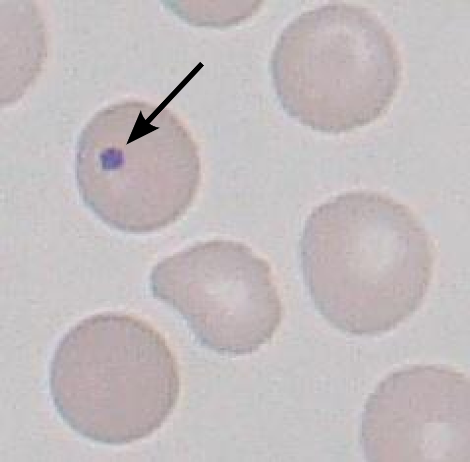
A Howell–Jolly body is a cytopathology, cytopathological finding of basophilic cell nucleus, nuclear remnants (clusters of DNA) in circulating erythrocytes. During maturation in the bone marrow, late erythroblasts normally expel their nuclei; but, in some cases, a small portion of DNA remains. The presence of Howell–Jolly bodies usually signifies a damaged or absent spleen, because a healthy spleen would normally filter such erythrocytes. The Howell–Jolly body is named after William Henry Howell and Justin Marie Jolly. Appearance This DNA appears as a basophilic (purple) spot on the otherwise eosinophilic (pink) erythrocyte on a standard H&E stained blood smear. These inclusions are normally removed by the spleen during erythrocyte circulation, but will persist in individuals with functional hyposplenia or asplenia. Causes Howell–Jolly bodies are seen with markedly decreased splenic function. Common causes include asplenia (post-splenectomy) or congenital absence of s ... [...More Info...] [...Related Items...] OR: [Wikipedia] [Google] [Baidu] |
Asplenia
Asplenia is the absence of normal spleen function and is associated with some serious infection risks. Hyposplenism is the condition of reduced ('hypo-'), but not absent, splenic functioning. ''Functional'' asplenia occurs when splenic tissue is present but does not work well (e.g. sickle-cell disease, polysplenia) such patients are managed as if asplenic while in ''anatomic'' asplenia, the spleen itself is absent. Causes Congenital * Congenital asplenia is rare. There are two distinct types of genetic disorders: heterotaxy syndromeOnline Mendelian Inheritance in Man. OMIM entry 208530: Right atrial isomerism; RAI. Johns Hopkins University/ref> and isolated congenital asplenia.Online Mendelian Inheritance in Man. Johns Hopkins UniversityOMIM entry 271400: Asplenia, isolated congenital; ICAS. * polysplenia Acquired Acquired asplenia occurs for several reasons: * Following splenectomy due to splenic rupture from trauma or because of tumor * After splenectomy with the ''goal'' ... [...More Info...] [...Related Items...] OR: [Wikipedia] [Google] [Baidu] |
Megaloblastic Anemia
Megaloblastic anemia is a type of macrocytic anemia. An anemia is a red blood cell defect that can lead to an undersupply of oxygen. Megaloblastic anemia results from inhibition of DNA replication, DNA synthesis during red blood cell production. When DNA synthesis is impaired, the cell cycle cannot progress from the G2 growth stage to the mitosis (M) stage. This leads to continuing cell growth without division, which presents as macrocytosis. Megaloblastic anemia has a rather slow onset, especially when compared to that of other anemias. The defect in red cell DNA synthesis is most often due to hypovitaminosis, specifically vitamin B12 deficiency or folate deficiency. Loss of micronutrients may also be a cause. Megaloblastic anemia which is not caused due to hypovitaminosis may be caused by antimetabolites that poison DNA production directly, such as some chemotherapeutic or antimicrobial agents (for example azathioprine or trimethoprim). The pathological state of megaloblastosi ... [...More Info...] [...Related Items...] OR: [Wikipedia] [Google] [Baidu] |
Hemolytic Anemia
Hemolytic anemia or haemolytic anaemia is a form of anemia due to hemolysis, the abnormal breakdown of red blood cells (RBCs), either in the blood vessels (intravascular hemolysis) or elsewhere in the human body (extravascular). This most commonly occurs within the spleen, but also can occur in the reticuloendothelial system or mechanically (prosthetic valve damage). Hemolytic anemia accounts for 5% of all existing anemias. It has numerous possible consequences, ranging from general symptoms to life-threatening systemic effects. The general classification of hemolytic anemia is either intrinsic or extrinsic. Treatment depends on the type and cause of the hemolytic anemia. Symptoms of hemolytic anemia are similar to other forms of anemia (fatigue and shortness of breath), but in addition, the breakdown of red cells leads to jaundice and increases the risk of particular long-term complications, such as gallstones and pulmonary hypertension. Signs and symptoms Symptoms of hemolytic ... [...More Info...] [...Related Items...] OR: [Wikipedia] [Google] [Baidu] |
Amyloidosis
Amyloidosis is a group of diseases in which abnormal proteins, known as amyloid fibrils, build up in tissue. There are several non-specific and vague signs and symptoms associated with amyloidosis. These include fatigue, peripheral edema, weight loss, shortness of breath, palpitations, and Orthostatic hypotension, feeling faint with standing. In AL amyloidosis, specific indicators can include enlargement of the tongue and periorbital purpura. In wild-type ATTR amyloidosis, non-cardiac symptoms include: bilateral carpal tunnel syndrome, lumbar spinal stenosis, biceps tendon rupture, Small fiber peripheral neuropathy, small fiber neuropathy, and autonomic dysfunction. There are about 36 different types of amyloidosis, each due to a specific Proteopathy, protein misfolding. Within these 36 proteins, 19 are grouped into Organ-limited amyloidosis, localized forms, 14 are grouped as Systemic disease, systemic forms, and three proteins can identify as either. These proteins can become ... [...More Info...] [...Related Items...] OR: [Wikipedia] [Google] [Baidu] |
Hodgkin Lymphoma
Hodgkin lymphoma (HL) is a type of lymphoma in which cancer originates from a specific type of white blood cell called lymphocytes, where multinucleated Reed–Sternberg cells (RS cells) are present in the lymph nodes. The condition was named after the English physician Thomas Hodgkin, who first described it in 1832. Symptoms may include fever, night sweats, and weight loss. Often, non-painful lymphadenopathy, enlarged lymph nodes occur in the neck, under the arm, or in the groin. Persons affected may feel tired or be itchy. The two major types of Hodgkin lymphoma are classic Hodgkin lymphoma and nodular lymphocyte-predominant Hodgkin lymphoma. About half of cases of Hodgkin lymphoma are due to Epstein–Barr virus (EBV) and these are generally the classic form. Other risk factors include a family history of the condition and having HIV/AIDS. Diagnosis is conducted by confirming the presence of cancer and identifying Reed–Sternberg cells in lymph node biopsies. The virus-posi ... [...More Info...] [...Related Items...] OR: [Wikipedia] [Google] [Baidu] |
Radiation Therapy
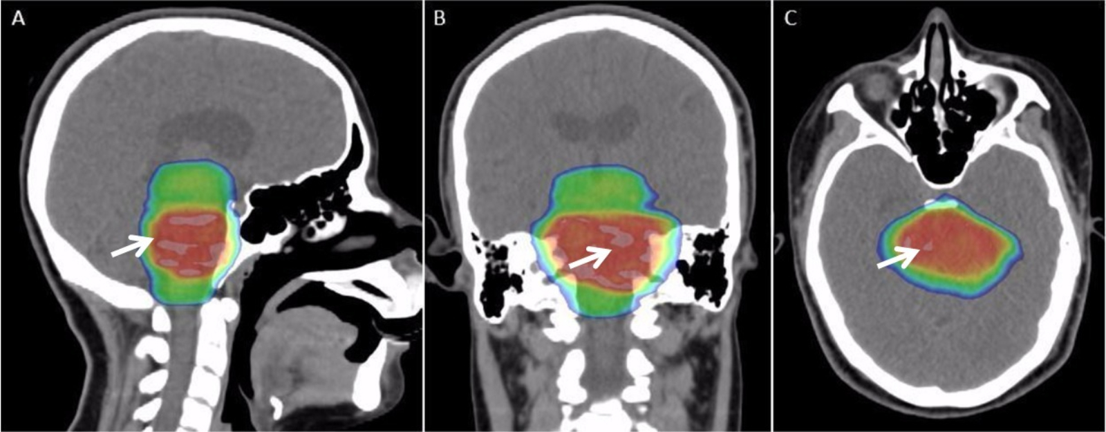
Radiation therapy or radiotherapy (RT, RTx, or XRT) is a therapy, treatment using ionizing radiation, generally provided as part of treatment of cancer, cancer therapy to either kill or control the growth of malignancy, malignant cell (biology), cells. It is normally delivered by a linear particle accelerator. Radiation therapy may be cure, curative in a number of types of cancer if they are localized to one area of the body, and have not metastasis, spread to other parts. It may also be used as part of adjuvant therapy, to prevent tumor recurrence after surgery to remove a primary malignant tumor (for example, early stages of breast cancer). Radiation therapy is synergistic with chemotherapy, and has been used before, during, and after chemotherapy in susceptible cancers. The subspecialty of oncology concerned with radiotherapy is called radiation oncology. A physician who practices in this subspecialty is a radiation oncologist. Radiation therapy is commonly applied to the canc ... [...More Info...] [...Related Items...] OR: [Wikipedia] [Google] [Baidu] |
Sickle Cell Anemia
Sickle cell disease (SCD), also simply called sickle cell, is a group of inherited haemoglobin-related blood disorders. The most common type is known as sickle cell anemia. Sickle cell anemia results in an abnormality in the oxygen-carrying protein haemoglobin found in red blood cells. This leads to the red blood cells adopting an abnormal sickle-like shape under certain circumstances; with this shape, they are unable to deform as they pass through capillaries, causing blockages. Problems in sickle cell disease typically begin around 5 to 6 months of age. A number of health problems may develop, such as attacks of pain (known as a sickle cell crisis) in joints, anemia, swelling in the hands and feet, bacterial infections, dizziness and stroke. The probability of severe symptoms, including long-term pain, increases with age. Without treatment, people with SCD rarely reach adulthood but with good healthcare, median life expectancy is between 58 and 66 years. All of the ma ... [...More Info...] [...Related Items...] OR: [Wikipedia] [Google] [Baidu] |
Autosplenectomy
An autosplenectomy (from'' 'auto-' ''self,'' '-splen-' ''spleen,'' 'List of -ectomies, -ectomy' ''removal) is a negative outcome of disease and occurs when a disease damages the spleen to such an extent that it becomes shrunken and non-functional. The spleen is an important immunological organ that acts as a filter for red blood cells, triggers phagocytosis of invaders, and mounts an immunological response when necessary. Lack of a spleen, called asplenia, can occur by autosplenectomy or the surgical counterpart, splenectomy. Asplenia can increase susceptibility to infection. Autosplenectomy can occur in cases of sickle-cell disease where the misshapen cells block blood flow to the spleen, causing fibrosis, scarring and eventual atrophy of the organ. Autosplenectomy is a rare condition that is linked to certain diseases but is not a common occurrence. It is also seen in systemic lupus erythematosus (SLE). Consequences Absence of effective splenic function or absence of the whole s ... [...More Info...] [...Related Items...] OR: [Wikipedia] [Google] [Baidu] |
Hereditary Spherocytosis
Hereditary spherocytosis (HS) is a congenital hemolytic disorder wherein a genetic genetic mutation, mutation coding for a structural membrane protein phenotype causes the red blood cells to be sphere-shaped (spherocytosis), rather than the normal biconcave disk shape. This abnormal shape interferes with the cells' ability to flex during blood circulation, and also makes them more prone to hemolysis, rupture under osmotic stress, mechanical stress, or both. Cells with the dysfunctional proteins are degraded in the spleen, which leads to a shortage of erythrocytes and results in hemolytic anemia. HS was first described in 1871, and is the most common cause of inherited hemolysis in populations of northern European descent, with an incidence of 1 in 5000 births. The clinical severity of HS varies from mild (symptom-free carrier), to moderate (anemic, jaundiced, and with splenomegaly), to severe (hemolytic crisis, in-utero hydrops fetalis), because HS is caused by genetic mutations i ... [...More Info...] [...Related Items...] OR: [Wikipedia] [Google] [Baidu] |
Hyposplenia
Asplenia is the absence of normal spleen function and is associated with some serious infection risks. Hyposplenism is the condition of reduced ('hypo-'), but not absent, splenic functioning. ''Functional'' asplenia occurs when splenic tissue is present but does not work well (e.g. sickle-cell disease, polysplenia) such patients are managed as if asplenic while in ''anatomic'' asplenia, the spleen itself is absent. Causes Congenital * Congenital asplenia is rare. There are two distinct types of genetic disorders: heterotaxy syndromeOnline Mendelian Inheritance in Man. OMIM entry 208530: Right atrial isomerism; RAI. Johns Hopkins University/ref> and isolated congenital asplenia.Online Mendelian Inheritance in Man. Johns Hopkins UniversityOMIM entry 271400: Asplenia, isolated congenital; ICAS. * polysplenia Acquired Acquired asplenia occurs for several reasons: * Following splenectomy due to splenic rupture from trauma or because of tumor * After splenectomy with the ''goal'' ... [...More Info...] [...Related Items...] OR: [Wikipedia] [Google] [Baidu] |


