|
High Pressure Receptor
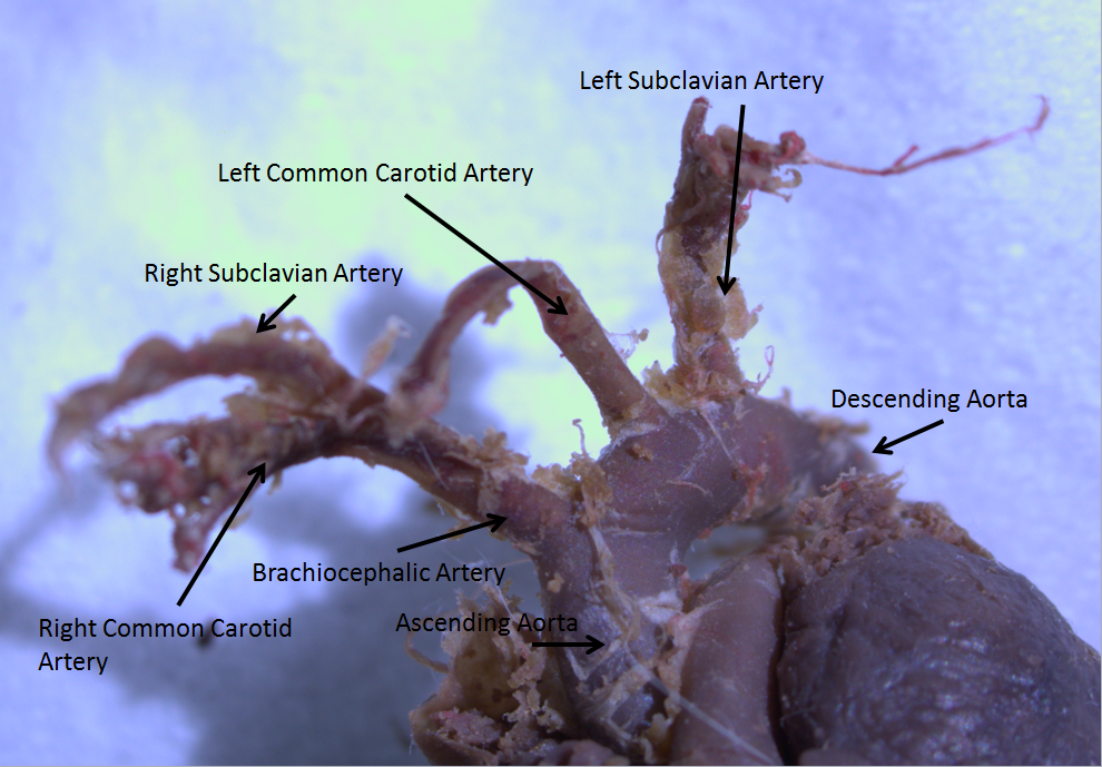
High pressure receptors or high pressure baroreceptors are the baroreceptors found within the aortic arch and carotid sinus. They are only sensitive to blood pressures above 60 mmHg. When these receptors are activated they elicit a depressor response; which decreases the heart rate and causes a general vasodilation. An increase in arterial blood pressure reflexively elicits an increase in vagal neuronal activity to the heart (i.e. the resulting decreased heart rate). The afferent nerves from the baroreceptors are called buffer nerves. See also * Low pressure receptors * Bainbridge reflex The Bainbridge reflex (or Bainbridge effect or atrial reflex) is a cardiovascular reflex causing an increase in heart rate in response to increased stretching of the wall of the right atrium and/or the inferior vena cava as a result of increased ... References *"Principles of medical physiology" by A Fonyo page 577 {{neuroanatomy-stub Sensory receptors ... [...More Info...] [...Related Items...] OR: [Wikipedia] [Google] [Baidu] |
Baroreceptor
Baroreceptors (or archaically, pressoreceptors) are stretch receptors that sense blood pressure. Thus, increases in the pressure of blood vessel triggers increased action potential generation rates and provides information to the central nervous system. This sensory information is used primarily in autonomic reflexes that in turn influence the heart cardiac output and vascular smooth muscle to influence vascular resistance. Baroreceptors act immediately as part of a negative feedback system called the baroreflex as soon as there is a change from the usual mean arterial blood pressure, returning the pressure toward a normal level. These reflexes help regulate short-term blood pressure. The solitary nucleus in the medulla oblongata of the brain recognizes changes in the firing rate of action potentials from the baroreceptors, and influences cardiac output and systemic vascular resistance. Baroreceptors can be divided into two categories based on the type of blood vessel in whic ... [...More Info...] [...Related Items...] OR: [Wikipedia] [Google] [Baidu] |
Aortic Arch
The aortic arch, arch of the aorta, or transverse aortic arch () is the part of the aorta between the ascending and descending aorta. The arch travels backward, so that it ultimately runs to the left of the trachea. Structure The aorta begins at the level of the upper border of the second/third sternocostal articulation of the right side, behind the ventricular outflow tract and pulmonary trunk. The right atrial appendage overlaps it. The first few centimeters of the ascending aorta and pulmonary trunk lies in the same pericardial sheath and runs at first upward, arches over the pulmonary trunk, right pulmonary artery, and right main bronchus to lie behind the right second coastal cartilage. The right lung and sternum lies anterior to the aorta at this point. The aorta then passes posteriorly and to the left, anterior to the trachea, and arches over left main bronchus and left pulmonary artery, and reaches to the left side of the T4 vertebral body. Apart from T4 vertebral ... [...More Info...] [...Related Items...] OR: [Wikipedia] [Google] [Baidu] |
Carotid Sinus
In human anatomy, the carotid sinus is a dilated area at the base of the internal carotid artery just superior to the bifurcation of the internal carotid and external carotid at the level of the superior border of thyroid cartilage. The carotid sinus extends from the bifurcation to the "true" internal carotid artery. The carotid sinuses are sensitive to pressure changes in the arterial blood at this level. They are two out of the four baroreception sites in humans and most mammals. Structure The carotid sinus is the reflex area of the carotid artery, consisting of baroreceptors which monitor blood pressure. Function The carotid sinus contains numerous baroreceptors which function as a "sampling area" for many homeostatic mechanisms for maintaining blood pressure. The carotid sinus baroreceptors are innervated by the carotid sinus nerve, which is a branch of the glossopharyngeal nerve (CN IX). The neurons which innervate the carotid sinus centrally project to the ... [...More Info...] [...Related Items...] OR: [Wikipedia] [Google] [Baidu] |
Blood Pressure
Blood pressure (BP) is the pressure of Circulatory system, circulating blood against the walls of blood vessels. Most of this pressure results from the heart pumping blood through the circulatory system. When used without qualification, the term "blood pressure" refers to the pressure in a brachial artery, where it is most commonly measured. Blood pressure is usually expressed in terms of the systolic pressure (maximum pressure during one Cardiac cycle, heartbeat) over diastolic pressure (minimum pressure between two heartbeats) in the cardiac cycle. It is measured in Millimetre of mercury, millimetres of mercury (mmHg) above the surrounding atmospheric pressure, or in Pascal (unit), kilopascals (kPa). The difference between the systolic and diastolic pressures is known as pulse pressure, while the average pressure during a cardiac cycle is known as mean arterial pressure. Blood pressure is one of the vital signs—together with respiratory rate, heart rate, Oxygen saturation (me ... [...More Info...] [...Related Items...] OR: [Wikipedia] [Google] [Baidu] |
MmHg
A millimetre of mercury is a manometric unit of pressure, formerly defined as the extra pressure generated by a column of mercury one millimetre high. Currently, it is defined as exactly , or approximately 1 torr = atmosphere = pascals.Council Directive 80/181/EEC of 20 December 1979 on the approximation of the laws of the Member States relating to units of measurement and on the repeal of Directive 71/354/EEC of the It is denoted mmHg or mm Hg ... [...More Info...] [...Related Items...] OR: [Wikipedia] [Google] [Baidu] |
Heart Rate
Heart rate is the frequency of the cardiac cycle, heartbeat measured by the number of contractions of the heart per minute (''beats per minute'', or bpm). The heart rate varies according to the body's Human body, physical needs, including the need to absorb oxygen and excrete carbon dioxide. It is also modulated by numerous factors, including (but not limited to) genetics, physical fitness, Psychological stress, stress or psychological status, diet, drugs, hormonal status, environment, and disease/illness, as well as the interaction between these factors. It is usually equal or close to the pulse rate measured at any peripheral point. The American Heart Association states the normal resting adult human heart rate is 60–100 bpm. An ultra-trained athlete would have a resting heart rate of 37–38 bpm. ''Tachycardia'' is a high heart rate, defined as above 100 bpm at rest. ''Bradycardia'' is a low heart rate, defined as below 60 bpm at rest. When a human sleeps, a heartbeat with ra ... [...More Info...] [...Related Items...] OR: [Wikipedia] [Google] [Baidu] |
Vasodilation
Vasodilation, also known as vasorelaxation, is the widening of blood vessels. It results from relaxation of smooth muscle cells within the vessel walls, in particular in the large veins, large arteries, and smaller arterioles. Blood vessel walls are composed of endothelial tissue and a basal membrane lining the lumen of the vessel, concentric smooth muscle layers on top of endothelial tissue, and an adventitia over the smooth muscle layers. Relaxation of the smooth muscle layer allows the blood vessel to dilate, as it is held in a semi-constricted state by sympathetic nervous system activity. Vasodilation is the opposite of vasoconstriction, which is the narrowing of blood vessels. When blood vessels dilate, the flow of blood is increased due to a decrease in vascular resistance and increase in cardiac output. Vascular resistance is the amount of force circulating blood must overcome in order to allow perfusion of body tissues. Narrow vessels create more vascular resista ... [...More Info...] [...Related Items...] OR: [Wikipedia] [Google] [Baidu] |
Vagal
The vagus nerve, also known as the tenth cranial nerve (CN X), plays a crucial role in the autonomic nervous system, which is responsible for regulating involuntary functions within the human body. This nerve carries both sensory and motor fibers and serves as a major pathway that connects the brain to various organs, including the heart, lungs, and digestive tract. As a key part of the parasympathetic nervous system, the vagus nerve helps regulate essential involuntary functions like heart rate, breathing, and digestion. By controlling these processes, the vagus nerve contributes to the body's "rest and digest" response, helping to calm the body after stress, lower heart rate, improve digestion, and maintain homeostasis. The vagus nerve consists of two branches: the right and left vagus nerves. In the neck, the right vagus nerve contains approximately 105,000 fibers, while the left vagus nerve has about 87,000 fibers, according to one source. However, other sources report sl ... [...More Info...] [...Related Items...] OR: [Wikipedia] [Google] [Baidu] |
Heart
The heart is a muscular Organ (biology), organ found in humans and other animals. This organ pumps blood through the blood vessels. The heart and blood vessels together make the circulatory system. The pumped blood carries oxygen and nutrients to the tissue, while carrying metabolic waste such as carbon dioxide to the lungs. In humans, the heart is approximately the size of a closed fist and is located between the lungs, in the middle compartment of the thorax, chest, called the mediastinum. In humans, the heart is divided into four chambers: upper left and right Atrium (heart), atria and lower left and right Ventricle (heart), ventricles. Commonly, the right atrium and ventricle are referred together as the right heart and their left counterparts as the left heart. In a healthy heart, blood flows one way through the heart due to heart valves, which prevent cardiac regurgitation, backflow. The heart is enclosed in a protective sac, the pericardium, which also contains a sma ... [...More Info...] [...Related Items...] OR: [Wikipedia] [Google] [Baidu] |
Afferent Nerves
A sensory nerve, or afferent nerve, is a nerve that contains exclusively afferent nerve fibers. Nerves containing also motor fibers are called mixed. Afferent nerve fibers in a sensory nerve carry sensory information toward the central nervous system (CNS) from different sensory receptors of sensory neurons in the peripheral nervous system (PNS). A motor nerve carries information from the CNS to the PNS. Afferent nerve fibers link the sensory neurons throughout the body, in pathways to the relevant processing circuits in the central nervous system. Afferent nerve fibers are often paired with efferent nerve fibers from the motor neurons (that travel from the CNS to the PNS), in mixed nerves. Stimuli cause nerve impulses in the receptors and alter the potentials, which is known as sensory transduction. Spinal cord entry Afferent nerve fibers leave the sensory neuron from the dorsal root ganglia of the spinal cord, and motor commands carried by the efferent fibers leave the c ... [...More Info...] [...Related Items...] OR: [Wikipedia] [Google] [Baidu] |
Low Pressure Receptors
Low pressure baroreceptors or low pressure receptors are baroreceptors that relay information derived from blood pressure within the autonomic nervous system. They are stimulated by stretching of the vessel wall. They are located in large systemic veins and in the walls of the atria of the heart, and pulmonary vasculature. Low pressure baroreceptors are also referred to as volume receptors, cardiopulmonary baroreceptors,Armstrong, Maggie, et al. ''Physiology, Baroreceptors - Statpearls - NCBI Bookshelf''. 9 Mar. 2022, https://www.ncbi.nlm.nih.gov/books/NBK538172/ . and veno-atrial stretch receptors Structure There are two types of cardiopulmonary baroreceptors, both of which are found within the atrial endocardium. Type A receptors are activated by wall tension, which occurs via atrial contraction during ventricular diastole. Type B receptors are activated by wall stretch, which occurs via atrial filling during ventricular systole. In the right atrium, the stretch receptors ... [...More Info...] [...Related Items...] OR: [Wikipedia] [Google] [Baidu] |
Bainbridge Reflex
The Bainbridge reflex (or Bainbridge effect or atrial reflex) is a cardiovascular reflex causing an increase in heart rate in response to increased stretching of the wall of the right atrium and/or the inferior vena cava as a result of increased venous filling (i.e., increased preload). It is detected by stretch receptors in the wall of the right atrium, the afferent limb is ''via'' the vagus nerve, it is regulated by a center in the medulla oblongata of the brain, and the efferent limb involves reduced vagal activity and increased sympathetic nervous system outflow. Mechanistically, the increased heart rate evoked by the Bainbridge reflex acts to match heart rate (and hence cardiac output) to effective circulating blood volume on a beat-to-beat basis. This, in combination with other cardiovascular reflexes, helps maintain homeostatic equilibrium of the circulation. The Bainbridge reflex may also contribute to respiratory sinus arrhythmia as intrathoracic pressure decreases dur ... [...More Info...] [...Related Items...] OR: [Wikipedia] [Google] [Baidu] |


