|
Glycerol-3-phosphate Shuttle
The glycerol-3-phosphate shuttle is a mechanism used in skeletal muscle and the brain that regenerates NAD+ from NADH, a by-product of glycolysis. NADH is a reducing equivalent that stores electrons generated in the cytoplasm during glycolysis. NADH must be transported into the mitochondria to enter the oxidative phosphorylation pathway. However, the inner mitochondrial membrane is impermeable to NADH and only contains a transport system for NAD+. Depending on the type of tissue either the glycerol-3-phosphate shuttle pathway or the malate–aspartate shuttle pathway is used to transport electrons from cytoplasmic NADH into the mitochondria. The shuttle consists of two proteins acting in sequence. Cytoplasmic glycerol-3-phosphate dehydrogenase (cGPD) transfers an electron pair from NADH to dihydroxyacetone phosphate (DHAP), forming glycerol-3-phosphate (G3P) and regenerating the NAD+ needed to generate energy via glycolysis. Mitochondrial glycerol-3-phosphate dehydrogenase (mGP ... [...More Info...] [...Related Items...] OR: [Wikipedia] [Google] [Baidu] |
Calliphoridae
The Calliphoridae (commonly known as blowflies, blow flies, blow-flies, carrion flies, bluebottles, or greenbottles) are a family of insects in the order Diptera, with almost 1,900 known species. The maggot larvae, often used as fishing bait, are known as gentles. The family is known to be polyphyletic, but much remains disputed regarding proper treatment of the constituent taxa, some of which are occasionally accorded family status (e.g., Bengaliidae and Helicoboscidae). Description Characteristics Calliphoridae adults are commonly shiny with metallic colouring, often with blue, green, or black thoraces and abdomens. Antennae are three-segmented and aristate. The aristae are plumose their entire length, and the second antennal segment is distinctly grooved. Members of Calliphoridae have branched Rs 2 veins, frontal sutures are present, and calypters are well developed. The characteristics and arrangements of hairlike bristles are used to differentiate among members of ... [...More Info...] [...Related Items...] OR: [Wikipedia] [Google] [Baidu] |
Mitochondrial Shuttle
The mitochondrial shuttles are biochemical transport systems used to transport reducing agents across the inner mitochondrial membrane. NADH as well as NAD+ cannot cross the membrane, but it can reduce another molecule like FAD and H2that can cross the membrane, so that its electrons can reach the electron transport chain. The two main systems in humans are the glycerol phosphate shuttle and the malate-aspartate shuttle. The malate/ a-ketoglutarate antiporter functions move electrons while the aspartate/glutamate antiporter moves amino groups. This allows the mitochondria to receive the substrates that it needs for its functionality in an efficient manner. Shuttles In humans, the glycerol phosphate shuttle is primarily found in brown adipose tissue, as the conversion is less efficient, thus generating heat, which is one of the main purposes of brown fat. It is primarily found in babies, though it is present in small amounts in adults around the kidneys and on the back of our ... [...More Info...] [...Related Items...] OR: [Wikipedia] [Google] [Baidu] |
Electron Transport Chain
An electron transport chain (ETC) is a series of protein complexes and other molecules which transfer electrons from electron donors to electron acceptors via redox reactions (both reduction and oxidation occurring simultaneously) and couples this electron transfer with the transfer of protons (H+ ions) across a membrane. Many of the enzymes in the electron transport chain are embedded within the membrane. The flow of electrons through the electron transport chain is an exergonic process. The energy from the redox reactions creates an electrochemical proton gradient that drives the synthesis of adenosine triphosphate (ATP). In aerobic respiration, the flow of electrons terminates with molecular oxygen as the final electron acceptor. In anaerobic respiration, other electron acceptors are used, such as sulfate. In an electron transport chain, the redox reactions are driven by the difference in the Gibbs free energy of reactants and products. The free energy released when ... [...More Info...] [...Related Items...] OR: [Wikipedia] [Google] [Baidu] |
Coenzyme Q
Coenzyme Q10 (CoQ10 ), also known as ubiquinone, is a naturally occurring biochemical cofactor (coenzyme) and an antioxidant produced by the human body. It can also be obtained from dietary sources, such as meat, fish, seed oils, vegetables, and dietary supplements. CoQ10 is found in many organisms, including animals and bacteria. CoQ10 plays a role in mitochondrial oxidative phosphorylation, aiding in the production of adenosine triphosphate (ATP), which is involved in energy transfer within cells. The structure of CoQ10 consists of a benzoquinone moiety and an isoprenoid side chain, with the "10" referring to the number of isoprenyl chemical subunits in its tail. Although a ubiquitous molecule in human tissues, CoQ10 is not a dietary nutrient and does not have a recommended intake level, and its use as a supplement is not approved in the United States for any health or anti-disease effect. Biological functions CoQ10 is a component of the mitochondrial electron transpor ... [...More Info...] [...Related Items...] OR: [Wikipedia] [Google] [Baidu] |
Flavin Adenine Dinucleotide
In biochemistry, flavin adenine dinucleotide (FAD) is a redox-active coenzyme associated with various proteins, which is involved with several enzymatic reactions in metabolism. A flavoprotein is a protein that contains a flavin group, which may be in the form of FAD or flavin mononucleotide (FMN). Many flavoproteins are known: components of the succinate dehydrogenase complex, α-ketoglutarate dehydrogenase, and a component of the pyruvate dehydrogenase complex. FAD can exist in four redox states, which are the flavin-N(5)-oxide, quinone, semiquinone, and hydroquinone. FAD is converted between these states by accepting or donating electrons. FAD, in its fully oxidized form, or quinone form, accepts two electrons and two protons to become FADH2 (hydroquinone form). The semiquinone (FADH·) can be formed by either reduction of FAD or oxidation of FADH2 by accepting or donating one electron and one proton, respectively. Some proteins, however, generate and maintain a super ... [...More Info...] [...Related Items...] OR: [Wikipedia] [Google] [Baidu] |
Dihydroxyacetone Phosphate To Glycerol 3-phosphate En
Dihydroxyacetone (; DHA), also known as glycerone, is a simple saccharide (a triose) with formula . DHA is primarily used as an ingredient in sunless tanning products. It is often derived from plant sources such as sugar beets and sugar cane, and by the fermentation of glycerin. Chemistry DHA is a hygroscopic white crystalline powder. It has a sweet cooling taste and a characteristic odor. It is the simplest of all ketoses and has no chiral center. The normal form is a dimer (2,5-bis(hydroxymethyl)-1,4-dioxane-2,5-diol). The dimer slowly dissolves in water, whereupon it converts to the monomer. These solutions are stable at pH's between 4 and 6. In more basic solution, it degrades to brown product. : This skin browning effect is attributed to a Maillard reaction. DHA condenses with the amino acid residues in the protein keratin, the major component of the skin surface. When injected, no pigmentation occurs, consistent with a role for oxygen in color development. The resul ... [...More Info...] [...Related Items...] OR: [Wikipedia] [Google] [Baidu] |
Glycerol 3-phosphate
''sn''-Glycerol 3-phosphate is the organic ion with the formula HOCH2CH(OH)CH2OPO32-. It is one of two stereoisomers of the ester of dibasic phosphoric acid (HOPO32-) and glycerol. It is a component of bacterial and eukaryotic glycerophospholipids. From a historical reason, it is also known as -glycerol 3-phosphate, -glycerol 1-phosphate, -α-glycerophosphoric acid. Biosynthesis Glycerol 3-phosphate is synthesized by reducing dihydroxyacetone phosphate (DHAP), an intermediate in glycolysis. The reduction is catalyzed by glycerol-3-phosphate dehydrogenase. DHAP and thus glycerol 3-phosphate can also be synthesized from amino acids and citric acid cycle intermediates via the glyceroneogenesis pathway. : + NAD(P)H + H+ → + NAD(P)+ It is also synthesized by the phosphorylation of glycerol, which is generated by hydrolysis of fats. This esterification is catalyzed by glycerol kinase. : + ATP → + ADP Metabolism and biological function Glycerol 3-phosphate is a start ... [...More Info...] [...Related Items...] OR: [Wikipedia] [Google] [Baidu] |
Beta Cell
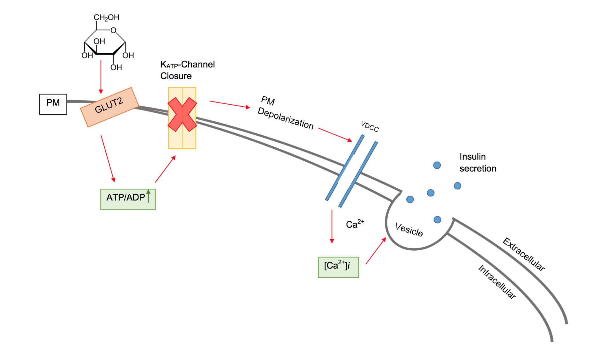
Beta cells (β-cells) are specialized endocrine cells located within the pancreatic islets of Langerhans responsible for the production and release of insulin and amylin. Constituting ~50–70% of cells in human islets, beta cells play a vital role in maintaining blood glucose levels. Problems with beta cells can lead to disorders such as diabetes. Function The function of beta cells is primarily centered around the synthesis and secretion of hormones, particularly insulin and amylin. Both hormones work to keep blood glucose levels within a narrow, healthy range by different mechanisms. Insulin facilitates the uptake of glucose by cells, allowing them to use it for energy or store it for future use. Amylin helps regulate the rate at which glucose enters the bloodstream after a meal, slowing down the absorption of nutrients by inhibit gastric emptying. Insulin synthesis Beta cells are the only site of insulin synthesis in mammals. As glucose stimulates insulin secretion, ... [...More Info...] [...Related Items...] OR: [Wikipedia] [Google] [Baidu] |
Brown Adipose Tissue
Brown adipose tissue (BAT) or brown fat makes up the adipose organ together with white adipose tissue (or white fat). Brown adipose tissue is found in almost all mammals. Classification of brown fat refers to two distinct cell populations with similar functions. The first shares a common embryological origin with muscle cells, found in larger "classic" deposits. The second develops from white adipocytes that are stimulated by the sympathetic nervous system. These adipocytes are found interspersed in white adipose tissue and are also named 'beige' or 'brite' (for "brown in white"). Brown adipose tissue is especially abundant in newborns and in hibernation, hibernating mammals. It is also present and metabolically active in adult humans, but its prevalence decreases as humans age. Its primary function is thermoregulation. In addition to heat produced by shivering muscle, brown adipose tissue produces heat by non-shivering thermogenesis. The therapeutic targeting of brown fat for the ... [...More Info...] [...Related Items...] OR: [Wikipedia] [Google] [Baidu] |
Glycerol-3-phosphate Dehydrogenase 1
Glycerol-3-phosphate dehydrogenase 1 is an enzyme that is encoded by the GPD1 gene in humans. Function This gene encodes a member of the NAD-dependent glycerol-3-phosphate dehydrogenase family. The encoded protein plays a critical role in carbohydrate and lipid metabolism by catalyzing the reversible conversion of dihydroxyacetone phosphate (DHAP) and reduced nicotine adenine dinucleotide (NADH) to glycerol 3-phosphate (G3P) and NAD+. The encoded cytosolic protein and mitochondrial glycerol-3-phosphate dehydrogenase also form a glycerol phosphate shuttle that facilitates the transfer of reducing equivalents from the cytosol to Mitochondrion, mitochondria. Mutations in this gene are a cause of transient infantile hypertriglyceridemia. Alternatively spliced transcript variants encoding multiple isoforms have been observed for this gene. References Further reading * * * * * * * * * {{gene-12-stub ... [...More Info...] [...Related Items...] OR: [Wikipedia] [Google] [Baidu] |


