|
Gastrula
Gastrulation is the stage in the early embryonic development of most animals, during which the blastula (a single-layered hollow sphere of Cell (biology), cells), or in mammals, the blastocyst, is reorganized into a two-layered or three-layered embryo known as the gastrula. Before gastrulation, the embryo is a continuous Epithelium, epithelial sheet of cells; by the end of gastrulation, the embryo has begun Cellular differentiation, differentiation to establish distinct cell lineages, set up the basic axes of the body (e.g. Anatomical terms of location#Dorsal and ventral, dorsal–ventral, Anatomical terms of location#Anterior and posterior, anterior–posterior), and internalized one or more cell types, including the prospective Gastrointestinal tract, gut. Gastrula layers In Triploblasty, triploblastic organisms, the gastrula is trilaminar (three-layered). These three germ layers are the ectoderm (outer layer), mesoderm (middle layer), and endoderm (inner layer).Mundlos 2009p ... [...More Info...] [...Related Items...] OR: [Wikipedia] [Google] [Baidu] |
Embryonic Development
In developmental biology, animal embryonic development, also known as animal embryogenesis, is the developmental stage of an animal embryo. Embryonic development starts with the fertilization of an egg cell (ovum) by a sperm, sperm cell (spermatozoon). Once fertilized, the ovum becomes a single diploid cell known as a zygote. The zygote undergoes mitosis, mitotic cell division, divisions with no significant growth (a process known as cleavage (embryo), cleavage) and cellular differentiation, leading to development of a multicellular embryo after passing through an organizational checkpoint during mid-embryogenesis. In mammals, the term refers chiefly to the early stages of prenatal development, whereas the terms fetus and fetal development describe later stages. The main stages of animal embryonic development are as follows: * The zygote undergoes a series of cell divisions (called cleavage) to form a structure called a morula. * The morula develops into a structure called a bla ... [...More Info...] [...Related Items...] OR: [Wikipedia] [Google] [Baidu] |
Blastocoel
The blastocoel (), also spelled blastocoele and blastocele, and also called cleavage cavity, or segmentation cavity is a fluid-filled or yolk-filled cavity that forms in the blastula during very early embryonic development. At this stage in mammals the blastula is called the blastocyst, which consists of an outer epithelium, the trophectoderm, enveloping the inner cell mass and the blastocoel . It develops following cleavage of the zygote after fertilization. It is the first fluid-filled cavity or lumen formed as the embryo enlarges, and is the essential precursor for the differentiated gastrula. In the ''Xenopus'' a very small cavity has been described in the two-cell stage of development. In mammals After fertilization, the zygote undergoes several rounds of cleavage divisions forming daughter cells known as blastomeres. At the 8- or 16-cell stage, the embryo undergoes compaction and forms the morula. Eventually, the morula is a solid ball of cells that has a small gr ... [...More Info...] [...Related Items...] OR: [Wikipedia] [Google] [Baidu] |
Archenteron
The archenteron, also called the gastrocoel, is the internal cavity formed in the gastrulation stage in early embryonic development that becomes the cavity of the primitive gut. Formation in sea urchins As primary mesenchyme cells detach from the vegetal pole in the gastrula and enter the fluid-filled cavity in the center (the blastocoel), the remaining cells at the vegetal pole flatten to form a vegetal plate. This buckles inwards towards the blastocoel in a process called invagination. The cells continue to be rearranged until the shallow dip formed by invagination transforms into a deeper, narrower pouch formed by the gastrula's endoderm. This pouch narrows and lengthens to become the archenteron, a process driven by convergent extension. The open end of the archenteron is called the blastopore. The filopodia—thin fibers formed by the mesenchyme cells, found in late gastrulation—contract to drag the tip of the archenteron across the blastocoel. The endoderm of ... [...More Info...] [...Related Items...] OR: [Wikipedia] [Google] [Baidu] |
Germ Layers
A germ layer is a primary layer of cell (biology), cells that forms during embryonic development. The three germ layers in vertebrates are particularly pronounced; however, all eumetazoans (animals that are sister taxa to the sponges) produce two or three primary germ layers. Some animals, like cnidarians, produce two germ layers (the ectoderm and endoderm) making them diploblastic. Other animals such as bilaterians produce a third layer (the mesoderm) between these two layers, making them triploblastic. Germ layers eventually give rise to all of an animal's Tissue (biology), tissues and organ (anatomy), organs through the process of organogenesis. History Caspar Friedrich Wolff observed organization of the early embryo in leaf-like layers. In 1817, Heinz Christian Pander discovered three primordial germ layers while studying chick embryos. Between 1850 and 1855, Robert Remak had further refined the germ cell layer (''Keimblatt'') concept, stating that the external, internal ... [...More Info...] [...Related Items...] OR: [Wikipedia] [Google] [Baidu] |
Mesoderm
The mesoderm is the middle layer of the three germ layers that develops during gastrulation in the very early development of the embryo of most animals. The outer layer is the ectoderm, and the inner layer is the endoderm.Langman's Medical Embryology, 11th edition. 2010. The mesoderm forms mesenchyme, mesothelium and coelomocytes. Mesothelium lines coeloms. Mesoderm forms the muscles in a process known as myogenesis, septa (cross-wise partitions) and mesenteries (length-wise partitions); and forms part of the gonads (the rest being the gametes). Myogenesis is specifically a function of mesenchyme. The mesoderm differentiates from the rest of the embryo through intercellular signaling, after which the mesoderm is polarized by an organizing center. The position of the organizing center is in turn determined by the regions in which beta-catenin is protected from degradation by GSK-3. Beta-catenin acts as a co-factor that alters the activity of the transcription facto ... [...More Info...] [...Related Items...] OR: [Wikipedia] [Google] [Baidu] |
Blastocyst
The blastocyst is a structure formed in the early embryonic development of mammals. It possesses an inner cell mass (ICM) also known as the ''embryoblast'' which subsequently forms the embryo, and an outer layer of trophoblast cells called the trophectoderm. This layer surrounds the inner cell mass and a fluid-filled cavity or lumen known as the blastocoel. In the late blastocyst, the trophectoderm is known as the trophoblast. The trophoblast gives rise to the chorion and amnion, the two fetal membranes that surround the embryo. The placenta derives from the embryonic chorion (the portion of the chorion that develops villi) and the underlying uterine tissue of the mother. The corresponding structure in non-mammalian animals is an undifferentiated ball of cells called the blastula. In humans, blastocyst formation begins about five days after fertilization when a fluid-filled cavity opens up in the morula, the early embryonic stage of a ball of 16 cells. The blastocyst ... [...More Info...] [...Related Items...] OR: [Wikipedia] [Google] [Baidu] |
Embryo
An embryo ( ) is the initial stage of development for a multicellular organism. In organisms that reproduce sexually, embryonic development is the part of the life cycle that begins just after fertilization of the female egg cell by the male sperm cell. The resulting fusion of these two cells produces a single-celled zygote that undergoes many cell divisions that produce cells known as blastomeres. The blastomeres (4-cell stage) are arranged as a solid ball that when reaching a certain size, called a morula, (16-cell stage) takes in fluid to create a cavity called a blastocoel. The structure is then termed a blastula, or a blastocyst in mammals. The mammalian blastocyst hatches before implantating into the endometrial lining of the womb. Once implanted the embryo will continue its development through the next stages of gastrulation, neurulation, and organogenesis. Gastrulation is the formation of the three germ layers that will form all of the different parts of t ... [...More Info...] [...Related Items...] OR: [Wikipedia] [Google] [Baidu] |
Blastula
Blastulation is the stage in early animal embryonic development that produces the blastula. In mammalian development, the blastula develops into the blastocyst with a differentiated inner cell mass and an outer trophectoderm. The blastula (from Greek '' βλαστός'' ( meaning ''sprout'')) is a hollow sphere of cells known as blastomeres surrounding an inner fluid-filled cavity called the blastocoel. Embryonic development begins with a sperm fertilizing an egg cell to become a zygote, which undergoes many cleavages to develop into a ball of cells called a morula. Only when the blastocoel is formed does the early embryo become a blastula. The blastula precedes the formation of the gastrula in which the germ layers of the embryo form. A common feature of a vertebrate blastula is that it consists of a layer of blastomeres, known as the blastoderm, which surrounds the blastocoel. In mammals, the blastocyst contains an embryoblast (or inner cell mass) that will eventuall ... [...More Info...] [...Related Items...] OR: [Wikipedia] [Google] [Baidu] |
Nervous System
In biology, the nervous system is the complex system, highly complex part of an animal that coordinates its behavior, actions and sense, sensory information by transmitting action potential, signals to and from different parts of its body. The nervous system detects environmental changes that impact the body, then works in tandem with the endocrine system to respond to such events. Nervous tissue first arose in Ediacara biota, wormlike organisms about 550 to 600 million years ago. In Vertebrate, vertebrates, it consists of two main parts, the central nervous system (CNS) and the peripheral nervous system (PNS). The CNS consists of the brain and spinal cord. The PNS consists mainly of nerves, which are enclosed bundles of the long fibers, or axons, that connect the CNS to every other part of the body. Nerves that transmit signals from the brain are called motor nerves (efferent), while those nerves that transmit information from the body to the CNS are called sensory nerves (aff ... [...More Info...] [...Related Items...] OR: [Wikipedia] [Google] [Baidu] |
Organogenesis
Organogenesis is the phase of embryonic development that starts at the end of gastrulation and continues until birth. During organogenesis, the three germ layers formed from gastrulation (the ectoderm, endoderm, and mesoderm) form the internal organs of the organism. The cells of each of the three germ layers undergo differentiation, a process where less-specialized cells become more-specialized through the expression of a specific set of genes. Cell differentiation is driven by cell signaling cascades. Differentiation is influenced by extracellular signals such as growth factors that are exchanged to adjacent cells which is called juxtracrine signaling or to neighboring cells over short distances which is called paracrine signaling. Intracellular signals – a cell signaling itself ( autocrine signaling) – also play a role in organ formation. These signaling pathways allow for cell rearrangement and ensure that organs form at specific sites within the organism. The organogen ... [...More Info...] [...Related Items...] OR: [Wikipedia] [Google] [Baidu] |
Ectoderm
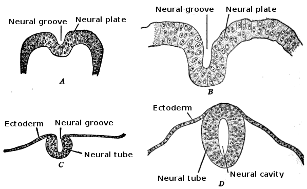
The ectoderm is one of the three primary germ layers formed in early embryonic development. It is the outermost layer, and is superficial to the mesoderm (the middle layer) and endoderm (the innermost layer). It emerges and originates from the outer layer of germ cells. The word ectoderm comes from the Greek language, Greek ''ektos'' meaning "outside", and ''derma'' meaning "skin".Gilbert, Scott F. Developmental Biology. 9th ed. Sunderland, MA: Sinauer Associates, 2010: 333-370. Print. Generally speaking, the ectoderm differentiates to form epithelial tissue, epithelial and nervous system, neural tissues (spinal cord, nerves and brain). This includes the Epidermis (skin), skin, linings of the mouth, anus, nostrils, sweat glands, hair and nails, and tooth enamel. Other types of epithelium are derived from the endoderm. In vertebrate embryos, the ectoderm can be divided into two parts: the dorsal surface ectoderm also known as the external ectoderm, and the neural plate, which inv ... [...More Info...] [...Related Items...] OR: [Wikipedia] [Google] [Baidu] |


