|
Four Prism Dioptre Reflex Test
The Four Prism Dioptre Reflex Test (also known as the 4 PRT, or 4 Prism Dioptre Base-out Test) is an objective, non-dissociative test used to prove the alignment of both eyes (i.e. the presence of binocular single vision) by assessing motor fusion.Frantz, K. A., Cotter, S. A., Wick, B. (1992). Re-evaluation of the four prism dioptre base-out test. Optometry and Vision Science, 69(10), pp. 777-786. Through the use of a 4 dioptre base out prism, diplopia is induced which is the driving force for the eyes to change fixation and therefore re-gain bifoveal fixation meaning, they overcome that amount of power. Indications for use This test is performed on patients suspected to have small angle deviations of less than 10 prism dioptres, a microtropia, that may or may not have been observed on cover test because of subtle eye movements. The test determines whether the patient has bifoveal fixation or monofixation despite their eyes seeming straight. On the cover component of cover tes ... [...More Info...] [...Related Items...] OR: [Wikipedia] [Google] [Baidu] |
Binocular Single Vision
In biology, binocular vision is a type of vision in which an animal has two eyes capable of facing the same direction to perceive a single three-dimensional image of its surroundings. Binocular vision does not typically refer to vision where an animal has eyes on opposite sides of its head and shares no field of view between them, like in some animals. Neurological researcher Manfred Fahle has stated six specific advantages of having two eyes rather than just one: #It gives a creature a "spare eye" in case one is damaged. #It gives a wider field of view. For example, humans have a maximum horizontal field of view of approximately 190 degrees with two eyes, approximately 120 degrees of which makes up the binocular field of view (seen by both eyes) flanked by two uniocular fields (seen by only one eye) of approximately 40 degrees. #It can give stereopsis in which binocular disparity (or parallax) provided by the two eyes' different positions on the head gives precise depth percep ... [...More Info...] [...Related Items...] OR: [Wikipedia] [Google] [Baidu] |
Diplopia
Diplopia is the simultaneous perception of two images of a single object that may be displaced horizontally or vertically in relation to each other. Also called double vision, it is a loss of visual focus under regular conditions, and is often voluntary. However, when occurring involuntarily, it results in impaired function of the extraocular muscles, where both eyes are still functional, but they cannot turn to target the desired object. Problems with these muscles may be due to mechanical problems, disorders of the neuromuscular junction, disorders of the cranial nerves (III, IV, and VI) that innervate the muscles, and occasionally disorders involving the supranuclear oculomotor pathways or ingestion of toxins. Diplopia can be one of the first signs of a systemic disease, particularly to a muscular or neurological process, and it may disrupt a person's balance, movement, or reading abilities. Causes Diplopia has a diverse range of ophthalmologic, infectious, autoimmune ... [...More Info...] [...Related Items...] OR: [Wikipedia] [Google] [Baidu] |
Microtropia
Monofixation syndrome (MFS) (also: microtropia or microstrabismus) is an eye condition defined by less-than-perfect binocular vision. It is defined by a small angle deviation with suppression of the deviated eye and the presence of binocular peripheral fusion. That is, MFS implies peripheral fusion without central fusion. Aside the manifest small-angle deviation ("tropia"), subjects with MFS often also have a large-angle latent deviation (''phoria''). Their stereoacuity is often in the range of 3000 to 70 arcsecond, and a small central suppression scotoma of 2 to 5 deg. A rare condition, MFS is estimated to affect only 1% of the general population. There are three distinguishable forms of this condition: primary constant, primary decompensating and consecutive MFS. It is believed that primary MFS is a result of a primary sensorial defect, predisposing to anomalous retinal correspondence. Secondary MFS is a frequent outcome of surgical treatment of congenital esotropia. A st ... [...More Info...] [...Related Items...] OR: [Wikipedia] [Google] [Baidu] |
Worth 4 Dot Test
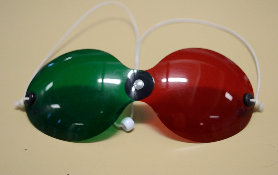
The Worth Four Light Test, also known as the Worth's Four Dot test or W4LT, is a clinical test mainly used for assessing a patient's degree of binocular vision and binocular single vision. Binocular vision involves an image being projected by each eye simultaneously into an area in space and being fused into a single image. The Worth Four Light Test is also used in detection of suppression of either the right or left eye. Suppression occurs during binocular vision when the brain does not process the information received from either of the eyes. This is a common adaptation to strabismus, amblyopia and aniseikonia. The W4LT can be performed by the examiner at two distances, at near (at 33 cm from the patient) and at far (at 6m from the patient). At both testing distances the patient is required to wear red-green goggles (with one red lens over one eye, usually the right, and one green lens over the left) When performing the test at far (distance) the W4LT instrument is compos ... [...More Info...] [...Related Items...] OR: [Wikipedia] [Google] [Baidu] |
Bagolini Striated Glasses Test
Bagolini striated glasses test, or BSGT, is a subjective clinical test to detect the presence or extent of binocular functions and is generally performed by an optometrist or orthoptist or ophthalmologist (medical/surgical eye doctor). It is mainly used in strabismus clinics. Through this test, suppression, microtropia, diplopia and manifest deviations can be noted. However this test should always be used in conjunction with other clinical tests, such as Worth 4 dot test, Cover test, Prism cover test and Maddox rod to come to a diagnosis. Equipment To perform the test you will need * Bagolini Striated Glasses * Pen torch or a distant light source. Alternatively, trial frames and lenses or a lorgnette can be used. In some cases, the use of prisms is necessary to measure a deviation and test for the presence of binocular functions. Principles Bagolini striated glasses are glasses of no dioptric power that have many narrow striations running parallel in one meridian. The ... [...More Info...] [...Related Items...] OR: [Wikipedia] [Google] [Baidu] |
Random Dot Stereogram
Random-dot stereogram (RDS) is stereo pair of images of random dots which, when viewed with the aid of a stereoscope, or with the eyes focused on a point in front of or behind the images, produces a sensation of depth, with objects appearing to be in front of or behind the display level. The random-dot stereogram technique, known since 1919, was elaborated on by Béla Julesz, described in his 1971 book, Foundations of Cyclopean Perception. Later concepts, involving single images, not necessarily consisting of random dots, and more well-known to the general public, are autostereograms. History In 1840, Sir Charles Wheatstone developed the stereoscope. Using it, two photographs, taken a small horizontal distance apart, could be viewed one to each eye so that the objects in the photograph appeared to be three-dimensional in a three-dimensional scene. Around 1956, Julesz began at Bell Labs on a project to detect patterns in the output of random number generators. He decided to try m ... [...More Info...] [...Related Items...] OR: [Wikipedia] [Google] [Baidu] |
Hering's Law Of Equal Innervation
Hering's law of equal innervation is used to explain the conjugacy of saccadic eye movement in stereoptic animals. The law proposes that conjugacy of saccades is due to innate connections in which the eye muscles responsible for each eye's movements are innervated equally. The law also states that apparent monocular eye movements are actually the summation of conjugate version and disjunctive (or vergence) eye movements. The law was put forward by Ewald Hering in the 19th century, though the underlying principles of the law date back considerably. Aristotle had commented upon this phenomenon and Ptolemy put forward a theory of why such a physiological law might be useful. It was clearly stated for the first time by Alhacen in his '' Book of Optics'' (1021). Hering's law of equal innervation is best understood with Johannes Peter Müller's stimulus where an observer refoveates a point that moved in one eye only. The least-effort way to refoveate is to move the misaligned eye o ... [...More Info...] [...Related Items...] OR: [Wikipedia] [Google] [Baidu] |
Anisometropia
Anisometropia refers to a condition when two eyes have unequal refractive power. Generally, a difference in power of one diopter (1D) or more is the accepted threshold to label the condition anisometropia. Patients can tolerate 3 D of anisometropia before it becomes clinically symptomatic with headaches, asthenopia, double vision and photophobia. In certain types of anisometropia, the visual cortex of the brain will not process images from both eyes together ( binocular summation), and will instead suppress the central vision of one of the eyes. If this occurs often enough during the first 10 years of life while the visual cortex is developing, it can result in amblyopia, a condition where even when correcting the refractive error properly, the person's vision in the affected eye is still not correctable to 20/20. The name is from four Greek components: ''an-'' "not," ''iso-'' "same," ''metr-'' "measure," ''ops'' "eye." Antimetropia is a rare sub-type of anisometropia, in which ... [...More Info...] [...Related Items...] OR: [Wikipedia] [Google] [Baidu] |
Amblyopia
Amblyopia, also called lazy eye, is a disorder of sight in which the brain fails to fully process input from one eye and over time favors the other eye. It results in decreased vision in an eye that typically appears normal in other aspects. Amblyopia is the most common cause of decreased vision in a single eye among children and younger adults. The cause of amblyopia can be any condition that interferes with focusing during early childhood. This can occur from poor alignment of the eyes (strabismic), an eye being irregularly shaped such that focusing is difficult, one eye being more nearsighted or farsighted than the other (refractive), or clouding of the lens of an eye (deprivational). After the underlying cause is addressed, vision is not restored right away, as the mechanism also involves the brain. Amblyopia can be difficult to detect, so vision testing is recommended for all children around the ages of four to five. Early detection improves treatment success. Glasse ... [...More Info...] [...Related Items...] OR: [Wikipedia] [Google] [Baidu] |
Stereopsis
Stereopsis () is the component of depth perception retrieved through binocular vision. Stereopsis is not the only contributor to depth perception, but it is a major one. Binocular vision happens because each eye receives a different image because they are in slightly different positions on one’s head (left and right eyes). These positional differences are referred to as "horizontal disparities" or, more generally, " binocular disparities". Disparities are processed in the visual cortex of the brain to yield depth perception. While binocular disparities are naturally present when viewing a real three-dimensional scene with two eyes, they can also be simulated by artificially presenting two different images separately to each eye using a method called stereoscopy. The perception of depth in such cases is also referred to as "stereoscopic depth". The perception of depth and three-dimensional structure is, however, possible with information visible from one eye alone, such as diff ... [...More Info...] [...Related Items...] OR: [Wikipedia] [Google] [Baidu] |
Horopter
The horopter was originally defined in geometric terms as the locus of points in space that make the same angle at each eye with the fixation point, although more recently in studies of binocular vision it is taken to be the locus of points in space that have the same disparity as fixation. This can be defined theoretically as the points in space that project on corresponding points in the two retinas, that is, on anatomically identical points. The horopter can be measured empirically in which it is defined using some criterion. The concept of horopter can then be extended as a geometrical locus of points in space where a specific condition is met: * the binocular horopter is the locus of iso-disparity points in space; * the oculomotor horopter is the locus of iso-vergence points in space. As other quantities that describe the functional principles of the visual system, it is possible to provide a theoretical description of the phenomenon. The measurement with psycho-physical e ... [...More Info...] [...Related Items...] OR: [Wikipedia] [Google] [Baidu] |


