|
Fibrous Hamartoma Of Infancy
Fibrous hamartoma of infancy (FHI) is a rare, typically painless, benign tumor that develops in the subcutaneous tissues of the axilla (i.e. armpit), arms, external genitalia, or, less commonly, various other areas. It is diagnosed in children who are usually less than 2 years old or, in up to 20% of cases, develops in utero and is diagnosed in an infant at birth. The cells involved in FHI include bland fibroblasts/myofibroblasts, mature fat cells, and primitive-appearing spindle-shaped and/or star-shaped cells. The tumor was first described by R.D. Reye in 1956 who termed the disorder "subdermal fibromatous tumor of infancy" and regarded it as a reactive lesion. In a 1964 study of 30 patients, F.M. Enzinger renamed the tumor hamartoma of infancy; he regarded it to be a hamartoma (i.e. a local malformation of various normal cell types due to a systemic genetic condition) rather than a reactive lesion. However, subsequent studies have found gene mutations in FHI tumor cells and con ... [...More Info...] [...Related Items...] OR: [Wikipedia] [Google] [Baidu] |
Dermatology
Dermatology is the branch of medicine dealing with the skin.''Random House Webster's Unabridged Dictionary.'' Random House, Inc. 2001. Page 537. . It is a speciality with both medical and surgical aspects. A List of dermatologists, dermatologist is a specialist medical doctor who manages diseases related to skin, hair, nails, and some cosmetic problems. Etymology Attested in English in 1819, the word "dermatology" derives from the Ancient Greek, Greek δέρματος (''dermatos''), genitive of δέρμα (''derma''), "skin" (itself from δέρω ''dero'', "to flay") and -λογία ''wikt:-logia, -logia''. Neo-Latin ''dermatologia'' was coined in 1630, an anatomical term with various French and German uses attested from the 1730s. History In 1708, the first great school of dermatology became a reality at the famous Hôpital Saint-Louis in Paris, and the first textbooks (Willan's, 1798–1808) and atlases (Jean-Louis-Marc Alibert, Alibert's, 1806–1816) appeared in print ar ... [...More Info...] [...Related Items...] OR: [Wikipedia] [Google] [Baidu] |
Fibroblastic And Myofibroblastic Tumors
Fibroblastic and myofibroblastic tumors (FMTs) develop from the mesenchymal stem cells which differentiate into fibroblasts (the most common cell type in connective tissue) and/or the myocytes/ myoblasts that differentiate into muscle cells. FMTs are a heterogeneous group of soft tissue neoplasms (i.e. abnormal and excessive tissue growths). The World Health Organization (2020) defined tumors as being FMTs based on their morphology and, more importantly, newly discovered abnormalities in the expression levels of key gene products made by these tumors' neoplastic cells. Histopathologically, FMTs consist of neoplastic connective tissue cells which have differented into cells that have microscopic appearances resembling fibroblasts and/or myofibroblasts. The fibroblastic cells are characterized as spindle-shaped cells with inconspicuous nucleoli that express vimentin, an intracellular protein typically found in mesenchymal cells, and CD34, a cell surface membrane glycopr ... [...More Info...] [...Related Items...] OR: [Wikipedia] [Google] [Baidu] |
Actin
Actin is a protein family, family of Globular protein, globular multi-functional proteins that form microfilaments in the cytoskeleton, and the thin filaments in myofibril, muscle fibrils. It is found in essentially all Eukaryote, eukaryotic cells, where it may be present at a concentration of over 100 micromolar, μM; its mass is roughly 42 kDa, with a diameter of 4 to 7 nm. An actin protein is the monomeric Protein subunit, subunit of two types of filaments in cells: microfilaments, one of the three major components of the cytoskeleton, and thin filaments, part of the Muscle contraction, contractile apparatus in muscle cells. It can be present as either a free monomer called G-actin (globular) or as part of a linear polymer microfilament called F-actin (filamentous), both of which are essential for such important cellular functions as the Motility, mobility and contraction of cell (biology), cells during cell division. Actin participates in many important cellular pr ... [...More Info...] [...Related Items...] OR: [Wikipedia] [Google] [Baidu] |
Immunohistochemical
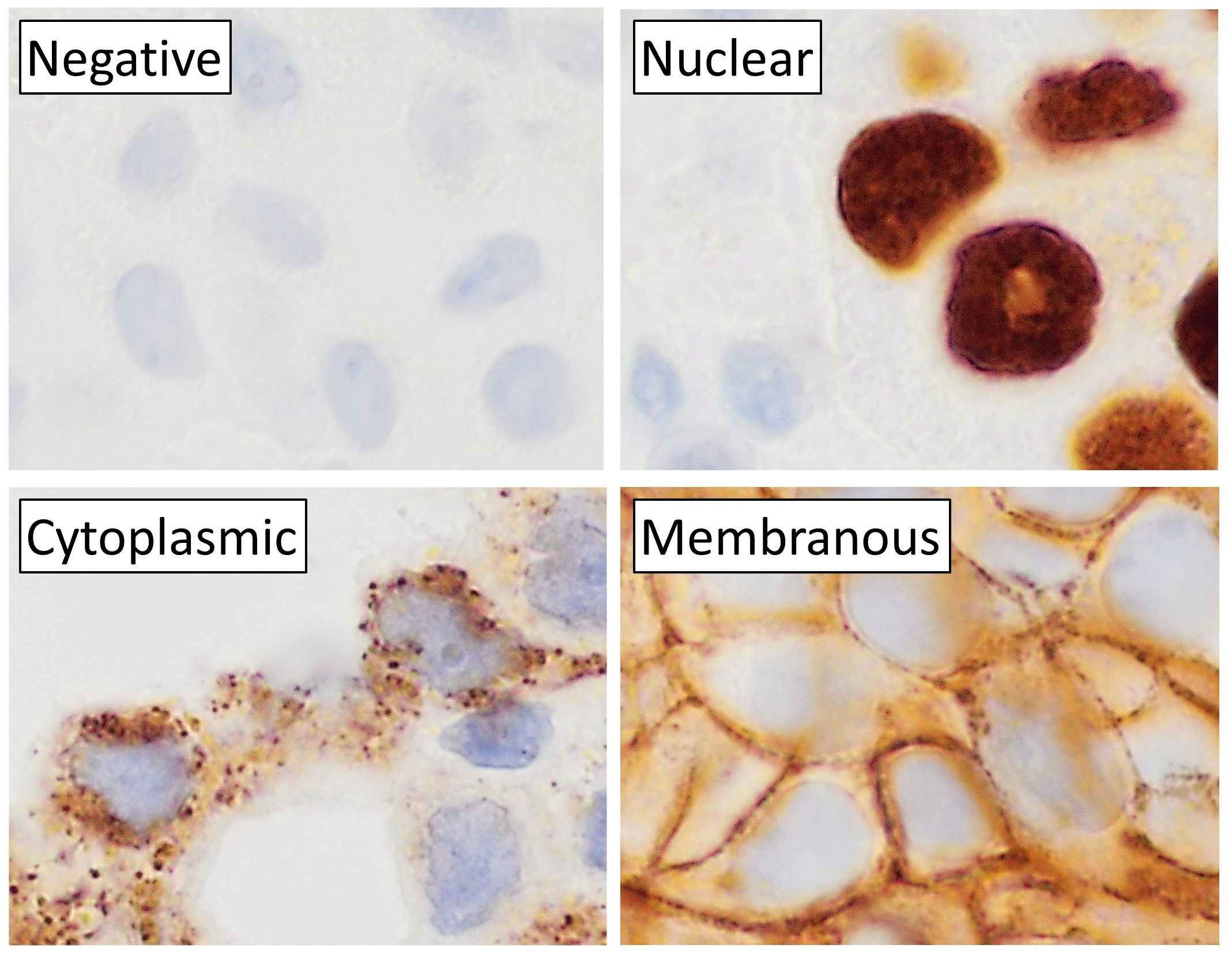
Immunohistochemistry (IHC) is the most common application of immunostaining. It involves the process of selectively identifying antigens (proteins) in cells of a tissue section by exploiting the principle of antibodies binding specifically to antigens in biological tissues. IHC takes its name from the roots "immuno", in reference to antibodies used in the procedure, and "histo", meaning tissue (compare to immunocytochemistry). Albert Coons conceptualized and first implemented the procedure in 1941. Visualising an antibody-antigen interaction can be accomplished in a number of ways, mainly either of the following: * ''Chromogenic immunohistochemistry'' (CIH), wherein an antibody is conjugated to an enzyme, such as peroxidase (the combination being termed immunoperoxidase), that can catalyse a colour-producing reaction. * ''Immunofluorescence'', where the antibody is tagged to a fluorophore, such as fluorescein or rhodamine. Immunohistochemical staining is widely used in the dia ... [...More Info...] [...Related Items...] OR: [Wikipedia] [Google] [Baidu] |
Plasma Cells
Plasma cells, also called plasma B cells or effector B cells, are white blood cells that originate in the lymphoid organs as B lymphocytes and secrete large quantities of proteins called antibodies in response to being presented specific substances called antigens. These antibodies are transported from the plasma cells by the blood plasma and the lymphatic system to the site of the target antigen (foreign substance), where they initiate its neutralization or destruction. B cells differentiate into plasma cells that produce antibody molecules closely modeled after the receptors of the precursor B cell. Structure Plasma cells are large lymphocytes with abundant cytoplasm and a characteristic appearance on light microscopy. They have basophilic cytoplasm and an eccentric nucleus with heterochromatin in a characteristic cartwheel or clock face arrangement. Their cytoplasm also contains a pale zone that on electron microscopy contains an extensive Golgi apparatus and centrioles ... [...More Info...] [...Related Items...] OR: [Wikipedia] [Google] [Baidu] |
Macrophages
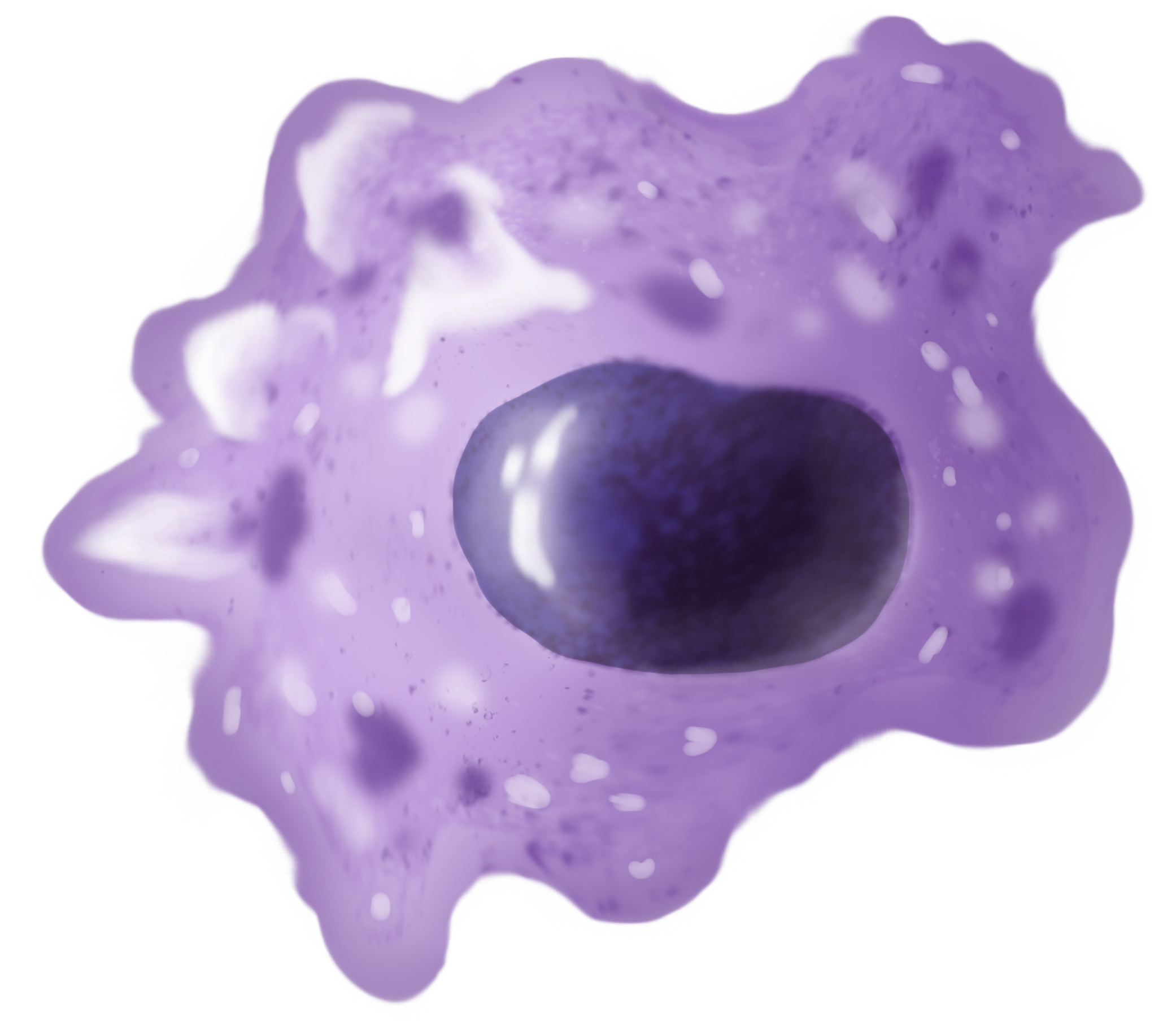
Macrophages (abbreviated as M φ, MΦ or MP) ( el, large eaters, from Greek ''μακρός'' (') = large, ''φαγεῖν'' (') = to eat) are a type of white blood cell of the immune system that engulfs and digests pathogens, such as cancer cells, microbes, cellular debris, and foreign substances, which do not have proteins that are specific to healthy body cells on their surface. The process is called phagocytosis, which acts to defend the host against infection and injury. These large phagocytes are found in essentially all tissues, where they patrol for potential pathogens by amoeboid movement. They take various forms (with various names) throughout the body (e.g., histiocytes, Kupffer cells, alveolar macrophages, microglia, and others), but all are part of the mononuclear phagocyte system. Besides phagocytosis, they play a critical role in nonspecific defense ( innate immunity) and also help initiate specific defense mechanisms ( adaptive immunity) by recruiting oth ... [...More Info...] [...Related Items...] OR: [Wikipedia] [Google] [Baidu] |
Lymphocytes
A lymphocyte is a type of white blood cell (leukocyte) in the immune system of most vertebrates. Lymphocytes include natural killer cells (which function in cell-mediated, cytotoxic innate immunity), T cells (for cell-mediated, cytotoxic adaptive immunity), and B cells (for humoral, antibody-driven adaptive immunity). They are the main type of cell found in lymph, which prompted the name "lymphocyte". Lymphocytes make up between 18% and 42% of circulating white blood cells. Types The three major types of lymphocyte are T cells, B cells and natural killer (NK) cells. Lymphocytes can be identified by their large nucleus. T cells and B cells T cells ( thymus cells) and B cells ( bone marrow- or bursa-derived cells) are the major cellular components of the adaptive immune response. T cells are involved in cell-mediated immunity, whereas B cells are primarily responsible for humoral immunity (relating to antibodies). The function of T cells and B cells is to recogn ... [...More Info...] [...Related Items...] OR: [Wikipedia] [Google] [Baidu] |
Adipose Tissue
Adipose tissue, body fat, or simply fat is a loose connective tissue composed mostly of adipocytes. In addition to adipocytes, adipose tissue contains the stromal vascular fraction (SVF) of cells including preadipocytes, fibroblasts, vascular endothelial cells and a variety of immune cells such as adipose tissue macrophages. Adipose tissue is derived from preadipocytes. Its main role is to store energy in the form of lipids, although it also cushions and insulates the body. Far from being hormonally inert, adipose tissue has, in recent years, been recognized as a major endocrine organ, as it produces hormones such as leptin, estrogen, resistin, and cytokines (especially TNFα). In obesity, adipose tissue is also implicated in the chronic release of pro-inflammatory markers known as adipokines, which are responsible for the development of metabolic syndrome, a constellation of diseases including, but not limited to, type 2 diabetes, cardiovascular disease and atheroscler ... [...More Info...] [...Related Items...] OR: [Wikipedia] [Google] [Baidu] |
Myofibroblast
A myofibroblast is a cell phenotype that was first described as being in a state between a fibroblast and a smooth muscle cell. Structure Myofibroblasts are contractile web-like fusiform cells that are identifiable by their expression of α-smooth muscle actin within their cytoplasmic stress fibers. In the gastrointestinal and genitourinary tracts, myofibroblasts are found subepithelially in mucosal surfaces. Here they not only act as a regulator of the shape of the crypts and villi, but also act as stem-niche cells in the intestinal crypts and as parts of atypical antigen-presenting cells. They have both support as well as paracrine function in most places. Location Myofibroblasts were first identified in granulation tissue during skin wound healing. Typically, these cells are found in granulation tissue, scar tissue (fibrosis) and the stroma of tumours. They also line the gastrointestinal tract, wherein they regulate the shapes of crypts and villi. Markers Myofibroblasts ... [...More Info...] [...Related Items...] OR: [Wikipedia] [Google] [Baidu] |
Fibroblast
A fibroblast is a type of biological cell that synthesizes the extracellular matrix and collagen, produces the structural framework ( stroma) for animal tissues, and plays a critical role in wound healing. Fibroblasts are the most common cells of connective tissue in animals. Structure Fibroblasts have a branched cytoplasm surrounding an elliptical, speckled nucleus having two or more nucleoli. Active fibroblasts can be recognized by their abundant rough endoplasmic reticulum. Inactive fibroblasts (called fibrocytes) are smaller, spindle-shaped, and have a reduced amount of rough endoplasmic reticulum. Although disjointed and scattered when they have to cover a large space, fibroblasts, when crowded, often locally align in parallel clusters. Unlike the epithelial cells lining the body structures, fibroblasts do not form flat monolayers and are not restricted by a polarizing attachment to a basal lamina on one side, although they may contribute to basal lamina components in so ... [...More Info...] [...Related Items...] OR: [Wikipedia] [Google] [Baidu] |
Hematoxylin And Eosin Stain
Hematoxylin and eosin stain ( or haematoxylin and eosin stain or hematoxylin-eosin stain; often abbreviated as H&E stain or HE stain) is one of the principal tissue stains used in histology. It is the most widely used stain in medical diagnosis and is often the gold standard. For example, when a pathologist looks at a biopsy of a suspected cancer, the histological section is likely to be stained with H&E. H&E is the combination of two histological stains: hematoxylin and eosin. The hematoxylin stains cell nuclei a purplish blue, and eosin stains the extracellular matrix and cytoplasm pink, with other structures taking on different shades, hues, and combinations of these colors. Hence a pathologist can easily differentiate between the nuclear and cytoplasmic parts of a cell, and additionally, the overall patterns of coloration from the stain show the general layout and distribution of cells and provides a general overview of a tissue sample's structure. Thus, pattern re ... [...More Info...] [...Related Items...] OR: [Wikipedia] [Google] [Baidu] |
Histopathological
Histopathology (compound of three Greek words: ''histos'' "tissue", πάθος ''pathos'' "suffering", and -λογία ''-logia'' "study of") refers to the microscopic examination of tissue in order to study the manifestations of disease. Specifically, in clinical medicine, histopathology refers to the examination of a biopsy or surgical specimen by a pathologist, after the specimen has been processed and histological sections have been placed onto glass slides. In contrast, cytopathology examines free cells or tissue micro-fragments (as "cell blocks"). Collection of tissues Histopathological examination of tissues starts with surgery, biopsy, or autopsy. The tissue is removed from the body or plant, and then, often following expert dissection in the fresh state, placed in a fixative which stabilizes the tissues to prevent decay. The most common fixative is 10% neutral buffered formalin (corresponding to 3.7% w/v formaldehyde in neutral buffered water, such as phosphate ... [...More Info...] [...Related Items...] OR: [Wikipedia] [Google] [Baidu] |


.jpg)

