|
Femoral Pulse
The femoral artery is a large artery in the thigh and the main arterial supply to the thigh and leg. The femoral artery gives off the deep femoral artery and descends along the anteromedial part of the thigh in the femoral triangle. It enters and passes through the adductor canal, and becomes the popliteal artery as it passes through the adductor hiatus in the adductor magnus near the junction of the middle and distal thirds of the thigh. The femoral artery proximal to the origin of the deep femoral artery is referred to as the ''common femoral artery'', whereas the femoral artery distal to this origin is referred to as the ''superficial femoral artery''. Structure The femoral artery represents the continuation of the external iliac artery beyond the inguinal ligament underneath which the vessel passes to enter the thigh. The vessel passes under the inguinal ligament just medial of the midpoint of this ligament, midway between the anterior superior iliac spine and the symphysi ... [...More Info...] [...Related Items...] OR: [Wikipedia] [Google] [Baidu] |
External Iliac Artery
The external iliac arteries are two major Artery, arteries which bifurcate off the common iliac arteries anterior to the sacroiliac joint of the pelvis. Structure The external iliac artery arises from the bifurcation of the common iliac artery. They proceed anterior and inferior along the medial border of the psoas major muscles. They exit the Pelvis, pelvic girdle posterior and inferior to the inguinal ligament. This occurs about one third laterally from the insertion point of the inguinal ligament on the pubic tubercle. At this point they are referred to as the femoral arteries. Branches Function The external iliac artery provides the main blood supply to the legs. It passes down along the brim of the pelvis and gives off two large branches - the "inferior epigastric artery" and a "deep circumflex artery." These vessels supply blood to the muscles and skin in the lower abdominal wall. The external iliac artery passes beneath the inguinal ligament in the lower part of the ... [...More Info...] [...Related Items...] OR: [Wikipedia] [Google] [Baidu] |
Physician
A physician, medical practitioner (British English), medical doctor, or simply doctor is a health professional who practices medicine, which is concerned with promoting, maintaining or restoring health through the Medical education, study, Medical diagnosis, diagnosis, prognosis and therapy, treatment of disease, injury, and other physical and mental impairments. Physicians may focus their practice on certain disease categories, types of patients, and methods of treatment—known as Specialty (medicine), specialities—or they may assume responsibility for the provision of continuing and comprehensive medical care to individuals, families, and communities—known as general practitioner, general practice. Medical practice properly requires both a detailed knowledge of the Discipline (academia), academic disciplines, such as anatomy and physiology, pathophysiology, underlying diseases, and their treatment, which is the science of medicine, and a decent Competence (human resources ... [...More Info...] [...Related Items...] OR: [Wikipedia] [Google] [Baidu] |
Subsartorial Canal
The adductor canal (also known as the subsartorial canal or Hunter's canal) is an aponeurotic tunnel in the middle third of the thigh giving passage to parts of the femoral artery, vein, and nerve. It extends from the apex of the femoral triangle to the adductor hiatus. Structure The adductor canal extends from the apex of the femoral triangle to the adductor hiatus. It is an intermuscular cleft situated on the medial aspect of the middle third of the anterior compartment of the thigh, and has the following boundaries: * medial wall - sartorius. * posterior wall - adductor longus and adductor magnus. * anterior wall - vastus medialis. It is covered by a strong aponeurosis which extends from the vastus medialis, across the femoral vessels to the adductor longus and magnus. Lying on the aponeurosis is the sartorius (tailor's) muscle. Contents The canal contains the femoral artery, femoral vein, and branches of the femoral nerve (specifically, the saphenous nerve ... [...More Info...] [...Related Items...] OR: [Wikipedia] [Google] [Baidu] |
Femoral Sheath
The femoral sheath (also called the crural sheath) is a funnel-shaped downward extension of abdominal fascia within which the femoral artery and femoral vein pass between the abdomen and the thigh. The femoral sheath is subdivided by two vertical partitions to form three compartments (medial, intermediate, and lateral); the medial compartment is known as the femoral canal and contains lymphatic vessels and a lymph node, whereas the intermediate canal and the lateral canal accommodate the femoral vein and the femoral artery (respectively). Some neurovascular structures perforate the femoral sheath. Topographically, the femoral sheath is contained within the femoral triangle. Structure The femoral sheath is funnel-shaped fascial structure, with the wide end directed superior-ward. The femoral sheath is formed by an inferior-ward prolongation - posterior to the inguinal ligament - of abdominal fascia, with transverse fascia being continued down anterior to the femoral vessels ... [...More Info...] [...Related Items...] OR: [Wikipedia] [Google] [Baidu] |
Femoral Vein
In the human body, the femoral vein is the vein that accompanies the femoral artery in the femoral sheath. It is a deep vein that begins at the adductor hiatus (an opening in the adductor magnus muscle) as the continuation of the popliteal vein. The great saphenous vein (a superficial vein), and the deep femoral vein drain into the femoral vein in the femoral triangle when it becomes known as the common femoral vein. It ends at the inferior margin of the inguinal ligament where it becomes the external iliac vein. Its major tributaries are the deep femoral vein, and the great saphenous vein. The femoral vein contains valves. Structure The femoral vein bears valves which are mostly bicuspid and whose number is variable between individuals and often between left and right leg. Course The femoral vein continues into the thigh as the continuation from the popliteal vein at the back of the knee. It drains blood from the deep thigh muscles and thigh bone. Proximal to th ... [...More Info...] [...Related Items...] OR: [Wikipedia] [Google] [Baidu] |
Federative International Programme On Anatomical Terminology
The Federative International Programme for Anatomical Terminology (FIPAT) is a group of experts who review, analyze, and discuss the terms of the morphological structures of the human body. It was created by the International Federation of Associations of Anatomists (IFAA) and was previously known as the Federative Committee on Anatomical Terminology (FCAT) and the Federative International Committee on Anatomical Terminology (FICAT). Origins and history This Committee was created in 1989, at the XIII International Congress of Anatomists, held in Rio de Janeiro (Brazil). It followed the old International Anatomical Nomenclature Committee (IANC). The professionals involved are renowned professors and researchers with knowledge of medical terminology. They hold periodic meetings in different countries on a rotating basis, where they study morphological terminology: anatomical, histological and embryology of the human being. The results of this committee were published in 1998 ... [...More Info...] [...Related Items...] OR: [Wikipedia] [Google] [Baidu] |
Human Anatomy
Human anatomy (gr. ἀνατομία, "dissection", from ἀνά, "up", and τέμνειν, "cut") is primarily the scientific study of the morphology of the human body. Anatomy is subdivided into gross anatomy and microscopic anatomy. Gross anatomy (also called macroscopic anatomy, topographical anatomy, regional anatomy, or anthropotomy) is the study of anatomical structures that can be seen by the naked eye. Microscopic anatomy is the study of minute anatomical structures assisted with microscopes, which includes histology (the study of the organization of tissues), and cytology (the study of cells). Anatomy, human physiology (the study of function), and biochemistry (the study of the chemistry of living structures) are complementary basic medical sciences that are generally together (or in tandem) to students studying medical sciences. In some of its facets human anatomy is closely related to embryology, comparative anatomy and comparative embryology, through common ... [...More Info...] [...Related Items...] OR: [Wikipedia] [Google] [Baidu] |
Terminologia Anatomica
''Terminologia Anatomica'' (commonly abbreviated TA) is the international standard for human anatomy, human anatomical terminology. It is developed by the Federative International Programme on Anatomical Terminology (FIPAT) a program of the International Federation of Associations of Anatomists (IFAA). History The sixth edition of the previous standard, ''Nomina Anatomica'', was released in 1989. The first edition of ''Terminologia Anatomica'', superseding Nomina Anatomica, was developed by the Federative Committee on Anatomical Terminology (FCAT) and the International Federation of Associations of Anatomists (IFAA) and released in 1998. In April 2011, this edition was published online by the Federative International Programme on Anatomical Terminologies (FIPAT), the successor of FCAT. The first edition contained 7635 Latin items. The second edition was released online by FIPAT in 2019 and approved and adopted by the IFAA General Assembly in 2020. The latest errata is dated Au ... [...More Info...] [...Related Items...] OR: [Wikipedia] [Google] [Baidu] |
Vascular Surgery
Vascular surgery is a surgical subspecialty in which vascular diseases involving the arteries, veins, or lymphatic vessels, are managed by medical therapy, minimally-invasive catheter procedures and surgical reconstruction. The specialty evolved from general surgery, general and cardiac surgery, cardiovascular surgery where it refined the management of just the vessels, no longer treating the heart or other organs. Modern vascular surgery includes open surgery techniques, endovascular (minimally invasive) techniques and medical management of vascular diseases - unlike the parent specialities. The vascular surgeon is trained in the diagnosis and management of diseases affecting all parts of the vascular system excluding the coronaries and intracranial vasculature. Vascular surgeons also are called to assist other physicians to carry out surgery near vessels, or to salvage vascular injuries that include hemorrhage control, dissection, occlusion or simply for safe exposure of vascul ... [...More Info...] [...Related Items...] OR: [Wikipedia] [Google] [Baidu] |
Angiology
Angiology (from Greek , ''angeīon'', "vessel"; and , ''-logia'') is the medical specialty dedicated to studying the circulatory system and of the lymphatic system, i.e., arteries, veins and lymphatic vessels. In the UK, this field is more often termed ''angiology'', and in the United States the term vascular medicine is more frequent. The field of vascular medicine (angiology) is the field that deals with preventing, diagnosing, and treating lymphatic and blood vessel related diseases. Overview Arterial diseases include the aorta ( aneurysms/dissection) and arteries supplying the legs, hands, kidneys, brain, intestines. It also covers arterial thrombosis and embolism; vasculitides; and vasospastic disorders. Naturally, it deals with preventing cardiovascular diseases such as heart attack and stroke. Venous diseases include venous thrombosis, chronic venous insufficiency, and varicose veins. Lymphatic diseases include primary and secondary forms of lymphedema. It ... [...More Info...] [...Related Items...] OR: [Wikipedia] [Google] [Baidu] |
Pubic Symphysis
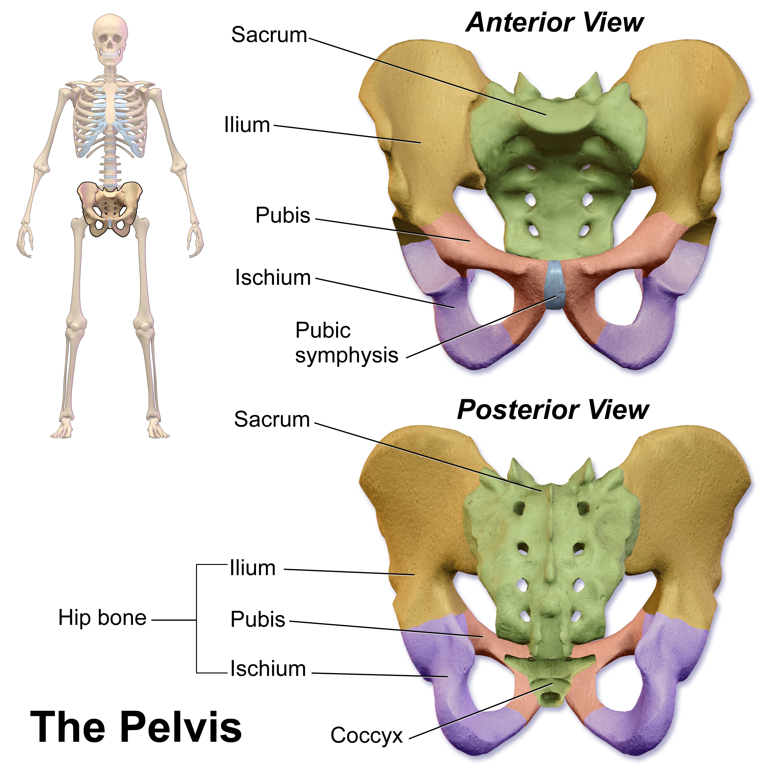
The pubic symphysis (: symphyses) is a secondary cartilaginous joint between the left and right superior rami of the pubis of the hip bones. It is in front of and below the urinary bladder. In males, the suspensory ligament of the penis attaches to the pubic symphysis. In females, the pubic symphysis is attached to the suspensory ligament of the clitoris. In most adults, it can be moved roughly 2 mm and with 1 degree rotation. This increases for women at the time of childbirth. The name comes from the Greek word ''symphysis'', meaning 'growing together'. Structure The pubic symphysis is a nonsynovial amphiarthrodial joint. The width of the pubic symphysis at the front is 3–5 mm greater than its width at the back. This joint is connected by fibrocartilage and may contain a fluid-filled cavity; the center is avascular, possibly due to the nature of the compressive forces passing through this joint, which may lead to harmful vascular disease. The ends of both pubi ... [...More Info...] [...Related Items...] OR: [Wikipedia] [Google] [Baidu] |


