|
Femoral Nerve Stretch Test
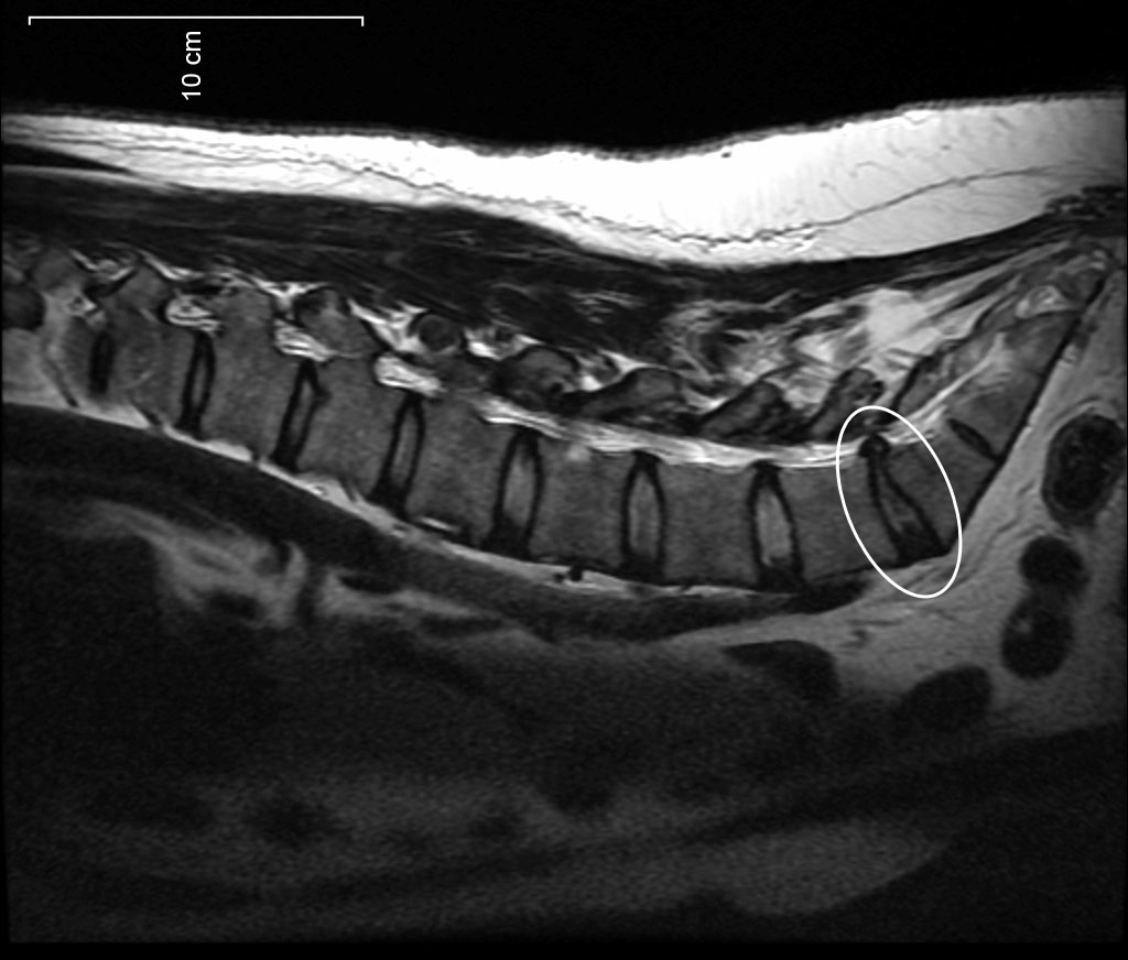
Femoral nerve stretch test, also known as Mackiewicz sign, is a test for spinal nerve root compression, which is associated with disc protrusion and femoral nerve injury. __TOC__ Uses The femoral nerve stretch test can identify spinal nerve root compression, which is associated with disc protrusion and femoral nerve injury. It can reliably identify spinal nerve root compression for L2, L3, and L4. It is usually positive for L2-L3 and L3-L4 (high lumbar) disc protrusions, slightly positive or negative in L4–L5 disc protrusions, and negative in cases of lumbosacral disc protrusion. Procedure To perform a femoral nerve stretch test, a patient lies prone, the knee is passively flexed to the thigh and the hip is passively extended (reverse Lasègues). The test is positive if the patient experiences anterior thigh pain Pain is a distressing feeling often caused by intense or damaging Stimulus (physiology), stimuli. The International Association for the Study of Pain defin ... [...More Info...] [...Related Items...] OR: [Wikipedia] [Google] [Baidu] |
Spinal Nerve Root
Spinal nerve root may refer to: * Posterior root of spinal nerve * Anterior root of spinal nerve Back anatomy Peripheral nervous system {{Short pages monitor ... [...More Info...] [...Related Items...] OR: [Wikipedia] [Google] [Baidu] |
Disc Protrusion
A disc protrusion is a medical condition that can occur in some vertebrates, including humans, in which the outermost layers of the ''anulus fibrosus'' of the intervertebral discs of the spine are intact but bulge when one or more of the discs are under pressure. Many disk abnormalities seen on MRI that are loosely referred to as "herniation" are actually just incidental findings. These may be unrelated to any symptoms and are just bulges of the ''anulus fibrosus''. Jensen and colleagues, in an MRI study of the lumbar spine in 98 ''asymptomatic'' adults, found that in more than half, there was a symmetrical extension of a disc (or discs) beyond the margins of the interspace (bulging). In 27 percent, there was a focal or asymmetrical extension of the disc beyond the margin of the interspace (protrusion), and in only 1 percent was there more extreme extension of the disc (extrusion or sequestration). These findings emphasize the importance of using precise terms in describing th ... [...More Info...] [...Related Items...] OR: [Wikipedia] [Google] [Baidu] |
Nerve Injury
Nerve injury is an injury to a nerve. There is no single classification system that can describe all the many variations of nerve injuries. In 1941, Herbert Seddon introduced a classification of nerve injuries based on three main types of nerve fiber injury and whether there is continuity of the nerve. Usually, however, nerve injuries are classified in five stages, based on the extent of damage to both the nerve and the surrounding connective tissue, since supporting glial cells may be involved. Unlike in the central nervous system, neuroregeneration in the peripheral nervous system is possible. The processes that occur in peripheral regeneration can be divided into the following major events: Wallerian degeneration, axon regeneration/growth, and reinnervation of nervous tissue. The events that occur in peripheral regeneration occur with respect to the axis of the nerve injury. The proximal stump refers to the end of the injured neuron that is still attached to the neuron cel ... [...More Info...] [...Related Items...] OR: [Wikipedia] [Google] [Baidu] |
Lumbar Nerves
The lumbar nerves are the five pairs of spinal nerves emerging from the lumbar vertebrae. They are divided into posterior and anterior divisions. Structure The lumbar nerves are five spinal nerves which arise from either side of the spinal cord below the thoracic spinal cord and above the sacral spinal cord. They arise from the spinal cord between each pair of lumbar spinal vertebrae and travel through the intervertebral foramina. The nerves then split into an anterior branch, which travels forward, and a posterior branch, which travels backwards and supplies the area of the back. Posterior divisions The middle divisions of the posterior branches run close to the articular processes of the vertebrae and end in the multifidus muscle. The outer branches supply the erector spinae muscles. The nerves give off branches to the skin. These pierce the aponeurosis of the greater trochanter. Anterior divisions The anterior divisions of the lumbar nerves () increase in size from ... [...More Info...] [...Related Items...] OR: [Wikipedia] [Google] [Baidu] |
Prone Position
Prone position () is a body position in which the person lies flat with the chest down and the back up. In anatomical terms of location, the dorsal side is up, and the ventral side is down. The supine position is the 180° contrast. Etymology The word ', meaning "naturally inclined to something, apt, liable," has been recorded in English since 1382; the meaning "lying face-down" was first recorded in 1578, but is also referred to as "lying down" or "going prone." ''Prone'' derives from the Latin ', meaning "bent forward, inclined to," from the adverbial form of the prefix ''pro-'' "forward." Both the original, literal, and the derived figurative sense were used in Latin, but the figurative is older in English. Anatomy In anatomy, the prone position is a position of the human body lying face down. It is opposed to the supine position which is face up. Using the terms defined in the anatomical position, the ventral side is down, and the dorsal side is up. Concerning the forea ... [...More Info...] [...Related Items...] OR: [Wikipedia] [Google] [Baidu] |
Knee
In humans and other primates, the knee joins the thigh with the leg and consists of two joints: one between the femur and tibia (tibiofemoral joint), and one between the femur and patella (patellofemoral joint). It is the largest joint in the human body. The knee is a modified hinge joint, which permits flexion and extension (kinesiology), extension as well as slight internal and external rotation. The knee is vulnerable to injury and to the development of osteoarthritis. It is often termed a ''compound joint'' having tibiofemoral and patellofemoral components. (The fibular collateral ligament is often considered with tibiofemoral components.) Structure The knee is a modified hinge joint, a type of synovial joint, which is composed of three functional compartments: the patellofemoral articulation, consisting of the patella, or "kneecap", and the patellar groove on the front of the femur through which it slides; and the medial and lateral tibiofemoral articulations linking the ... [...More Info...] [...Related Items...] OR: [Wikipedia] [Google] [Baidu] |
Thigh
In anatomy, the thigh is the area between the hip (pelvis) and the knee. Anatomically, it is part of the lower limb. The single bone in the thigh is called the femur. This bone is very thick and strong (due to the high proportion of bone tissue), and forms a ball and socket joint at the hip, and a modified hinge joint at the knee. Structure Bones The femur is the only bone in the thigh and serves as an attachment site for all thigh muscles. The head of the femur articulates with the acetabulum in the pelvic bone forming the hip joint, while the distal part of the femur articulates with the tibia and patella forming the knee. By most measures, the femur is the strongest and longest bone in the body. The femur is categorised as a long bone and comprises a diaphysis, the shaft (or body) and two epiphyses, the lower extremity and the upper extremity of femur, that articulate with adjacent bones in the hip and knee. Muscular compartments In cross-section, the thigh is d ... [...More Info...] [...Related Items...] OR: [Wikipedia] [Google] [Baidu] |
Lasègue Test
The straight leg raise is a test that can be performed during a physical examination, with the leg being lifted actively by the patient or passively by the clinician. If the straight leg raise is done actively by the patient, it is a test of functional leg strength, particularly the rectus femoris element of the quadriceps (checking both hip flexion and knee extension strength simultaneously). If carried out passively (also called Lasègue's sign, Lasègue test or Lazarević's sign), it is used to determine whether a patient with low back pain has an underlying nerve root sensitivity, often located at L5 (fifth lumbar spinal nerve). The rest of this article relates to the passive version of the test. Technique With the patient lying down on their back on an examination table or exam floor, the examiner lifts the patient's leg while the knee is straight. A variation is to lift the leg while the patient is sitting. However, this reduces the sensitivity of the test. In order t ... [...More Info...] [...Related Items...] OR: [Wikipedia] [Google] [Baidu] |
Pain
Pain is a distressing feeling often caused by intense or damaging Stimulus (physiology), stimuli. The International Association for the Study of Pain defines pain as "an unpleasant sense, sensory and emotional experience associated with, or resembling that associated with, actual or potential tissue damage." Pain motivates organisms to withdraw from damaging situations, to protect a damaged body part while it heals, and to avoid similar experiences in the future. Congenital insensitivity to pain may result in reduced life expectancy. Most pain resolves once the noxious stimulus is removed and the body has healed, but it may persist despite removal of the stimulus and apparent healing of the body. Sometimes pain arises in the absence of any detectable stimulus, damage or disease. Pain is the most common reason for physician consultation in most developed countries. It is a major symptom in many medical conditions, and can interfere with a person's quality of life and general fun ... [...More Info...] [...Related Items...] OR: [Wikipedia] [Google] [Baidu] |


