|
Extraperitoneal Fascia
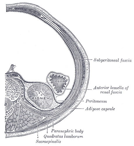
Extraperitoneal fascia (also: endoabdominal fascia or subperitoneal fascia) is a fascial plane – consisting mostly of loose areolar connective tissue – situated between the fascial linings of the walls of the abdominal and pelvic cavities (transversalis fascia, anterior layer of thoracolumbar fascia, iliac fascia, and psoas fascia) externally, and the parietal peritoneum internally. Its quality and quantity varies considerably. It occupies the extraperitoneal space. Preperitoneal space Anteriorly, it forms the thin and fibrous preperitoneal fascia that is interposed between the transversalis fascia, and the parietal peritoneum. The preperitoneal fascia contains a variable amount of fat, loose connective tissue Loose connective tissue, also known as areolar tissue, is a cellular connective tissue with thin and relatively sparse collagen fibers. They have a semi-fluid matrix with lesser proportions of fibers. Its ground substance occupies more vol ..., and membranous ... [...More Info...] [...Related Items...] OR: [Wikipedia] [Google] [Baidu] |
Loose Areolar Connective Tissue
Loose connective tissue, also known as areolar tissue, is a cellular connective tissue with thin and relatively sparse collagen fibers. They have a semi-fluid matrix with lesser proportions of fibers. Its ground substance occupies more volume than the fibers do. It has a viscous to gel-like consistency and plays an important role in the diffusion of oxygen and nutrients from the capillaries that course through this connective tissue as well as in the diffusion of carbon dioxide and metabolic wastes back to the vessels. Moreover, loose connective tissue is primarily located beneath the epithelium, epithelia that cover the body surfaces and line the internal surfaces of the body. It is also associated with the epithelium of glands and surrounds the smallest blood vessels. This tissue is thus the initial site where pathogenic agents, such as bacteria that have breached an epithelial surface, are challenged and destroyed by cells of the immune system. In the past, the designations ... [...More Info...] [...Related Items...] OR: [Wikipedia] [Google] [Baidu] |
Transversalis Fascia
The transversalis fascia (or transverse fascia) is the fascial lining of the anterolateral abdominal wall situated between the inner surface of the transverse abdominal muscle, and the preperitoneal fascia. It is directly continuous with the iliac fascia, the internal spermatic fascia, and pelvic fascia. Structure In the inguinal region, the transversalis fascia is thick and dense; here, it is joined by fibers of the aponeurosis of the transverse abdominal muscle. It becomes thin towards to the diaphragm, blending with the fascia covering the inferior surface of the diaphragm. Posteriorly, it is lost in Below, it has the following attachments: posteriorly, to the whole length of the iliac crest, between the attachments of the transverse abdominal and Iliacus; between the anterior superior iliac spine and the femoral vessels it is connected to the posterior margin of the inguinal ligament, and is there continuous with the iliac fascia. Medial to the femoral vessels it is t ... [...More Info...] [...Related Items...] OR: [Wikipedia] [Google] [Baidu] |
Thoracolumbar Fascia
The thoracolumbar fascia (lumbodorsal fascia or thoracodorsal fascia) is a complex, multilayer arrangement of fascial and aponeurotic layers forming a separation between the paraspinal muscles on one side, and the muscles of the posterior abdominal wall (quadratus lumborum, and psoas major) on the other. It spans the length of the back, extending between the neck superiorly and the sacrum inferiorly. It entails the fasciae and aponeuroses of the latissimus dorsi muscle, serratus posterior inferior muscle, abdominal internal oblique muscle, and transverse abdominal muscle. In the lumbar region, it is known as lumbar fascia and here consists of 3 layers (posterior, middle, and anterior) enclosing two muscular compartments. In the thoracic region, it consists of a single layer (an upward extension of the posterior layer of the lumbar fascia). The thoracolumbar fascia is most prominent at its lower end where its various layers fuse into a thick composite. Anatomy Thoracic reg ... [...More Info...] [...Related Items...] OR: [Wikipedia] [Google] [Baidu] |
Iliac Fascia
The iliac fascia (or Abernethy's fascia) is the fascia overlying the iliacus muscle. Superiorly and laterally, the iliac fascia is attached to the inner aspect of the iliac crest; inferiorly and laterally, it extends into the thigh to unite with the femoral sheath; medially, it attaches to the periosteum of the ilium and iliopubic eminence near the linea terminalis, and blends with the psoas fascia and - over the quadratus lumborum muscle - with the anterior layer of thoracolumbar fascia. The iliac fascia overlies the femoral nerve, and lateral femoral cutaneous nerve. Structure It has the following connections: * ''laterally'', to the whole length of the inner lip of the iliac crest. * ''medially'', to the linea terminalis of the lesser pelvis, where it is continuous with the periosteum. At the iliopectineal eminence it receives the tendon of insertion of the psoas minor, when that muscle exists. Lateral to the femoral vessels it is intimately connected to the poster ... [...More Info...] [...Related Items...] OR: [Wikipedia] [Google] [Baidu] |
Psoas Fascia
Psoas (from Greek ψόας) can refer to: * Psoas major muscle * Psoas minor muscle The psoas minor muscle ( or ; from ) is a long, slender skeletal muscle. When present, it is located anterior to the psoas major muscle.Tank (2005), p 93Gray (2008), p 1372 Structure The psoas minor muscle originates from the vertical fascicle ... * Psoas sign {{disambiguation ... [...More Info...] [...Related Items...] OR: [Wikipedia] [Google] [Baidu] |
Parietal Peritoneum
The peritoneum is the serous membrane forming the lining of the abdominal cavity or coelom in amniotes and some invertebrates, such as annelids. It covers most of the intra-abdominal (or coelomic) organs, and is composed of a layer of mesothelium supported by a thin layer of connective tissue. This peritoneal lining of the cavity supports many of the abdominal organs and serves as a conduit for their blood vessels, lymphatic vessels, and nerves. The abdominal cavity (the space bounded by the vertebrae, abdominal muscles, diaphragm, and pelvic floor) is different from the intraperitoneal space (located within the abdominal cavity but wrapped in peritoneum). The structures within the intraperitoneal space are called "intraperitoneal" (e.g., the stomach and intestines), the structures in the abdominal cavity that are located behind the intraperitoneal space are called " retroperitoneal" (e.g., the kidneys), and those structures below the intraperitoneal space are called "sub ... [...More Info...] [...Related Items...] OR: [Wikipedia] [Google] [Baidu] |
Extraperitoneal Space
The extraperitoneal space is the portion of the abdomen and pelvis which does not lie within the peritoneum. It includes: * Retroperitoneal space, situated posteriorly to the peritoneum * ''Preperitoneal space'', situated anteriorly to the peritoneum ** Retropubic space, deep to the pubic bone ** Retro-inguinal space, deep to the inguinal ligament The space in the pelvis is divided into the following components: * prevesical space * perivesical space * perirectal space See also * Retropubic space * Rectovesical pouch * Vesicouterine pouch * Rectouterine pouch (Pouch of Douglas The rectouterine pouch (rectovaginal pouch, pouch of Douglas or cul-de-sac) is the extension of the peritoneum into the space between the posterior wall of the uterus and the rectum in the human female. Structure In women, the rectouterine pouch ...) References Abdomen {{Anatomy-stub ... [...More Info...] [...Related Items...] OR: [Wikipedia] [Google] [Baidu] |
Loose Connective Tissue
Loose connective tissue, also known as areolar tissue, is a cellular connective tissue with thin and relatively sparse collagen fibers. They have a semi-fluid matrix with lesser proportions of fibers. Its ground substance occupies more volume than the fibers do. It has a viscous to gel-like consistency and plays an important role in the diffusion of oxygen and nutrients from the capillaries that course through this connective tissue as well as in the diffusion of carbon dioxide and metabolic wastes back to the vessels. Moreover, loose connective tissue is primarily located beneath the epithelia that cover the body surfaces and line the internal surfaces of the body. It is also associated with the epithelium of glands and surrounds the smallest blood vessels. This tissue is thus the initial site where pathogenic agents, such as bacteria that have breached an epithelial surface, are challenged and destroyed by cells of the immune system. In the past, the designations areola ... [...More Info...] [...Related Items...] OR: [Wikipedia] [Google] [Baidu] |
Pararenal Fat
The retroperitoneal space (retroperitoneum) is the anatomical space (sometimes a potential space) behind (''retro'') the peritoneum. It has no specific delineating anatomical structures. Organs are retroperitoneal if they have peritoneum on their anterior side only. Structures that are not suspended by mesentery in the abdominal cavity and that lie between the parietal peritoneum and abdominal wall are classified as retroperitoneal. This is different from organs that are not retroperitoneal, which have peritoneum on their posterior side and are suspended by mesentery in the abdominal cavity. The retroperitoneum can be further subdivided into the following: *Perirenal (or perinephric) space *Anterior pararenal (or paranephric) space *Posterior pararenal (or paranephric) space Retroperitoneal structures Structures that lie behind the peritoneum are termed "retroperitoneal". Organs that were once suspended within the abdominal cavity by mesentery but migrated posterior to the peri ... [...More Info...] [...Related Items...] OR: [Wikipedia] [Google] [Baidu] |

