|
Epidural Space
In anatomy, the epidural space is the potential space between the dura mater and vertebrae ( spine). The anatomy term "epidural space" has its origin in the Ancient Greek language; , "on, upon" + dura mater also known as "epidural cavity", "extradural space" or "peridural space". In humans the epidural space contains lymphatics, spinal nerve roots, loose connective tissue, adipose tissue, small arteries, dural venous sinuses and a network of internal vertebral venous plexuses. Cranial epidural space In the skull, the periosteal layer of the dura mater adheres to the inner surface of the skull bones while the meningeal layer lays over the arachnoid mater. Between them is the epidural space. The two layers of the dura mater separate at several places, with the meningeal layer projecting deeper into the brain parenchyma forming fibrous septa that compartmentalize the brain tissue. At these sites, the epidural space is wide enough to house the epidural venous sinuses. There are ... [...More Info...] [...Related Items...] OR: [Wikipedia] [Google] [Baidu] |
Epidural Administration
Epidural administration (from Ancient Greek ἐπί, "upon" + '' dura mater'') is a method of medication administration in which a medicine is injected into the epidural space around the spinal cord. The epidural route is used by physicians and nurse anesthetists to administer local anesthetic agents, analgesics, diagnostic medicines such as radiocontrast agents, and other medicines such as glucocorticoids. Epidural administration involves the placement of a catheter into the epidural space, which may remain in place for the duration of the treatment. The technique of intentional epidural administration of medication was first described in 1921 by the Spanish Aragonese military surgeon Fidel Pagés. Epidural anaesthesia causes a loss of sensation, including pain, by blocking the transmission of signals through nerve fibres in or near the spinal cord. For this reason, epidurals are commonly used for pain control during childbirth and surgery, for which the technique is c ... [...More Info...] [...Related Items...] OR: [Wikipedia] [Google] [Baidu] |
Cerebral Hemisphere
The vertebrate cerebrum (brain) is formed by two cerebral hemispheres that are separated by a groove, the longitudinal fissure. The brain can thus be described as being divided into left and right cerebral hemispheres. Each of these hemispheres has an outer layer of grey matter, the cerebral cortex, that is supported by an inner layer of white matter. In eutherian (placental) mammals, the hemispheres are linked by the corpus callosum, a very large bundle of axon, nerve fibers. Smaller commissures, including the anterior commissure, the posterior commissure and the fornix (neuroanatomy), fornix, also join the hemispheres and these are also present in other vertebrates. These commissures transfer information between the two hemispheres to coordinate localized functions. There are three known poles of the cerebral hemispheres: the ''occipital lobe, occipital pole'', the ''frontal lobe, frontal pole'', and the ''temporal lobe, temporal pole''. The central sulcus is a prominent fissu ... [...More Info...] [...Related Items...] OR: [Wikipedia] [Google] [Baidu] |
Occipital Sinus
The occipital sinus is the smallest of the dural venous sinuses. It is usually unpaired, and is sometimes altogether absent. It is situated in the attached margin of the falx cerebelli. It commences near the foramen magnum, and ends by draining into the confluence of sinuses. Occipital sinuses were discovered by Guichard Joseph Duverney. Anatomy The occipital sinus is present in around 65% of individuals. It is usually single, but occasionally paired. It is situated in the attached margin of the falx cerebelli. Course The occipital sinus commences around the margin of the foramen magnum by several small venous channels (one of which joins the terminal part of the sigmoid sinus The sigmoid sinuses (sigma- or s-shaped hollow curve), also known as the , are paired dural venous sinuses within the skull that receive blood from posterior transverse sinuses. Structure The sigmoid sinus is a dural venous sinus situated withi ...). It terminates by draining into the confluenc ... [...More Info...] [...Related Items...] OR: [Wikipedia] [Google] [Baidu] |
Falx Cerebelli
The falx cerebelli is a small sickle-shaped fold of dura mater projecting forwards into the posterior cerebellar notch as well as projecting into the vallecula of the cerebellum between the two cerebellar hemispheres. The name comes from two Latin words: ''falx'', meaning "curved blade or scythe", and ''cerebellum'', meaning "little brain". Anatomy The falx cerebelli is a small midline fold of dura mater projecting anterior-ward from the skull and into the space between the cerebellar hemispheres. It generally measures between 2.8 and 4.5 cm in length, and approximately 1–2 mm in thickness. Attachments Superiorly, it (with its upwardly directed base) attaches at the midline to the posterior portion of the inferior surface of the tentorium cerebelli. Posteriorly, it attaches to the internal occipital crest; the inferior-most extremity of its posterior attachment frequently divides into two small folds that terminate at either side of the foramen magnum. An ... [...More Info...] [...Related Items...] OR: [Wikipedia] [Google] [Baidu] |
Pituitary Gland
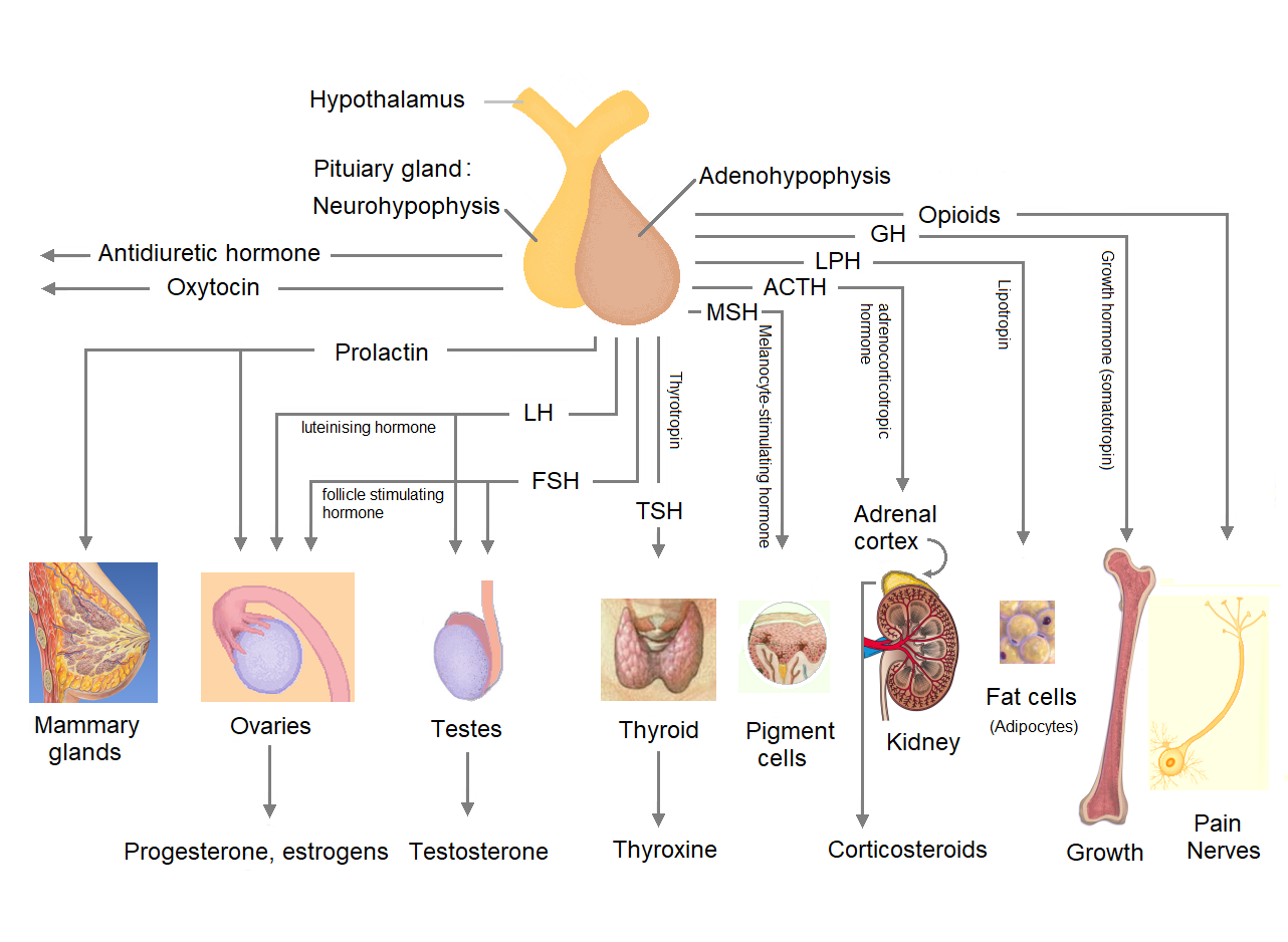
The pituitary gland or hypophysis is an endocrine gland in vertebrates. In humans, the pituitary gland is located at the base of the human brain, brain, protruding off the bottom of the hypothalamus. The pituitary gland and the hypothalamus control much of the body's endocrine system. It is seated in part of the sella turcica a fossa (anatomy), depression in the sphenoid bone, known as the hypophyseal fossa. The human pituitary gland is ovoid, oval shaped, about 1 cm in diameter, in weight on average, and about the size of a kidney bean. Digital version. There are two main lobes of the pituitary, an anterior pituitary, anterior lobe, and a posterior pituitary, posterior lobe joined and separated by a small intermediate lobe. The anterior lobe (adenohypophysis) is the glandular part that produces and secretes several hormones. The posterior lobe (neurohypophysis) secretes neurohypophysial hormones produced in the hypothalamus. Both lobes have different origins and they are both co ... [...More Info...] [...Related Items...] OR: [Wikipedia] [Google] [Baidu] |
Sella Turcica
The sella turcica (Latin for 'Turkish saddle') is a saddle-shaped depression in the body of the sphenoid bone of the human skull and of the skulls of other hominids including chimpanzees, gorillas and orangutans. It serves as a cephalometric landmark. The pituitary gland or hypophysis is located within the most inferior aspect of the sella turcica, the hypophyseal fossa. Structure The sella turcica is located in the sphenoid bone behind the chiasmatic groove and the tuberculum sellae. It belongs to the middle cranial fossa. The sella turcica's most inferior portion is known as the hypophyseal fossa (the "seat of the saddle"), and contains the pituitary gland (hypophysis). In front of the hypophyseal fossa is the tuberculum sellae. Completing the formation of the saddle posteriorly is the dorsum sellae, which is continuous with the clivus, inferoposteriorly. The dorsum sellae is terminated laterally by the posterior clinoid processes. Development It is widely believed th ... [...More Info...] [...Related Items...] OR: [Wikipedia] [Google] [Baidu] |
Diaphragma Sellae
The diaphragma sellae or sellar diaphragm is a small, circular sheet of dura mater forming an (incomplete) roof over the sella turcica and covering the pituitary gland lodged therein. The diaphragma sellae forms a central opening to accommodate the passage of the pituitary stalk (infundibulum) which interconnects the pituitary gland and the hypothalamus. The diaphragma sellae is an important neurosurgical landmark. Anatomy Boundaries The diaphragma sellae has a posterior boundary at the dorsum sellae and an anterior boundary at the tuberculum sellae along with the two small eminences (one on either side) called the middle clinoid processes. Variation The opening formed by the diaphragma sellae varies greatly in size between individuals. Clinical significance Pituitary tumours may grow to extend superiorly beyond the diaphragma sellae. Violation of the diaphragma sellae during an endoscopic endonasal transsphenoidal pituitary tumor resection will result in a cerebrosp ... [...More Info...] [...Related Items...] OR: [Wikipedia] [Google] [Baidu] |
Superior Petrosal Sinus
The superior petrosal sinus is one of the dural venous sinuses located beneath the brain. It receives blood from the cavernous sinus and passes backward and laterally to drain into the transverse sinus. The sinus receives superior petrosal veins, some cerebellar veins, some inferior cerebral veins, and veins from the tympanic cavity. They may be affected by arteriovenous malformation or arteriovenous fistula, usually treated with surgery. Structure The superior petrosal sinus is located beneath the brain. It originates from the cavernous sinus. It passes backward and laterally to drain into the transverse sinus. The sinus runs in the attached margin of the tentorium cerebelli, in a groove in the petrous part of the temporal bone formed by the sinus itself - the superior petrosal sulcus. Function The superior petrosal sinus drains many veins of the brain, including superior petrosal veins, some cerebellar veins, some inferior cerebral veins, and veins from the tympanic c ... [...More Info...] [...Related Items...] OR: [Wikipedia] [Google] [Baidu] |
Straight Sinus
The straight sinus, also known as tentorial sinus or the , is an area within the skull beneath the brain. It receives blood from the inferior sagittal sinus and the great cerebral vein, and drains into the confluence of sinuses. Structure The straight sinus is situated within the dura mater, where the falx cerebri meets the midline of tentorium cerebelli. It forms from the confluence of the inferior sagittal sinus and the great cerebral vein. It may also drain blood from the superior cerebellar veins and veins from the falx cerebri. In cross-section, it is triangular, contains a few transverse bands across its interior, and increases in size as it proceeds backward. It is usually around 5 cm long. Variation The straight sinus is usually an unpaired structure. However, there may be two straight sinuses, which may be one on top of the other or parallel. Function The straight sinus allows blood to drain from the inferior center of the head outwards posteriorly. It receive ... [...More Info...] [...Related Items...] OR: [Wikipedia] [Google] [Baidu] |
Transverse Sinuses
The transverse sinuses (left and right lateral sinuses), within the human head, are two areas beneath the brain which allow blood to drain from the back of the head. They run laterally in a groove along the interior surface of the occipital bone. They drain from the confluence of sinuses (by the internal occipital protuberance) to the sigmoid sinuses, which ultimately connect to the internal jugular vein. ''See diagram (at right)'': labeled under the brain as "" (for Latin: ''sinus transversus'' https://www.ncbi.nlm.nih.gov/books/NBK482257] Structure The transverse sinuses are of large size and begin at the internal occipital protuberance; one, generally the right, being the direct continuation of the |
Cerebellum
The cerebellum (: cerebella or cerebellums; Latin for 'little brain') is a major feature of the hindbrain of all vertebrates. Although usually smaller than the cerebrum, in some animals such as the mormyrid fishes it may be as large as it or even larger. In humans, the cerebellum plays an important role in motor control and cognition, cognitive functions such as attention and language as well as emotion, emotional control such as regulating fear and pleasure responses, but its movement-related functions are the most solidly established. The human cerebellum does not initiate movement, but contributes to motor coordination, coordination, precision, and accurate timing: it receives input from sensory systems of the spinal cord and from other parts of the brain, and integrates these inputs to fine-tune motor activity. Cerebellar damage produces disorders in fine motor skill, fine movement, sense of balance, equilibrium, list of human positions, posture, and motor learning in humans. ... [...More Info...] [...Related Items...] OR: [Wikipedia] [Google] [Baidu] |
Cerebellar Tentorium
The cerebellar tentorium or tentorium cerebelli (Latin for "tent of the cerebellum") is one of four dural folds that separate the cranial cavity into four (incomplete) compartments. The cerebellar tentorium separates the cerebellum from the cerebrum forming a supratentorial and an infratentorial region; the cerebrum is supratentorial and the cerebellum infratentorial. The free border of the tentorium gives passage to the midbrain (the upper-most part of the brainstem). Structure Free border The free border of the tentorium is U-shaped; it forms an aperture - the tentorial notch (tentorial incisure) - which gives passage to the midbrain. The free border of each side extends anteriorly beyond the medial end of the superior petrosal sinus (i.e. the apex of the petrous part of the temporal bone) to overlap the attached margin, thenceforth forming a ridge of dura matter upon the roof of the cavernous sinus, terminating anteriorly by attaching at the anterior clinoid process. ... [...More Info...] [...Related Items...] OR: [Wikipedia] [Google] [Baidu] |


