|
Endocarpon Maritimum
''Endocarpon'' is a genus of saxicolous (rock-dwelling), squamulose lichens in the family Verrucariaceae. It comprises 23 species. Taxonomy The genus was circumscribed by the German bryologist Johann Hedwig in 1789. '' Endocarpon pusillum'' was assigned as the type species. Description Lichens in the genus ''Endocarpon'' have a thallus, meaning their body is made up of small, scale-like lobes. These lobes typically lie flat against the surface they grow on but can sometimes be slightly raised. In rare cases, the thallus may take on a more leaf-like (subfoliose) form. The outer layer of the lichen, the , is composed of roughly spherical cells. Beneath this, the inner tissue (medulla) may contain similar cells or more elongated ones. Unlike some lichens, ''Endocarpon'' species often lack a distinct lower cortex, though they may have a loosely arranged layer of rounded or angular cells. Many species produce rhizines—small, root-like structures that help anchor the lichen to its ... [...More Info...] [...Related Items...] OR: [Wikipedia] [Google] [Baidu] |
Rhizine
In lichens, rhizines are multicellular root-like structures arising mainly from the lower surface. A lichen with rhizines is termed rhizinate, while a lichen lacking rhizines is termed erhizinate. Rhizines serve only to anchor the lichen to their substrate; they do not absorb nutrients, as plant roots do. Characteristics of the rhizines are used to identify lichens, for example, whether they are dense or sparse, uniformly distributed or clumped in specific areas, and straight or branched. Only foliose lichens may possess rhizines, not crustose or fruticose lichens, which lack a lower cortex. Rhizohyphae are a type of attachment structure on some lichens. Rhizohyphae are more slender than rhizines and are one cell thick in diameter. Rhizohyphae often occur as a felt-like hypha A hypha (; ) is a long, branching, filamentous structure of a fungus, oomycete, or actinobacterium. In most fungi, hyphae are the main mode of vegetative growth, and are collectively called a mycel ... [...More Info...] [...Related Items...] OR: [Wikipedia] [Google] [Baidu] |
Septum
In biology, a septum (Latin language, Latin for ''something that encloses''; septa) is a wall, dividing a Body cavity, cavity or structure into smaller ones. A cavity or structure divided in this way may be referred to as septate. Examples Human anatomy * Interatrial septum, the wall of tissue that is a sectional part of the left and right atria of the heart * Interventricular septum, the wall separating the left and right ventricles of the heart * Lingual septum, a vertical layer of fibrous tissue that separates the halves of the tongue *Nasal septum: the cartilage wall separating the nostrils of the nose * Alveolar septum: the thin wall which separates the Pulmonary alveolus, alveoli from each other in the lungs * Orbital septum, a palpebral ligament in the upper and lower eyelids * Septum pellucidum or septum lucidum, a thin structure separating two fluid pockets in the brain * Uterine septum, a malformation of the uterus * Septum of the penis, Penile septum, a fibrous w ... [...More Info...] [...Related Items...] OR: [Wikipedia] [Google] [Baidu] |
Ascus
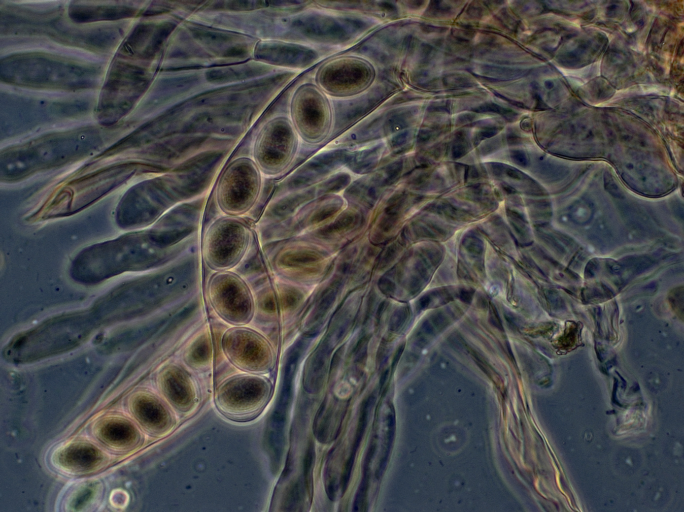
An ascus (; : asci) is the sexual spore-bearing cell produced in ascomycete fungi. Each ascus usually contains eight ascospores (or octad), produced by meiosis followed, in most species, by a mitotic cell division. However, asci in some genera or species can occur in numbers of one (e.g. '' Monosporascus cannonballus''), two, four, or multiples of four. In a few cases, the ascospores can bud off conidia that may fill the asci (e.g. '' Tympanis'') with hundreds of conidia, or the ascospores may fragment, e.g. some '' Cordyceps'', also filling the asci with smaller cells. Ascospores are nonmotile, usually single celled, but not infrequently may be coenocytic (lacking a septum), and in some cases coenocytic in multiple planes. Mitotic divisions within the developing spores populate each resulting cell in septate ascospores with nuclei. The term ocular chamber, or oculus, refers to the epiplasm (the portion of cytoplasm not used in ascospore formation) that is surrounded by the ... [...More Info...] [...Related Items...] OR: [Wikipedia] [Google] [Baidu] |
Spore
In biology, a spore is a unit of sexual reproduction, sexual (in fungi) or asexual reproduction that may be adapted for biological dispersal, dispersal and for survival, often for extended periods of time, in unfavourable conditions. Spores form part of the Biological life cycle, life cycles of many plants, algae, fungus, fungi and protozoa. They were thought to have appeared as early as the mid-late Ordovician period as an adaptation of early land plants. Bacterial spores are not part of a sexual cycle, but are resistant structures used for survival under unfavourable conditions. Myxozoan spores release amoeboid infectious germs ("amoebulae") into their hosts for parasitic infection, but also reproduce within the hosts through the pairing of two nuclei within the plasmodium, which develops from the amoebula. In plants, spores are usually haploid and unicellular and are produced by meiosis in the sporangium of a diploid sporophyte. In some rare cases, a diploid spore is also p ... [...More Info...] [...Related Items...] OR: [Wikipedia] [Google] [Baidu] |
Paraphyses
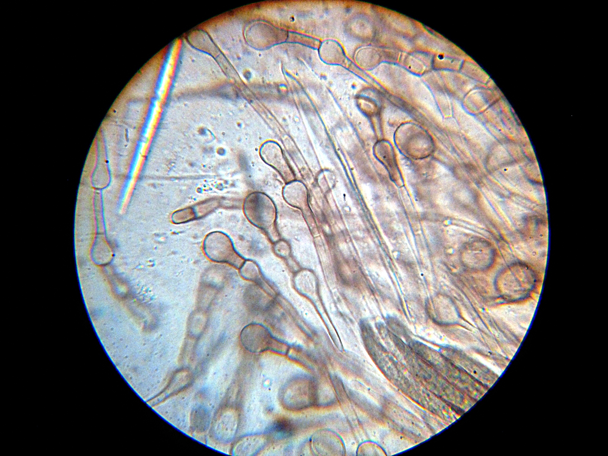
Paraphyses are erect sterile filament-like support structures occurring among the reproductive apparatuses of fungi, ferns, bryophytes and some thallophytes. The singular form of the word is paraphysis. In certain fungi, they are part of the fertile spore-bearing layer. More specifically, paraphyses are sterile filamentous hyphal end cells composing part of the hymenium of Ascomycota and Basidiomycota interspersed among either the asci or basidia respectively, and not sufficiently differentiated to be called cystidia A cystidium (: cystidia) is a relatively large cell found on the sporocarp of a basidiomycete (for example, on the surface of a mushroom gill), often between clusters of basidia. Since cystidia have highly varied and distinct shapes that are o ..., which are specialized, swollen, often protruding cells. The tips of paraphyses may contain the pigments which colour the hymenium. In ferns and mosses, they are filament-like structures that are found on sporangi ... [...More Info...] [...Related Items...] OR: [Wikipedia] [Google] [Baidu] |
Potassium Iodide
Potassium iodide is a chemical compound, medication, and dietary supplement. It is a medication used for treating hyperthyroidism, in radiation emergencies, and for protecting the thyroid gland when certain types of radiopharmaceuticals are used. It is also used for treating skin sporotrichosis and phycomycosis. It is a supplement used by people with low dietary intake of iodine. It is administered orally. Common side effects include vomiting, diarrhea, abdominal pain, rash, and swelling of the salivary glands. Other side effects include allergic reactions, headache, goitre, and depression. While use during pregnancy may harm the baby, its use is still recommended in radiation emergencies. Potassium iodide has the chemical formula K I. Commercially it is made by mixing potassium hydroxide with iodine. Potassium iodide has been used medically since at least 1820. It is on the World Health Organization's List of Essential Medicines. Potassium iodide is available as a g ... [...More Info...] [...Related Items...] OR: [Wikipedia] [Google] [Baidu] |
Staining
Staining is a technique used to enhance contrast in samples, generally at the Microscope, microscopic level. Stains and dyes are frequently used in histology (microscopic study of biological tissue (biology), tissues), in cytology (microscopic study of cell (biology), cells), and in the medical fields of histopathology, hematology, and cytopathology that focus on the study and diagnoses of diseases at the microscopic level. Stains may be used to define biological tissues (highlighting, for example, muscle fibers or connective tissue), cell (biology), cell populations (classifying different blood cells), or organelles within individual cells. In biochemistry, it involves adding a class-specific (DNA, proteins, lipids, carbohydrates) dye to a substrate to qualify or quantify the presence of a specific compound. Staining and fluorescent tagging can serve similar purposes. Biological staining is also used to mark cells in flow cytometry, and to flag proteins or nucleic acids in gel ... [...More Info...] [...Related Items...] OR: [Wikipedia] [Google] [Baidu] |
Iodine
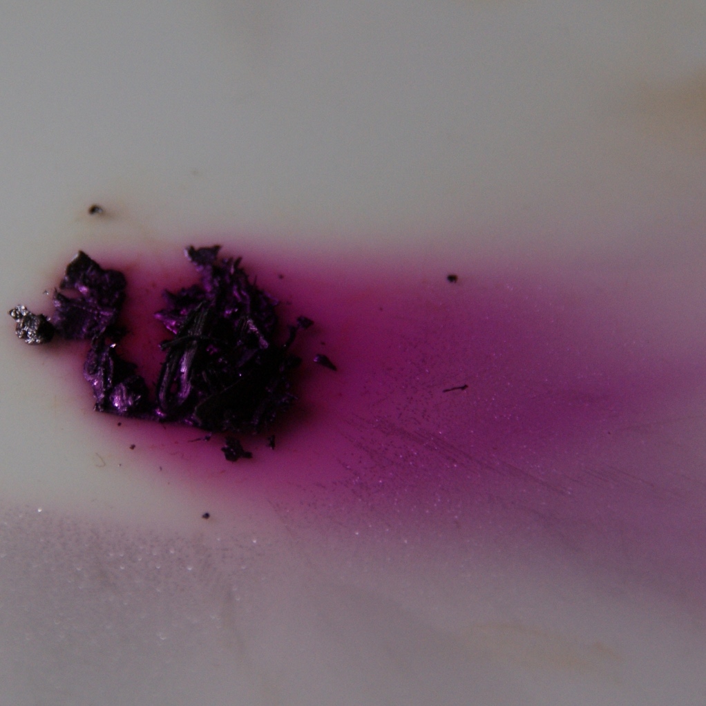
Iodine is a chemical element; it has symbol I and atomic number 53. The heaviest of the stable halogens, it exists at standard conditions as a semi-lustrous, non-metallic solid that melts to form a deep violet liquid at , and boils to a violet gas at . The element was discovered by the French chemist Bernard Courtois in 1811 and was named two years later by Joseph Louis Gay-Lussac, after the Ancient Greek , meaning 'violet'. Iodine occurs in many oxidation states, including iodide (I−), iodate (), and the various periodate anions. As the heaviest essential mineral nutrient, iodine is required for the synthesis of thyroid hormones. Iodine deficiency affects about two billion people and is the leading preventable cause of intellectual disabilities. The dominant producers of iodine today are Chile and Japan. Due to its high atomic number and ease of attachment to organic compounds, it has also found favour as a non-toxic radiocontrast material. Because of the spec ... [...More Info...] [...Related Items...] OR: [Wikipedia] [Google] [Baidu] |
Hymenium
The hymenium is the tissue layer on the hymenophore of a fungal fruiting body where the cells develop into basidia or asci, which produce spores. In some species all of the cells of the hymenium develop into basidia or asci, while in others some cells develop into sterile cells called cystidia ( basidiomycetes) or paraphyses ( ascomycetes). Cystidia are often important for microscopic identification. The subhymenium consists of the supportive hyphae from which the cells of the hymenium grow, beneath which is the hymenophoral trama, the hyphae that make up the mass of the hymenophore. The position of the hymenium is traditionally the first characteristic used in the classification and identification of mushrooms. Below are some examples of the diverse types which exist among the macroscopic Basidiomycota and Ascomycota. * In agarics, the hymenium is on the vertical faces of the gills. * In boletes and polypores, it is in a spongy mass of downward-pointing tubes ... [...More Info...] [...Related Items...] OR: [Wikipedia] [Google] [Baidu] |
Ostiole
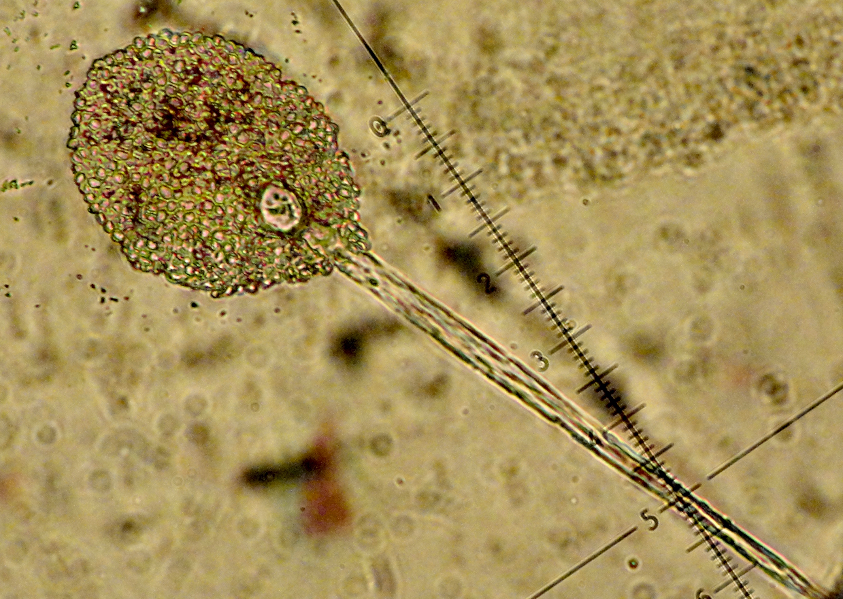
An ''ostiole'' is a small hole or opening through which algae or fungi release their mature spores. The word is a diminutive of wikt:ostium, "ostium", "opening". The term is also used in higher plants, for example to denote the opening of the involuted syconium (fig inflorescence) through which fig wasps enter to Pollination, pollinate and breed. The species pharamacosycea have an arrangement interlocking pattern but there is an exception because of insipdia because it is partly cover the ostiole. On the adaxial side of the bracts is made out of cubic cells, that has a staining reactions and contain phenolic compounds. Sometimes a stomatal aperture is called an "ostiole"."Synergistic Pectin Degradation and Guard Cell Pressurization Underlie Stomatal Pore Formation", See also *Ostium (other) References Castro-Cárdenas, N., Vázquez-Santana, S., Teixeira, S. P., & Ibarra-Manríquez, G. (2023). Correction to: The roles of the ostiole in the fig-fig wasp mutu ... [...More Info...] [...Related Items...] OR: [Wikipedia] [Google] [Baidu] |
Perithecia
An ascocarp, or ascoma (: ascomata), is the fruiting body ( sporocarp) of an ascomycete phylum fungus. It consists of very tightly interwoven hyphae and millions of embedded asci, each of which typically contains four to eight ascospores. Ascocarps are most commonly bowl-shaped (apothecia) but may take on a spherical or flask-like form that has a pore opening to release spores (perithecia) or no opening (cleistothecia). Classification The ascocarp is classified according to its placement (in ways not fundamental to the basic taxonomy). It is called ''epigeous'' if it grows above ground, as with the morels, while underground ascocarps, such as truffles, are termed ''hypogeous''. The structure enclosing the hymenium is divided into the types described below (apothecium, cleistothecium, etc.) and this character ''is'' important for the taxonomic classification of the fungus. Apothecia can be relatively large and fleshy, whereas the others are microscopic—about the size of ... [...More Info...] [...Related Items...] OR: [Wikipedia] [Google] [Baidu] |
