|
Elbow Fracture
Elbow fractures are any broken bone in or near the elbow joint and include olecranon fractures, supracondylar humerus fractures and radial head fractures. The two most common causes of elbow fractures are direct trauma to the elbow joint or bracing a fall with and extended arm. The elbow joint is formed by the articulation of three different bones: the ulna, radius (bone), radius, and humerus that permit the joint to move like a hinge and allow a person to straighten, bend their arm, and rotate their forearm. These bones are connected by tendons, ligaments, and muscle to form the joint. Due to the complexity of the elbow joint, mechanisms of injury, treatment strategies, and complications differ depending on which bones are affected. Adults Distal humerus In young healthy bone, Distal humeral fracture, distal humerus fractures require a high impact mechanism to break. However, Osteoporosis, osteoporotic, or weaker bone can break at the distal humerus with lower energy mechanism ... [...More Info...] [...Related Items...] OR: [Wikipedia] [Google] [Baidu] |
Elbow Joint
The elbow is the region between the upper arm and the forearm that surrounds the elbow joint. The elbow includes prominent landmarks such as the olecranon, the cubital fossa (also called the chelidon, or the elbow pit), and the lateral and the medial epicondyles of the humerus. The elbow joint is a hinge joint between the arm and the forearm; more specifically between the humerus in the upper arm and the radius and ulna in the forearm which allows the forearm and hand to be moved towards and away from the body. The term ''elbow'' is specifically used for humans and other primates, and in other vertebrates it is not used. In those cases, forelimb plus joint is used. The name for the elbow in Latin is ''cubitus'', and so the word cubital is used in some elbow-related terms, as in ''cubital nodes'' for example. Structure Joint The elbow joint has three different portions surrounded by a common joint capsule. These are joints between the three bones of the elbow, the humerus o ... [...More Info...] [...Related Items...] OR: [Wikipedia] [Google] [Baidu] |
Osteoarthritis
Osteoarthritis is a type of degenerative joint disease that results from breakdown of articular cartilage, joint cartilage and underlying bone. A form of arthritis, it is believed to be the fourth leading cause of disability in the world, affecting 1 in 7 adults in the United States alone. The most common symptoms are joint pain and Joint stiffness, stiffness. Usually the symptoms progress slowly over years. Other symptoms may include joint effusion, joint swelling, decreased range of motion, and, when the back is affected, weakness or numbness of the arms and legs. The most commonly involved joints are the two near the ends of the fingers and the joint at the base of the thumbs, the knee and hip joints, and the joints of the neck and lower back. The symptoms can interfere with work and normal daily activities. Unlike some other types of arthritis, only the joints, not internal organs, are affected. Possible causes include previous joint injury, abnormal joint or limb development ... [...More Info...] [...Related Items...] OR: [Wikipedia] [Google] [Baidu] |
Ulnar Nerve
The ulnar nerve is a nerve that runs near the ulna, one of the two long bones in the forearm. The ulnar collateral ligament of elbow joint is in relation with the ulnar nerve. The nerve is the largest in the human body unprotected by muscle or bone, so injury is common. This nerve is directly connected to the little finger, and the adjacent half of the ring finger, innervating the palmar aspect of these fingers, including both front and back of the tips, perhaps as far back as the fingernail beds. This nerve can cause an electric shock-like sensation by striking the medial epicondyle of the humerus posteriorly, or inferiorly with the elbow flexed. The ulnar nerve is trapped between the bone and the overlying skin at this point. This is commonly referred to as bumping one's "funny bone". This name is thought to be a pun, based on the sound resemblance between the name of the bone of the upper arm, the humerus, and the word " humorous". Alternatively, according to the Oxfor ... [...More Info...] [...Related Items...] OR: [Wikipedia] [Google] [Baidu] |
Brachial Artery
The brachial artery is the major blood vessel of the (upper) arm. It is the continuation of the axillary artery beyond the lower margin of teres major muscle. It continues down the ventral surface of the arm until it reaches the cubital fossa at the Elbow-joint, elbow. It then divides into the radial artery, radial and ulnar artery, ulnar artery, arteries which run down the forearm. In some individuals, the bifurcation occurs much earlier and the ulnar and radial arteries extend through the upper arm. The pulse of the brachial artery is palpation, palpable on the anterior aspect of the elbow, medial to the tendon of the Biceps brachii muscle, biceps, and, with the use of a stethoscope and sphygmomanometer (blood pressure cuff), often used to measure the blood pressure. The brachial artery is closely related to the median nerve; in proximal regions, the median nerve is immediately lateral to the brachial artery. Distally, the median nerve crosses the medial side of the brachial ... [...More Info...] [...Related Items...] OR: [Wikipedia] [Google] [Baidu] |
Anterior Interosseous Nerve
The anterior interosseous nerve (volar interosseous nerve) is a branch of the median nerve that supplies the deep muscles on the anterior of the forearm, except the ulnar (medial) half of the flexor digitorum profundus. Its nerve roots come from C8 and T1. It accompanies the anterior interosseous artery along the anterior of the interosseous membrane of the forearm, in the interval between the flexor pollicis longus and flexor digitorum profundus, supplying the whole of the former and (most commonly) the radial half of the latter, and ending below in the pronator quadratus and wrist joint. Note that the median nerve supplies all flexor muscles of the forearm except for the ulnar half of flexor digitorum profundus and the flexor carpi ulnaris, which is a superficial muscle of the forearm. Innervation The anterior interosseous nerve classically innervates 2.5 muscles: which are deep muscles of the forearm * flexor pollicis longus * pronator quadratus * the radial (lateral) h ... [...More Info...] [...Related Items...] OR: [Wikipedia] [Google] [Baidu] |
Supracondylar Humerus Fracture
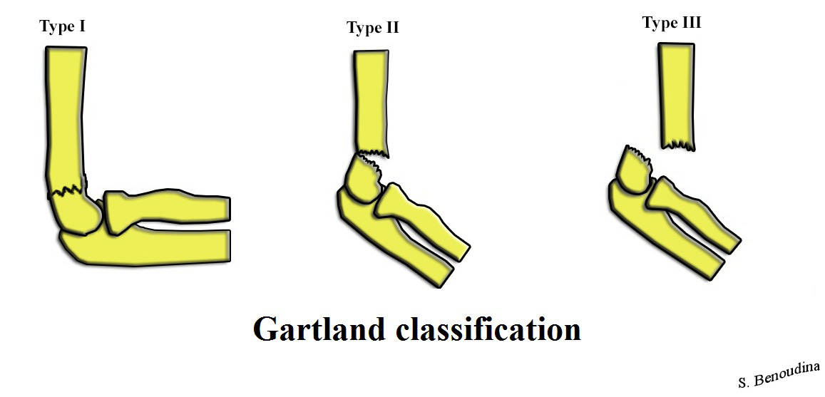
A supracondylar humerus fracture is a fracture of the Anatomical terms of location#Proximal and distal, distal humerus just above the elbow joint. The fracture is usually transverse or oblique and above the medial and lateral condyles and epicondyles. This fracture pattern is relatively rare in adults, but is the most common type of elbow fracture in children. In children, many of these fractures are non-displaced and can be treated with casting. Some are angulated or displaced and are best treated with surgery. In children, most of these fractures can be treated effectively with expectation for full recovery. Some of these injuries can be complicated by poor healing or by associated blood vessel or nerve injuries with serious complications. Signs and symptoms A child will complain of pain and swelling over the elbow immediately post trauma with loss of function of affected upper limb. Late onset of pain (hours after injury) could be due to muscle ischaemia (reduced oxygen supply). ... [...More Info...] [...Related Items...] OR: [Wikipedia] [Google] [Baidu] |
Little League Elbow
Little League elbow, technically termed medial epicondyle apophysitis, is a condition that is caused by repetitive overhand throwing motions in children. Little League elbow is most often seen in young pitchers under the age of sixteen. The pitching motion causes a valgus stress to be placed on the inside of the elbow joint which can cause damage to the structures of the elbow, resulting in an avulsion (separation) of the medial epiphyseal plate (growth plate). Adult pitchers do not experience the same injury because they do not have an open growth plate in the elbow. Instead, adult athletes have a fused growth plate, meaning that ligaments and tendons must bear the stress of the repeated throwing motion. A more common injury in adults is to the ulnar collateral ligament of the elbow, an injury that often requires Tommy John surgery in order for the athlete to resume high-level competitive throwing. "Little Leaguer's elbow" was coined by Brogdon and Crow in an eponymous 1960 arti ... [...More Info...] [...Related Items...] OR: [Wikipedia] [Google] [Baidu] |
Condyle Of Humerus
The condyle of humerus is the distal end of the humerus. It is made up of the capitulum capitulum (plural capitula) may refer to: *the Latin word for chapter ** an index or list of chapters at the head of a gospel manuscript ** a short reading in the Liturgy of the Hours *** derived from which, it is the Latin for the assembly known ... and the trochlea.xiphoid.biostr.washington.edu/fma/fmabrowser-hierarchy.html?search=Condyle of humerus References Anatomy {{musculoskeletal-stub ... [...More Info...] [...Related Items...] OR: [Wikipedia] [Google] [Baidu] |
Elbow
The elbow is the region between the upper arm and the forearm that surrounds the elbow joint. The elbow includes prominent landmarks such as the olecranon, the cubital fossa (also called the chelidon, or the elbow pit), and the lateral and the medial epicondyles of the humerus. The elbow joint is a hinge joint between the arm and the forearm; more specifically between the humerus in the upper arm and the radius and ulna in the forearm which allows the forearm and hand to be moved towards and away from the body. The term ''elbow'' is specifically used for humans and other primates, and in other vertebrates it is not used. In those cases, forelimb plus joint is used. The name for the elbow in Latin is ''cubitus'', and so the word cubital is used in some elbow-related terms, as in ''cubital nodes'' for example. Structure Joint The elbow joint has three different portions surrounded by a common joint capsule. These are joints between the three bones of the elbow, the ... [...More Info...] [...Related Items...] OR: [Wikipedia] [Google] [Baidu] |
Coronoid Process Of The Ulna
The coronoid process of the ulna is a triangular process projecting forward from the anterior proximal portion of the ulna. Structure Its ''base'' is continuous with the body of the bone, and of considerable strength. Its ''apex'' is pointed, slightly curved upward, and in flexion of the forearm is received into the coronoid fossa of the humerus. Its ''upper surface'' is smooth, convex, and forms the lower part of the semilunar notch. Its ''antero-inferior'' surface is concave, and marked by a rough impression for the insertion of the brachialis muscle. At the junction of this surface with the front of the body is a rough eminence, the tuberosity of the ulna, which gives insertion to a part of the brachialis; to the lateral border of this tuberosity the oblique cord is attached. Its ''lateral surface'' presents a narrow, oblong, articular depression, the radial notch. Its ''medial surface'', by its prominent, free margin, serves for the attachment of part of the ulnar ... [...More Info...] [...Related Items...] OR: [Wikipedia] [Google] [Baidu] |
Head Of Radius
The head of the radius has a cylindrical form, and on its upper surface is a shallow cup or fovea for articulation with the capitulum of the humerus. The circumference of the head is smooth; it is broad medially where it articulates with the radial notch of the ulna, narrow in the rest of its extent, which is embraced by the annular ligament.''Gray's Anatomy'' (1918), see infobox Articular surfaces The head of the radius is shaped to articulate with a complex of articular surfaces during both flexion-extension at the elbow and supination-pronation in the forearm: Humeroradial joint The head's proximal surface is concave and cup-shaped to correspond to the spherical surface of the capitulum of the humerus. The radius can thus glide on the capitulum during elbow flexion-extension while simultaneously rotate about its own main axis during supination-pronation. Between the capitulum and the trochlea of the humerus is the capitulotrochlear groove. A semi-lunar surface around th ... [...More Info...] [...Related Items...] OR: [Wikipedia] [Google] [Baidu] |
Terrible Triad
The unhappy triad, also known as a blown knee among other names, is an injury to the anterior cruciate ligament, medial collateral ligament, and meniscus. Analysis during the 1990s indicated that this 'classic' O'Donoghue triad is actually an unusual clinical entity among athletes with knee injuries. Some authors mistakenly believe that in this type of injury, "combined anterior cruciate and medial collateral ligament (ACL- MCL) disruptions that were incurred during athletic endeavors" always present with concomitant medial meniscus injury. However, the 1990 analysis showed that lateral meniscus tears are more common than medial meniscus tears in conjunction with sprains of the ACL. Symptoms *Pain in affected knee *Stiffness and swelling in affected knee *Catching or locking of the knee in affected knee *Instability of the knee with twisting or side-to-side movements (The sensation of the knee "giving out"). *Inability to move the knee through its full range of motion Cause The ... [...More Info...] [...Related Items...] OR: [Wikipedia] [Google] [Baidu] |


