|
Ectopic Ureter
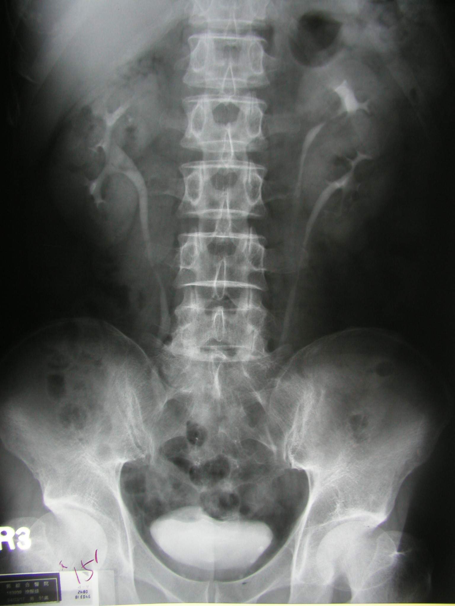
Ectopic ureter (or ureteral ectopia) is a medical condition where the ureter, rather than terminating at the urinary bladder, terminates at a different site. In males this site is usually the urethra, in females this is usually the urethra or vagina. It can be associated with renal dysplasia, frequent urinary tract infections, and urinary incontinence (usually continuous drip incontinence). Ectopic ureters are found in 1 of every 2000–4000 patients, and can be difficult to diagnose, but are most often seen on CT scans A computed tomography scan (CT scan; formerly called computed axial tomography scan or CAT scan) is a medical imaging technique used to obtain detailed internal images of the body. The personnel that perform CT scans are called radiographers .... Ectopic ureter is commonly a result of a duplicated renal collecting system, a duplex kidney with 2 ureters. In this case, usually one ureter drains correctly to the bladder, with the duplicated ureter presenting ... [...More Info...] [...Related Items...] OR: [Wikipedia] [Google] [Baidu] |
Ureteric Bud
The ureteric bud, also known as the metanephric diverticulum, is a protrusion from the mesonephric duct during the development of the urinary and reproductive organs. It later develops into a conduit for urine drainage from the kidneys, which, in contrast, originate from the metanephric blastema The metanephrogenic blastema or metanephric blastema (or metanephric mesenchyme, or metanephric mesoderm) is one of the two embryological structures that give rise to the kidney, the other being the ureteric bud. The metanephric blastema mostly de .... References {{Authority control Embryology of urogenital system ... [...More Info...] [...Related Items...] OR: [Wikipedia] [Google] [Baidu] |
Rectum
The rectum is the final straight portion of the large intestine in humans and some other mammals, and the gut in others. The adult human rectum is about long, and begins at the rectosigmoid junction (the end of the sigmoid colon) at the level of the third sacral vertebra or the sacral promontory depending upon what definition is used. Its diameter is similar to that of the sigmoid colon at its commencement, but it is dilated near its termination, forming the rectal ampulla. It terminates at the level of the anorectal ring (the level of the puborectalis sling) or the dentate line, again depending upon which definition is used. In humans, the rectum is followed by the anal canal which is about long, before the gastrointestinal tract terminates at the anal verge. The word rectum comes from the Latin '' rectum intestinum'', meaning ''straight intestine''. Structure The rectum is a part of the lower gastrointestinal tract. The rectum is a continuation of the sigmoi ... [...More Info...] [...Related Items...] OR: [Wikipedia] [Google] [Baidu] |
Duplex Kidney
The kidneys are two reddish-brown bean-shaped organs found in vertebrates. They are located on the left and right in the retroperitoneal space, and in adult humans are about in length. They receive blood from the paired renal arteries; blood exits into the paired renal veins. Each kidney is attached to a ureter, a tube that carries excreted urine to the bladder. The kidney participates in the control of the volume of various body fluids, fluid osmolality, acid–base balance, various electrolyte concentrations, and removal of toxins. Filtration occurs in the glomerulus: one-fifth of the blood volume that enters the kidneys is filtered. Examples of substances reabsorbed are solute-free water, sodium, bicarbonate, glucose, and amino acids. Examples of substances secreted are hydrogen, ammonium, potassium and uric acid. The nephron is the structural and functional unit of the kidney. Each adult human kidney contains around 1 million nephrons, while a mouse kidney contains only ... [...More Info...] [...Related Items...] OR: [Wikipedia] [Google] [Baidu] |
Duplicated Ureter
Duplicated ureter or Duplex Collecting System is a congenital condition in which the ureteric bud, the embryological origin of the ureter, splits (or arises twice), resulting in two ureters draining a single kidney. It is the most common renal abnormality, occurring in approximately 1% of the population.Siomou E. et alDuplex collecting system diagnosed during the first 6 years of life after a first urinary tract infection: a study of 63 children Journal of Urology, 2006; 175(2):678-81; discussion 681-2J. Gatti, J. Murphy, J. Williams, H. Koo, emedicine overviewUreteral Duplication, Ureteral Ectopia, and Ureterocele/ref> Pathophysiology Ureteral development begins in the human fetus around the 4th week of embryonic development. A ureteric bud, arising from the mesonephric (or Wolffian) duct, gives rise to the ureter, as well as other parts of the collective system. In the case of a duplicated ureter, the ureteric bud either splits or arises twice. In most cases, the kidney is di ... [...More Info...] [...Related Items...] OR: [Wikipedia] [Google] [Baidu] |
CT Scans
A computed tomography scan (CT scan; formerly called computed axial tomography scan or CAT scan) is a medical imaging technique used to obtain detailed internal images of the body. The personnel that perform CT scans are called radiographers or radiology technologists. CT scanners use a rotating X-ray tube and a row of detectors placed in a gantry to measure X-ray attenuations by different tissues inside the body. The multiple X-ray measurements taken from different angles are then processed on a computer using tomographic reconstruction algorithms to produce tomographic (cross-sectional) images (virtual "slices") of a body. CT scans can be used in patients with metallic implants or pacemakers, for whom magnetic resonance imaging (MRI) is contraindicated. Since its development in the 1970s, CT scanning has proven to be a versatile imaging technique. While CT is most prominently used in medical diagnosis, it can also be used to form images of non-living objects. The 1979 N ... [...More Info...] [...Related Items...] OR: [Wikipedia] [Google] [Baidu] |
Urinary Incontinence
Urinary incontinence (UI), also known as involuntary urination, is any uncontrolled leakage of urine. It is a common and distressing problem, which may have a large impact on quality of life. It has been identified as an important issue in geriatric health care. The term enuresis is often used to refer to urinary incontinence primarily in children, such as nocturnal enuresis (bed wetting). UI is an example of a stigmatized medical condition, which creates barriers to successful management and makes the problem worse. People may be too embarrassed to seek medical help, and attempt to self-manage the symptom in secrecy from others. Pelvic surgery, pregnancy, childbirth, and menopause are major risk factors. Urinary incontinence is often a result of an underlying medical condition but is under-reported to medical practitioners. There are four main types of incontinence: * Urge incontinence due to an overactive bladder * Stress incontinence due to "a poorly functioning urethral ... [...More Info...] [...Related Items...] OR: [Wikipedia] [Google] [Baidu] |
Urinary Tract Infections
A urinary tract infection (UTI) is an infection that affects part of the urinary tract. When it affects the lower urinary tract it is known as a bladder infection (cystitis) and when it affects the upper urinary tract it is known as a kidney infection (pyelonephritis). Symptoms from a lower urinary tract infection include pain with urination, frequent urination, and feeling the need to urinate despite having an empty bladder. Symptoms of a kidney infection include fever and flank pain usually in addition to the symptoms of a lower UTI. Rarely the urine may appear bloody. In the very old and the very young, symptoms may be vague or non-specific. The most common cause of infection is ''Escherichia coli'', though other bacteria or fungi may sometimes be the cause. Risk factors include female anatomy, sexual intercourse, diabetes, obesity, and family history. Although sexual intercourse is a risk factor, UTIs are not classified as sexually transmitted infections (STIs). Kidne ... [...More Info...] [...Related Items...] OR: [Wikipedia] [Google] [Baidu] |
Renal Dysplasia
Multicystic dysplastic kidney (MCDK) is a condition that results from the malformation of the kidney during fetal development. The kidney consists of irregular cysts of varying sizes. Multicystic dysplastic kidney is a common type of renal cystic disease, and it is a cause of an abdominal mass in infants. Signs and symptoms When a diagnosis of multicystic kidney is made in utero by ultrasound, the disease is found to be bilateral in many cases. Those with bilateral disease often have other severe deformities or polysystemic malformation syndromes. In bilateral cases, the newborn has the classic characteristic of Potter's syndrome. The bilateral condition is incompatible with survival, as the contralateral system frequently is abnormal as well. Contralateral ureteropelvic junction obstruction is found in 3% to 12% of infants with multicystic kidney and contralateral vesicoureteral reflux is seen even more often, in 18% to 43% of infants. Because the high incidence of reflux, voi ... [...More Info...] [...Related Items...] OR: [Wikipedia] [Google] [Baidu] |
Vagina
In mammals, the vagina is the elastic, muscular part of the female genital tract. In humans, it extends from the vestibule to the cervix. The outer vaginal opening is normally partly covered by a thin layer of mucosal tissue called the hymen. At the deep end, the cervix (neck of the uterus) bulges into the vagina. The vagina allows for sexual intercourse and birth. It also channels menstrual flow, which occurs in humans and closely related primates as part of the menstrual cycle. Although research on the vagina is especially lacking for different animals, its location, structure and size are documented as varying among species. Female mammals usually have two external openings in the vulva; these are the urethral opening for the urinary tract and the vaginal opening for the genital tract. This is different from male mammals, who usually have a single urethral opening for both urination and reproduction. The vaginal opening is much larger than the nearby urethral ... [...More Info...] [...Related Items...] OR: [Wikipedia] [Google] [Baidu] |
Urinary Bladder
The urinary bladder, or simply bladder, is a hollow organ in humans and other vertebrates that stores urine from the kidneys before disposal by urination. In humans the bladder is a distensible organ that sits on the pelvic floor. Urine enters the bladder via the ureters and exits via the urethra. The typical adult human bladder will hold between 300 and (10.14 and ) before the urge to empty occurs, but can hold considerably more. The Latin phrase for "urinary bladder" is ''vesica urinaria'', and the term ''vesical'' or prefix ''vesico -'' appear in connection with associated structures such as vesical veins. The modern Latin word for "bladder" – ''cystis'' – appears in associated terms such as cystitis (inflammation of the bladder). Structure In humans, the bladder is a hollow muscular organ situated at the base of the pelvis. In gross anatomy, the bladder can be divided into a broad , a body, an apex, and a neck. The apex (also called the vertex) is directed fo ... [...More Info...] [...Related Items...] OR: [Wikipedia] [Google] [Baidu] |
Hindgut
The hindgut (or epigaster) is the posterior (caudal) part of the alimentary canal. In mammals, it includes the distal one third of the transverse colon and the splenic flexure, the descending colon, sigmoid colon and up to the ano-rectal junction. In zoology, the term ''hindgut'' refers also to the cecum and ascending colon. Structure Blood supply Arterial supply is by the inferior mesenteric artery, and venous drainage is to the portal venous system. Lymphatic drainage is to the chyle cistern. Nerve supply The hindgut is innervated via the inferior mesenteric plexus. Sympathetic innervation is from the Lumbar splanchnic nerves (L1-L2), parasympathetic innervation is from S2-S4. Development Additional images File:Gray985.png, Abdominal part of digestive tube and its attachment to the primitive or common mesentery. Human embryo of six weeks. File:Gray1115.png, Tail end of human embryo twenty-five to twenty-nine days old. File:Illacme plenipes female with 170 segmen ... [...More Info...] [...Related Items...] OR: [Wikipedia] [Google] [Baidu] |
Ureter
The ureters are tubes made of smooth muscle that propel urine from the kidneys to the urinary bladder. In a human adult, the ureters are usually long and around in diameter. The ureter is lined by urothelial cells, a type of transitional epithelium, and has an additional smooth muscle layer that assists with peristalsis in its lowest third. The ureters can be affected by a number of diseases, including urinary tract infections and kidney stone. is when a ureter is narrowed, due to for example chronic inflammation. Congenital abnormalities that affect the ureters can include the development of two ureters on the same side or abnormally placed ureters. Additionally, reflux of urine from the bladder back up the ureters is a condition commonly seen in children. The ureters have been identified for at least two thousand years, with the word "ureter" stemming from the stem relating to urinating and seen in written records since at least the time of Hippocrates. It is, ho ... [...More Info...] [...Related Items...] OR: [Wikipedia] [Google] [Baidu] |


