|
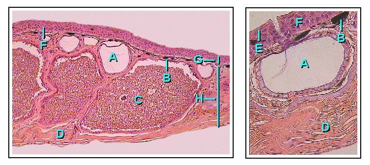
Ear Drum
In the anatomy of humans and various other tetrapods, the eardrum, also called the tympanic membrane or myringa, is a thin, cone-shaped membrane that separates the external ear from the middle ear. Its function is to transmit changes in pressure of sound from the air to the ossicles inside the middle ear, and thence to the oval window in the fluid-filled cochlea. The ear thereby converts and amplifies vibration in the air to vibration in cochlear fluid. The malleus bone bridges the gap between the eardrum and the other ossicles. Rupture or perforation of the eardrum can lead to conductive hearing loss. Collapse or retraction of the eardrum can cause conductive hearing loss or cholesteatoma. Structure Orientation and relations The tympanic membrane is oriented obliquely in the anteroposterior, mediolateral, and superoinferior planes. Consequently, its superoposterior end lies lateral to its anteroinferior end. Anatomically, it relates superiorly to the middle cranial fos ... [...More Info...] [...Related Items...] OR: [Wikipedia] [Google] [Baidu] |
Round Window
The round window is one of the two openings from the middle ear into the inner ear. It is sealed by the secondary tympanic membrane (round window membrane), which vibrates with opposite phase to vibrations entering the inner ear through the oval window. It allows fluid in the cochlea to move, which in turn ensures that hair cells of the basilar membrane will be stimulated and that audition will occur. Structure The round window is situated below (inferior to) and a little behind (posterior to) the oval window, from which it is separated by a rounded elevation, the promontory. It is located at the bottom of a funnel-shaped depression (the round window niche) and, in the macerated bone, opens into the cochlea of the internal ear; in the fresh state it is closed by a membrane, the secondary tympanic membrane (, or ) or round window membrane, which is a complex saddle point shape. The visible central portion is concave (curved inwards) toward the tympanic cavity and convex (curve ... [...More Info...] [...Related Items...] OR: [Wikipedia] [Google] [Baidu] |
Middle Cranial Fossa
The middle cranial fossa is formed by the sphenoid bones, and the temporal bones. It lodges the temporal lobes, and the pituitary gland. It is deeper than the anterior cranial fossa, is narrow medially and widens laterally to the sides of the skull. It is separated from the posterior cranial fossa by the clivus and the petrous crest. It is bounded in front by the posterior margins of the lesser wings of the sphenoid bone, the anterior clinoid processes, and the ridge forming the anterior margin of the chiasmatic groove; behind, by the superior angles of the petrous portions of the temporal bones and the dorsum sellae; laterally by the temporal squamae, sphenoidal angles of the parietals, and greater wings of the sphenoid. It is traversed by the squamosal, sphenoparietal, sphenosquamosal, and sphenopetrosal sutures. Anatomy Features Middle part The middle part of the fossa presents, in front, the chiasmatic groove and tuberculum sellae; the chiasmatic groove ends o ... [...More Info...] [...Related Items...] OR: [Wikipedia] [Google] [Baidu] |
Fibrocartilage
Fibrocartilage consists of a mixture of white fibrous tissue and cartilaginous tissue in various proportions. It owes its inflexibility and toughness to the former of these constituents, and its elasticity to the latter. It is the only type of cartilage that contains type I collagen in addition to the normal type II. Structure The extracellular matrix of fibrocartilage is mainly made from type I collagen secreted by chondroblasts. Locations of fibrocartilage in the human body * secondary cartilaginous joints: ** pubic symphysis ** annulus fibrosis of intervertebral discs ** manubriosternal joint * glenoid labrum of shoulder joint * acetabular labrum of hip joint * medial and lateral menisci of the knee joint * location where tendons and ligaments attach to bone * triangular fibrocartilage complex (UTFCC) Function Repair If hyaline cartilage is torn all the way down to the bone, the blood supply from inside the bone is sometimes enough to start some heal ... [...More Info...] [...Related Items...] OR: [Wikipedia] [Google] [Baidu] |
Mucosa
A mucous membrane or mucosa is a membrane that lines various cavities in the body of an organism and covers the surface of internal organs. It consists of one or more layers of epithelial cells overlying a layer of loose connective tissue. It is mostly of endodermal origin and is continuous with the skin at body openings such as the eyes, eyelids, ears, inside the nose, inside the mouth, lips, the genital areas, the urethral opening and the anus. Some mucous membranes secrete mucus, a thick protective fluid. The function of the membrane is to stop pathogens and dirt from entering the body and to prevent bodily tissues from becoming dehydrated. Structure The mucosa is composed of one or more layers of epithelial cells that secrete mucus, and an underlying lamina propria of loose connective tissue. The type of cells and type of mucus secreted vary from organ to organ and each can differ along a given tract. Mucous membranes line the digestive, respiratory and rep ... [...More Info...] [...Related Items...] OR: [Wikipedia] [Google] [Baidu] |
Fibrous Tissue
Connective tissue is one of the four primary types of animal tissue, a group of cells that are similar in structure, along with epithelial tissue, muscle tissue, and nervous tissue. It develops mostly from the mesenchyme, derived from the mesoderm, the middle embryonic germ layer. Connective tissue is found in between other tissues everywhere in the body, including the nervous system. The three meninges, membranes that envelop the brain and spinal cord, are composed of connective tissue. Most types of connective tissue consists of three main components: elastic and collagen fibers, ground substance, and cells. Blood and lymph are classed as specialized fluid connective tissues that do not contain fiber. All are immersed in the body water. The cells of connective tissue include fibroblasts, adipocytes, macrophages, mast cells and leukocytes. The term "connective tissue" (in German, ) was introduced in 1830 by Johannes Peter Müller. The tissue was already recognized as a distinc ... [...More Info...] [...Related Items...] OR: [Wikipedia] [Google] [Baidu] |
Skin
Skin is the layer of usually soft, flexible outer tissue covering the body of a vertebrate animal, with three main functions: protection, regulation, and sensation. Other animal coverings, such as the arthropod exoskeleton, have different developmental origin, structure and chemical composition. The adjective cutaneous means "of the skin" (from Latin ''cutis'' 'skin'). In mammals, the skin is an organ of the integumentary system made up of multiple layers of ectodermal tissue and guards the underlying muscles, bones, ligaments, and internal organs. Skin of a different nature exists in amphibians, reptiles, and birds. Skin (including cutaneous and subcutaneous tissues) plays crucial roles in formation, structure, and function of extraskeletal apparatus such as horns of bovids (e.g., cattle) and rhinos, cervids' antlers, giraffids' ossicones, armadillos' osteoderm, and os penis/ os clitoris. All mammals have some hair on their skin, even marine mammals like whales, ... [...More Info...] [...Related Items...] OR: [Wikipedia] [Google] [Baidu] |
Eustachian Tube
The Eustachian tube (), also called the auditory tube or pharyngotympanic tube, is a tube that links the nasopharynx to the middle ear, of which it is also a part. In adult humans, the Eustachian tube is approximately long and in diameter. It is named after the sixteenth-century Italian anatomist Bartolomeo Eustachi. In humans and other tetrapods, both the middle ear and the ear canal are normally filled with air. Unlike the air of the ear canal, however, the air of the middle ear is not in direct contact with the atmosphere outside the body; thus, a pressure difference can develop between the atmospheric pressure of the ear canal and the middle ear. Normally, the Eustachian tube is collapsed, but it gapes open with swallowing and with positive pressure, allowing the middle ear's pressure to adjust to the atmospheric pressure. When taking off in an aircraft, the ambient air pressure goes from higher (on the ground) to lower (in the sky). The air in the middle ear expands as ... [...More Info...] [...Related Items...] OR: [Wikipedia] [Google] [Baidu] |
Notch Of Rivinus
{{Technical, date=November 2013 The Notch of Rivinus is a small defect in the posterior edge of the bony annular tympanic ring. The defect is located just superior to the tympano-mastoid suture line in the posterior ear canal The ear canal (external acoustic meatus, external auditory meatus, EAM) is a pathway running from the outer ear to the middle ear. The adult human ear canal extends from the auricle to the eardrum and is about in length and in diameter. S .... Following identification of the spine of Henle it is possible to follow the tympano-mastoid suture line medially towards the annular ring. At this location the Chorda Tympani Nerve is often identified. Just superior to this the Notch of Rivinus can be seen and the neck of the malleus occupies the notch and often is the superior limit of a tympanomeatal flap. Etymology: Augustus Q. Rivinus, German anatomist, 1652–1723 a deficiency in the tympanic sulcus of the ear that forms an attachment for the flaccid ... [...More Info...] [...Related Items...] OR: [Wikipedia] [Google] [Baidu] |
Process (anatomy)
In anatomy, a process () is a projection or outgrowth of tissue from a larger body. For instance, in a vertebra, a process may serve for muscle attachment and leverage (as in the case of the transverse and spinous processes), or to fit (forming a synovial joint A synovial joint, also known as diarthrosis, joins bones or cartilage with a fibrous joint capsule that is continuous with the periosteum of the joined bones, constitutes the outer boundary of a synovial cavity, and surrounds the bones' articulati ...), with another vertebra (as in the case of the articular processes).Moore, Keith L. et al. (2010) ''Clinically Oriented Anatomy'', 6th Ed, p.442 fig. 4.2 The word is also used at the microanatomic level, where cells can have processes such as cilia or pedicels. Depending on the tissue, processes may also be called by other terms, such as ''apophysis'', '' tubercle'', or ''protuberance''. Examples Examples of processes include: *The many processes of the human skull: ... [...More Info...] [...Related Items...] OR: [Wikipedia] [Google] [Baidu] |
Pars Tensa Of Tympanic Membrane
Pars may refer to: * Fars province of Iran, also known as Pars Province * Pars (Sasanian province), a province roughly corresponding to the present-day Fars, 224–651 * ''Pars'', for ''Persia'' or ''Iran'', in the Persian language * Pars News Agency, former name of Iranian news agency * Pars-e Jonubi (other), villages in Iran * FNSS Pars, a Turkish wheeled armoured vehicle * Pars (surname) * Pars interarticularis, in spinal anatomy * ''The Pars'', nickname for Dunfermline Athletic Football Club PARS may refer to: * Point-a-rally scoring in the game of squash * Pakistan Amateur Radio Society * Programmed Airline Reservations System * Russian Mission Airport. Alaska, US, ICAO location indicator * Pre-arrival Review System for import into Canada * PARS 3 LR, a German anti-tank missile See also * Parsa (other) * Fars (other) *Persia (other) Persia, or Iran, is a country in Western Asia. Persia or Persias, may also refer to: Places * ... [...More Info...] [...Related Items...] OR: [Wikipedia] [Google] [Baidu] |
Pars Flaccida Of Tympanic Membrane
In human anatomy, the pars flaccida of tympanic membrane or Shrapnell's membrane (also known as Rivinus' ligament) is the small, triangular, flaccid portion of the tympanic membrane, or eardrum. It lies above the malleolar folds attached directly to the petrous bone at the notch of Rivinus. On the inner surface of the tympanic membrane, the chorda tympani Chorda tympani is a branch of the facial nerve that carries gustatory (taste) sensory innervation from the front of the tongue and parasympathetic ( secretomotor) innervation to the submandibular and sublingual salivary glands. Chorda tymp ... crosses this area. The name ''Shrapnell's membrane'' refers to Henry Jones Shrapnell, and the name ''Rivinus' ligament'' to Augustus Quirinus Rivinus. References Auditory system {{anatomy-stub ... [...More Info...] [...Related Items...] OR: [Wikipedia] [Google] [Baidu] |
Temporomandibular Joint
In anatomy, the temporomandibular joints (TMJ) are the two joints connecting the jawbone to the skull. It is a bilateral Synovial joint, synovial articulation between the temporal bone of the skull above and the condylar process of mandible below; it is from these bones that its name is derived. The joints are unique in their bilateral function, being connected via the mandible. Structure The main components are the joint capsule, articular disc, mandibular condyles, articular surface of the temporal bone, temporomandibular ligament, stylomandibular ligament, sphenomandibular ligament, and lateral pterygoid muscle. Capsule The articular capsule (capsular ligament) is a thin, loose envelope, attached above to the circumference of the mandibular fossa and the articular tubercle immediately in front; below, to the neck of the condyle of the mandible. Its loose attachment to the neck of the mandible allows for free movement. Articular disc The unique feature of the temporomand ... [...More Info...] [...Related Items...] OR: [Wikipedia] [Google] [Baidu] |


