|
Dorsal Interossei Of The Hand
In human anatomy, the dorsal interossei (DI) are four muscles in the back of the hand that act to abduct (spread) the index, middle, and ring fingers away from the hand's midline (ray of middle finger) and assist in flexion at the metacarpophalangeal The metacarpophalangeal joints (MCP) are situated between the metacarpal bones and the proximal phalanges of the fingers. These joints are of the condyloid kind, formed by the reception of the rounded heads of the metacarpal bones into shallow ... joints and extension at the interphalangeal joints of the index, middle and ring fingers. Structure There are four dorsal interossei in each hand. They are specified as 'dorsal' to contrast them with the palmar interossei, which are located on the anterior side of the metacarpals. The dorsal interosseous muscles are bipennate, with each muscle arising by two heads from the adjacent sides of the metacarpal bones, but more extensively from the metacarpal bone of the finger into whic ... [...More Info...] [...Related Items...] OR: [Wikipedia] [Google] [Baidu] |
Metacarpals
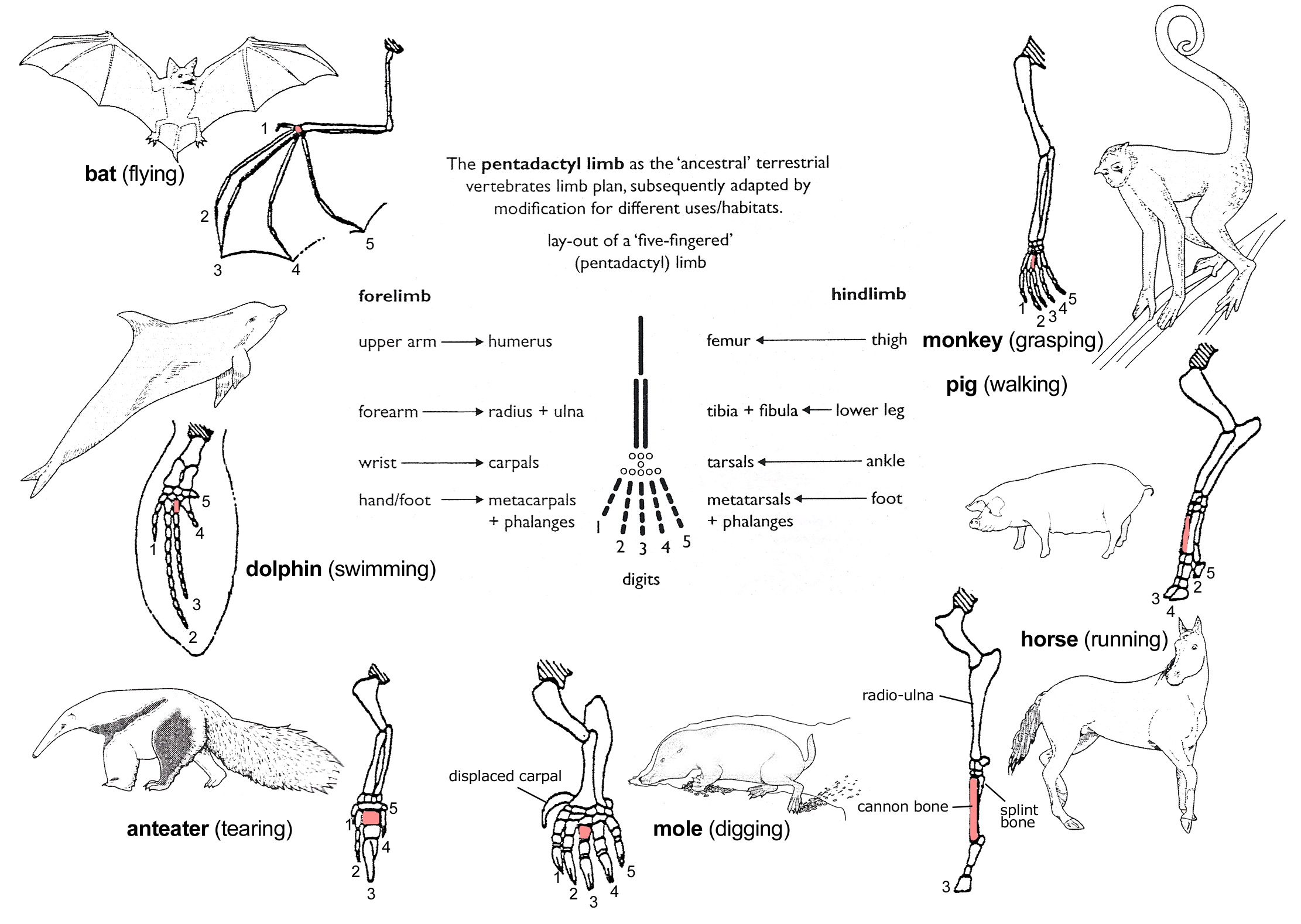
In human anatomy, the metacarpal bones or metacarpus, also known as the "palm bones", are the appendicular skeleton, appendicular bones that form the intermediate part of the hand between the phalanges (fingers) and the carpal bones (wrist, wrist bones), which joint, articulate with the forearm. The metacarpal bones are homologous to the metatarsal bones in the foot. Structure The metacarpals form a transverse arch to which the rigid row of distal carpal bones are fixed. The peripheral metacarpals (those of the thumb and little finger) form the sides of the cup of the palmar gutter and as they are brought together they deepen this concavity. The index metacarpal is the most firmly fixed, while the thumb metacarpal articulates with the trapezium and acts independently from the others. The middle metacarpals are tightly united to the carpus by intrinsic interlocking bone elements at their bases. The ring metacarpal is somewhat more mobile while the fifth metacarpal is semi-indepen ... [...More Info...] [...Related Items...] OR: [Wikipedia] [Google] [Baidu] |
Phalanges
The phalanges (: phalanx ) are digit (anatomy), digital bones in the hands and foot, feet of most vertebrates. In primates, the Thumb, thumbs and Hallux, big toes have two phalanges while the other Digit (anatomy), digits have three phalanges. The phalanges are classed as long bones. Structure The phalanges are the bones that make up the fingers of the hand and the toes of the foot. There are 56 phalanges in the human body, with fourteen on each hand and foot. Three phalanges are present on each finger and toe, with the exception of the thumb and hallux, big toe, which possess only two. The middle and far phalanges of the fifth toes are often fused together (symphalangism). The phalanges of the hand are commonly known as the finger bones. The phalanges of the foot differ from the hand in that they are often shorter and more compressed, especially in the proximal phalanges, those closest to the torso. A phalanx is named according to whether it is Anatomical terms of locatio ... [...More Info...] [...Related Items...] OR: [Wikipedia] [Google] [Baidu] |
Adductor Pollicis Muscle
In human anatomy, the adductor pollicis muscle is a muscle in the hand that functions to adduct the thumb. It has two heads: transverse and oblique. It is a fleshy, flat, triangular, and fan-shaped muscle deep in the thenar compartment beneath the long flexor tendons and the lumbrical muscles at the center of the palm. It overlies the metacarpal bones and the interosseous muscles. Structure Oblique head The oblique head (Latin: ''adductor obliquus pollicis'') arises by several slips from the capitate bone, the bases of the second and third metacarpals, the intercarpal ligaments, and the sheath of the tendon of the flexor carpi radialis. Gray's Anatomy 1918. (See infobox) From this origin the greater number of fibers pass obliquely downward and converge to a tendon, which, uniting with the tendons of the medial portion of the flexor pollicis brevis and the transverse head of the adductor pollicis, is inserted into the ulnar side of the base of the proximal phalanx ... [...More Info...] [...Related Items...] OR: [Wikipedia] [Google] [Baidu] |
Ulnar Nerve
The ulnar nerve is a nerve that runs near the ulna, one of the two long bones in the forearm. The ulnar collateral ligament of elbow joint is in relation with the ulnar nerve. The nerve is the largest in the human body unprotected by muscle or bone, so injury is common. This nerve is directly connected to the little finger, and the adjacent half of the ring finger, innervating the palmar aspect of these fingers, including both front and back of the tips, perhaps as far back as the fingernail beds. This nerve can cause an electric shock-like sensation by striking the medial epicondyle of the humerus posteriorly, or inferiorly with the elbow flexed. The ulnar nerve is trapped between the bone and the overlying skin at this point. This is commonly referred to as bumping one's "funny bone". This name is thought to be a pun, based on the sound resemblance between the name of the bone of the upper arm, the humerus, and the word " humorous". Alternatively, according to the Oxfor ... [...More Info...] [...Related Items...] OR: [Wikipedia] [Google] [Baidu] |
Deep Branch Of Ulnar Nerve
The deep branch of the ulnar nerve is a terminal, primarily motor branch of the ulnar nerve. It is accompanied by the deep palmar branch of ulnar artery. Structure It passes between the abductor digiti minimi and the flexor digiti minimi brevis. It then perforates the opponens digiti minimi and follows the course of the deep palmar arch beneath the flexor tendons. As the deep ulnar nerve passes across the palm, it lies in a fibrous tunnel formed between the hook of the hamate and the pisiform ( Guyon's canal). Function At its origin it innervates the hypothenar muscles. As it crosses the deep part of the hand, it innervates all the interosseous muscles and the third and fourth lumbricals. It ends by innervating the adductor pollicis and the medial (deep) head of the flexor pollicis brevis The flexor pollicis brevis is a muscle in the hand that flexes the thumb. It is one of three thenar muscles. It has both a superficial part and a deep part. Origin and insertion The ... [...More Info...] [...Related Items...] OR: [Wikipedia] [Google] [Baidu] |
Fifth Metacarpal
The fifth metacarpal bone (metacarpal bone of the little finger or pinky finger) is the most medial and second-shortest of the metacarpal bones. Surfaces It presents on its base one facet on its superior surface, which is concavo-convex and articulates with the hamate, and one on its radial side, which articulates with the fourth metacarpal. On its ulnar side is a prominent tubercle for the insertion of the tendon of the extensor carpi ulnaris muscle. The dorsal surface of the body is divided by an oblique ridge, which extends from near the ulnar side of the base to the radial side of the head. The lateral part of this surface serves for the attachment of the fourth interosseus dorsalis; the medial part is smooth, triangular, and covered by the extensor tendons of the little finger. The palmar surface is similarly divided: Its lateral side (facing the fourth metacarpal) provides the origin for the third palmar interosseus, its medial side contains the insertion of opponens di ... [...More Info...] [...Related Items...] OR: [Wikipedia] [Google] [Baidu] |
Fourth Metacarpal
The fourth metacarpal bone (metacarpal bone of the ring finger) is shorter and smaller than the third. The base is small and quadrilateral; its superior surface presents two facets, a large one medially for articulation with the hamate, and a small one laterally for the capitate. On the radial side are two oval facets, for articulation with the third metacarpal; and on the ulnar side a single concave facet, for the fifth metacarpal. Clinical relevance A shortened fourth metacarpal bone can be a symptom of Kallmann syndrome, a genetic condition which results in the failure to commence or the non-completion of puberty. A short fourth metacarpal bone can also be found in Turner syndrome, a disorder involving sex chromosomes. A fracture of the fourth and/or fifth metacarpal bones transverse neck secondary due to axial loading is known as a boxer's fracture.Shultz, S. J., Houglum, P. A., Perrin, D. H. (2010). Examination of Musculoskeletal Injuries. Chicago: Human Kinetics Ossific ... [...More Info...] [...Related Items...] OR: [Wikipedia] [Google] [Baidu] |
Third Metacarpal
The third metacarpal bone (metacarpal bone of the middle finger) is a little smaller than the second. The dorsal aspect of its base presents on its radial side a pyramidal eminence, the Third metacarpal styloid process, styloid process, which extends upward behind the capitate; immediately distal to this is a rough surface for the attachment of the extensor carpi radialis brevis muscle. The carpal articular facet is concave behind, flat in front, and articulates with the capitate. On the radial side is a smooth, concave facet for articulation with the second metacarpal, and on the ulnar side two small oval facets for the fourth metacarpal. Ossification The ossification process begins in the shaft during prenatal life, and in the head between the 11th and 27th months. Additional images File:Third metacarpal bone (left hand) - animation01.gif, Third metacarpal bone of the left hand (shown in red). Animation. File:Third metacarpal bone (left hand) - animation02.gif, Third met ... [...More Info...] [...Related Items...] OR: [Wikipedia] [Google] [Baidu] |
First Metacarpal
The first metacarpal bone or the metacarpal bone of the thumb is the first bone proximal to the thumb. It is connected to the trapezium of the carpus at the first carpometacarpal joint and to the proximal thumb phalanx at the first metacarpophalangeal joint. Characteristics The first metacarpal bone is short and thick with a shaft thicker and broader than those of the other metacarpal bones. Its narrow shaft connects its widened base and rounded head; the former consisting of a thick cortical bone surrounding the open medullary canal; the latter two consisting of cancellous bone surrounded by a thin cortical shell. Head The head is less rounded and less spherical than those of the other metacarpals, making it better suited for a hinge-like articulation. The distal articular surface is quadrilateral, wide, and flat; thicker and broader transversely and extends much further palmarly than dorsally. On the palmar aspect of the articular surface there is a pair of eminences ... [...More Info...] [...Related Items...] OR: [Wikipedia] [Google] [Baidu] |
Second Metacarpal
The second metacarpal bone (metacarpal bone of the index finger) is the longest, and its base the largest, of all the metacarpal bones.''Gray's Anatomy'' (1918). See infobox. Human anatomy Its base is prolonged upward and medialward, forming a prominent ridge. It presents four articular facets, three on the upper surface and one on the ulnar side: * Of the facets on the upper surface: ** the ''intermediate'' is the largest and is concave from side to side, convex from before backward for articulation with the lesser multangular; ** the ''lateral'' is small, flat and oval for articulation with the greater multangular; ** the ''medial'', on the summit of the ridge, is long and narrow for articulation with the capitate. * The facet on the ulnar side articulates with the third metacarpal. The extensor carpi radialis longus muscle is inserted on the dorsal surface and the flexor carpi radialis muscle on the volar surface of the base. The shaft gives origin to the first palma ... [...More Info...] [...Related Items...] OR: [Wikipedia] [Google] [Baidu] |
Deep Palmar Arch
The deep palmar arch (deep volar arch) is an arterial network found in the palm. It is usually primarily formed from the terminal part of the radial artery. The ulnar artery also contributes through an anastomosis. This is in contrast to the superficial palmar arch, which is formed predominantly by the ulnar artery. Structure The deep palmar arch is usually primarily formed from the radial artery. The ulnar artery also contributes through an anastomosis. The deep palmar arch lies upon the bases of the metacarpal bones and on the interossei of the hand. It is deep to the oblique head of the adductor pollicis muscle, the flexor tendons of the fingers, and the lumbricals of the hand. Alongside of it, but running in the opposite direction—toward the radial side of the hand—is the deep branch of the ulnar nerve. The superficial palmar arch is more distally located than the deep palmar arch. If one were to fully extend the thumb and draw a line from the distal border of the thu ... [...More Info...] [...Related Items...] OR: [Wikipedia] [Google] [Baidu] |
Radial Artery
In human anatomy, the radial artery is the main artery of the lateral aspect of the forearm. Structure The radial artery arises from the bifurcation of the brachial artery in the antecubital fossa. It runs distally on the anterior part of the forearm. There, it serves as a landmark for the division between the anterior compartment of the forearm, anterior and posterior compartment of the forearm, posterior compartments of the forearm, with the posterior compartment beginning just lateral to the artery. The artery winds laterally around the wrist, passing through the anatomical snuff box and between the heads of the first dorsal interossei of the hand, dorsal interosseous muscle. It passes anteriorly between the heads of the adductor pollicis, and becomes the deep palmar arch, which joins with the deep branch of the ulnar artery. Along its course, it is accompanied by a similarly named vein, the radial vein. Branches The named branches of the radial artery may be divided int ... [...More Info...] [...Related Items...] OR: [Wikipedia] [Google] [Baidu] |


