|
Diaphragma Sellae
The diaphragma sellae or sellar diaphragm is a small, circular sheet of dura mater forming an (incomplete) roof over the sella turcica and covering the pituitary gland lodged therein. The diaphragma sellae forms a central opening to accommodate the passage of the pituitary stalk (infundibulum) which interconnects the pituitary gland and the hypothalamus. The diaphragma sellae is an important neurosurgical landmark. Anatomy Boundaries The diaphragma sellae has a posterior boundary at the dorsum sellae and an anterior boundary at the tuberculum sellae along with the two small eminences (one on either side) called the middle clinoid processes. Variation The opening formed by the diaphragma sellae varies greatly in size between individuals. Clinical significance Pituitary tumours may grow to extend superiorly beyond the diaphragma sellae. Violation of the diaphragma sellae during an endoscopic endonasal transsphenoidal pituitary tumor resection will result in a cerebrosp ... [...More Info...] [...Related Items...] OR: [Wikipedia] [Google] [Baidu] |
Tentorium Cerebelli
The cerebellar tentorium or tentorium cerebelli (Latin for "tent of the cerebellum") is one of four dural folds that separate the cranial cavity into four (incomplete) compartments. The cerebellar tentorium separates the cerebellum from the cerebrum forming a supratentorial and an infratentorial region; the cerebrum is supratentorial and the cerebellum infratentorial. The free border of the tentorium gives passage to the midbrain (the upper-most part of the brainstem). Structure Free border The free border of the tentorium is U-shaped; it forms an aperture - the tentorial notch (tentorial incisure) - which gives passage to the midbrain. The free border of each side extends anteriorly beyond the medial end of the superior petrosal sinus (i.e. the apex of the petrous part of the temporal bone) to overlap the attached margin, thenceforth forming a ridge of dura matter upon the roof of the cavernous sinus, terminating anteriorly by attaching at the anterior clinoid process. ... [...More Info...] [...Related Items...] OR: [Wikipedia] [Google] [Baidu] |
Sella Turcica
The sella turcica (Latin for 'Turkish saddle') is a saddle-shaped depression in the body of the sphenoid bone of the human skull and of the skulls of other hominids including chimpanzees, gorillas and orangutans. It serves as a cephalometric landmark. The pituitary gland or hypophysis is located within the most inferior aspect of the sella turcica, the hypophyseal fossa. Structure The sella turcica is located in the sphenoid bone behind the chiasmatic groove and the tuberculum sellae. It belongs to the middle cranial fossa. The sella turcica's most inferior portion is known as the hypophyseal fossa (the "seat of the saddle"), and contains the pituitary gland (hypophysis). In front of the hypophyseal fossa is the tuberculum sellae. Completing the formation of the saddle posteriorly is the dorsum sellae, which is continuous with the clivus, inferoposteriorly. The dorsum sellae is terminated laterally by the posterior clinoid processes. Development It is widely believed th ... [...More Info...] [...Related Items...] OR: [Wikipedia] [Google] [Baidu] |
Pituitary Gland
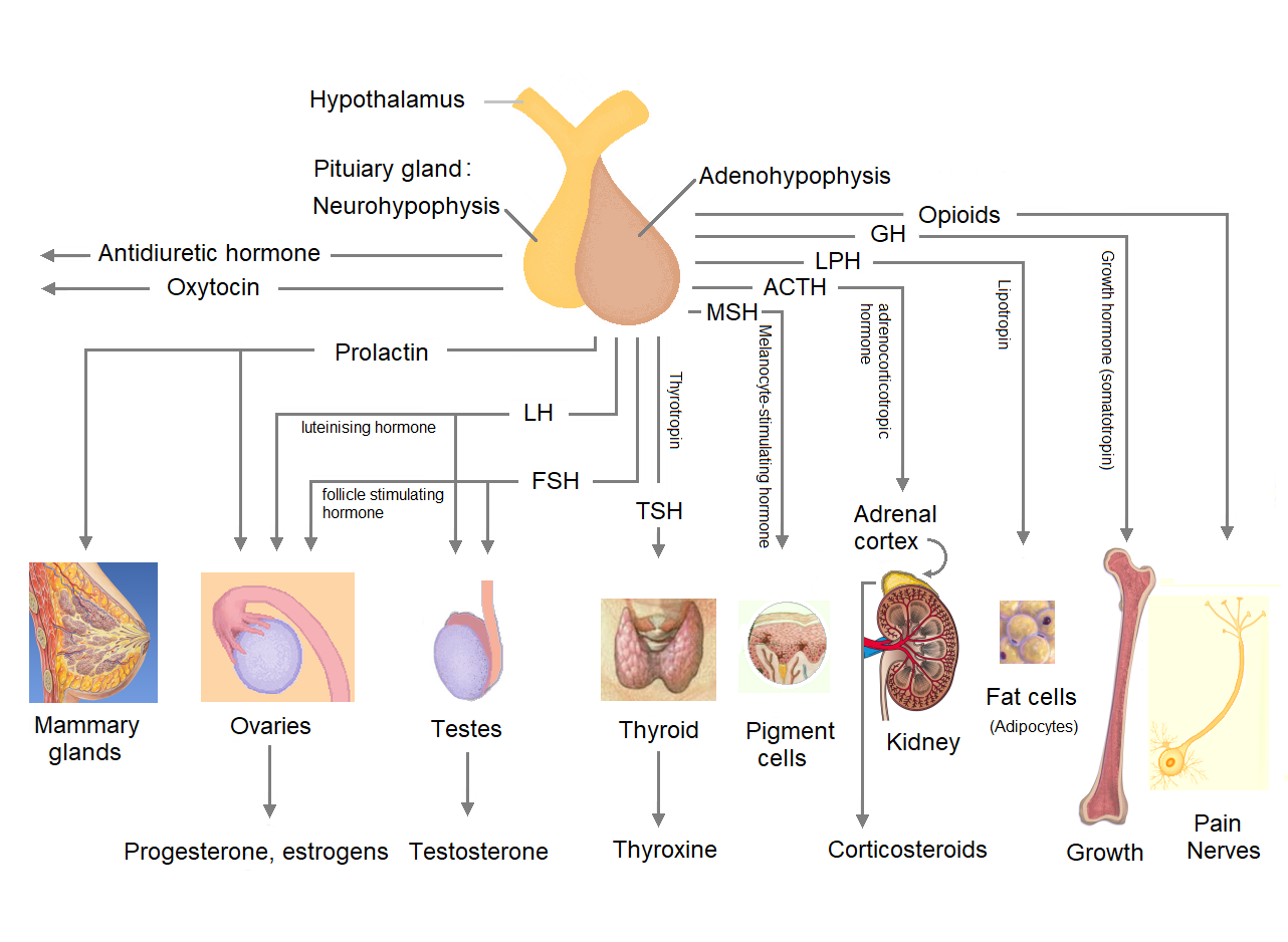
The pituitary gland or hypophysis is an endocrine gland in vertebrates. In humans, the pituitary gland is located at the base of the human brain, brain, protruding off the bottom of the hypothalamus. The pituitary gland and the hypothalamus control much of the body's endocrine system. It is seated in part of the sella turcica a fossa (anatomy), depression in the sphenoid bone, known as the hypophyseal fossa. The human pituitary gland is ovoid, oval shaped, about 1 cm in diameter, in weight on average, and about the size of a kidney bean. Digital version. There are two main lobes of the pituitary, an anterior pituitary, anterior lobe, and a posterior pituitary, posterior lobe joined and separated by a small intermediate lobe. The anterior lobe (adenohypophysis) is the glandular part that produces and secretes several hormones. The posterior lobe (neurohypophysis) secretes neurohypophysial hormones produced in the hypothalamus. Both lobes have different origins and they are both co ... [...More Info...] [...Related Items...] OR: [Wikipedia] [Google] [Baidu] |
Hypothalamus
The hypothalamus (: hypothalami; ) is a small part of the vertebrate brain that contains a number of nucleus (neuroanatomy), nuclei with a variety of functions. One of the most important functions is to link the nervous system to the endocrine system via the pituitary gland. The hypothalamus is located below the thalamus and is part of the limbic system. It forms the Basal (anatomy), basal part of the diencephalon. All vertebrate brains contain a hypothalamus. In humans, it is about the size of an Almond#Nut, almond. The hypothalamus has the function of regulating certain metabolic biological process, processes and other activities of the autonomic nervous system. It biosynthesis, synthesizes and secretes certain neurohormones, called releasing hormones or hypothalamic hormones, and these in turn stimulate or inhibit the secretion of hormones from the pituitary gland. The hypothalamus controls thermoregulation, body temperature, hunger (physiology), hunger, important aspects o ... [...More Info...] [...Related Items...] OR: [Wikipedia] [Google] [Baidu] |
Dorsum Sellae
The dorsum sellae is part of the sphenoid bone in the skull. Together with the basilar part of the occipital bone it forms the clivus. In the sphenoid bone, the anterior boundary of the sella turcica is completed by two small eminences, one on either side, called the middle clinoid processes, while the posterior boundary is formed by a square-shaped plate of bone, the dorsum sellae, ending at its superior angles in two tubercles, the posterior clinoid processes The posterior clinoid processes are the tubercles of the sphenoid bone situated at the superior angles of the dorsum sellae (one on each angle) which represents the posterior boundary of the sella turcica. They vary considerably in size and form. ..., the size and form of which vary considerably in different individuals. Additional images File:Gray569.png, Tentorium cerebelli from above. File:Slide2iiii.JPG, Dorsum sellae References External links * * Bones of the head and neck {{musculoskeletal-s ... [...More Info...] [...Related Items...] OR: [Wikipedia] [Google] [Baidu] |
Tuberculum Sellae
The tuberculum sellae (or the tubercle of the sella turcica) is a slight median elevation upon the superior aspect of the body of sphenoid bone (that forms the floor of the middle cranial fossa) at the anterior boundary of the sella turcica ( hypophyseal (pituitary) fossa) and posterior boundary of the chiasmatic groove. A middle clinoid process The middle clinoid process is a small, bilaterally paired elevation on either side of the tuberculum sellae, at the anterior boundary of the sella turcica. A (larger) anterior clinoid process is situated lateral to each middle clinoid process. ... flanks the tuberculum sellae on either side. Additional images File:Slide1iiii.JPG, Tuberculum sellae References External links * Diagram at uni-mainz.de Bones of the head and neck {{musculoskeletal-stub ... [...More Info...] [...Related Items...] OR: [Wikipedia] [Google] [Baidu] |
Middle Clinoid Processes
The middle clinoid process is a small, bilaterally paired elevation on either side of the tuberculum sellae, at the anterior boundary of the sella turcica. A (larger) anterior clinoid process is situated lateral to each middle clinoid process. The diaphragma sellae (i.e. the dura forming the roof of the cavernous sinus) and the dura of the floor of the hypophyseal fossa (sella turcica) attach onto the middle clinoid processes. On each side of the body, the internal carotid artery passes between the anterior and middle clinoid processes. Etymology Clinoid likely comes from the Greek root ''klinein'' or the Latin Latin ( or ) is a classical language belonging to the Italic languages, Italic branch of the Indo-European languages. Latin was originally spoken by the Latins (Italic tribe), Latins in Latium (now known as Lazio), the lower Tiber area aroun ... ''clinare'', both meaning "sloped" as in "inclined." References Bones of the head and neck {{musculoskelet ... [...More Info...] [...Related Items...] OR: [Wikipedia] [Google] [Baidu] |
Pituitary Tumor
Pituitary adenomas are tumors that occur in the pituitary gland. Most pituitary tumors are benign, approximately 35% are invasive and just 0.1% to 0.2% are carcinomas.Pituitary Tumors Treatment (PDQ®)–Health Professional Version NIH National Cancer Institute Pituitary adenomas represent from 10% to 25% of all intracranial , with an estimated prevalence rate in the general population of approximately 17%. Non-invasive and non-secreting pituitary adenomas are considered to be |
Endoscopic Endonasal Surgery
Endoscopic endonasal surgery is a minimally invasive technique used mainly in neurosurgery and otolaryngology. A neurosurgeon or an otolaryngologist, using an endoscope that is entered through the nose, fixes or removes brain defects or tumors in the anterior skull base. Normally an otolaryngologist performs the initial stage of surgery through the nasal cavity and sphenoid bone; a neurosurgeon performs the rest of the surgery involving drilling into any cavities containing a neural organ such as the pituitary gland. The use of endoscope was first introduced in Transsphenoidal Pituitary Surgery by R Jankowsky, J Auque, C Simon et al. in 1992 G (Laryngoscope. 1992 Feb;102(2):198-202). Introduction History of endoscopic endonasal surgery Antonin Jean Desomeaux, a urologist from Paris, was the first person to use the term, endoscope. However, the precursor to the modern endoscope was invented in the 1800s when a physician in Frankfurt, Germany by the name of Philipp Bozzini, dev ... [...More Info...] [...Related Items...] OR: [Wikipedia] [Google] [Baidu] |
Cerebrospinal Fluid
Cerebrospinal fluid (CSF) is a clear, colorless Extracellular fluid#Transcellular fluid, transcellular body fluid found within the meninges, meningeal tissue that surrounds the vertebrate brain and spinal cord, and in the ventricular system, ventricles of the brain. CSF is mostly produced by specialized Ependyma, ependymal cells in the choroid plexuses of the ventricles of the brain, and absorbed in the arachnoid granulations. It is also produced by ependymal cells in the lining of the ventricles. In humans, there is about 125 mL of CSF at any one time, and about 500 mL is generated every day. CSF acts as a shock absorber, cushion or buffer, providing basic mechanical and immune system, immunological protection to the brain inside the Human skull, skull. CSF also serves a vital function in the cerebral autoregulation of cerebral blood flow. CSF occupies the subarachnoid space (between the arachnoid mater and the pia mater) and the ventricular system around and inside t ... [...More Info...] [...Related Items...] OR: [Wikipedia] [Google] [Baidu] |


