|
Development Of Joints
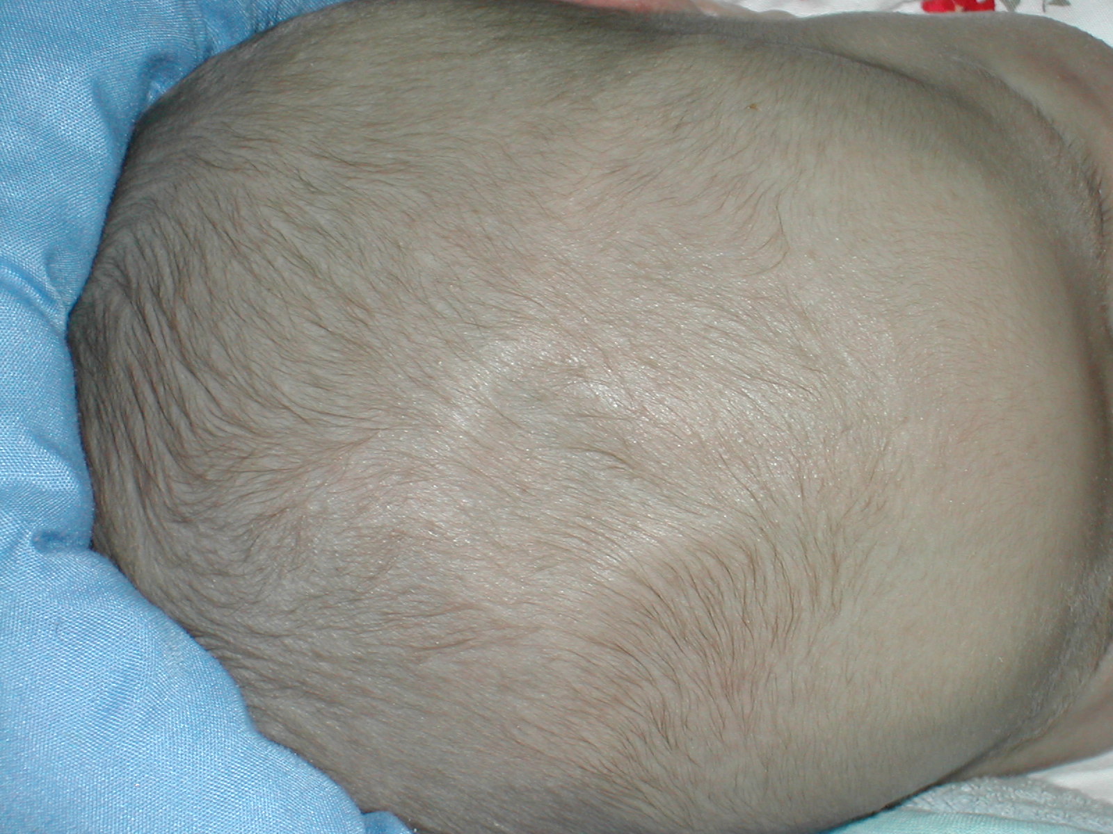
Joints form during embryonic development in conjunction with the formation and growth of the associated bones. The joints and bones are developed from the embryonic tissue called mesenchyme. Head and face In the head, mesenchyme will accumulate at those areas that will become the bones that form the top and sides of the skull. The mesenchyme in these areas will develop directly into bone through the process of intramembranous ossification, in which mesenchymal cells differentiate into bone-producing cells that then generate bone tissue. The mesenchyme between the areas of bone production will become the fibrous connective tissue that fills the spaces between the developing bones. Initially, the connective tissue-filled gaps between the bones are wide, and are called fontanelles. After birth, as the skull bones grow and enlarge, the gaps between them decrease in width and the fontanelles are reduced to suture joints in which the bones are united by a narrow layer of fibrous connec ... [...More Info...] [...Related Items...] OR: [Wikipedia] [Google] [Baidu] |
Mesenchyme
Mesenchyme () is a type of loosely organized animal embryonic connective tissue of undifferentiated cells that give rise to most tissues, such as skin, blood, or bone. The interactions between mesenchyme and epithelium help to form nearly every organ in the developing embryo. Vertebrates Structure Mesenchyme is characterized morphologically by a prominent ground substance matrix containing a loose aggregate of reticular fibers and unspecialized mesenchymal stem cells. Mesenchymal cells can migrate easily (in contrast to epithelial cells, which lack mobility, are organized into closely adherent sheets, and are polarized in an apical- basal orientation). Development The mesenchyme originates from the mesoderm. From the mesoderm, the mesenchyme appears as an embryologically primitive "soup". This "soup" exists as a combination of the mesenchymal cells plus serous fluid plus the many different tissue proteins. Serous fluid is typically stocked with the many serous elements, ... [...More Info...] [...Related Items...] OR: [Wikipedia] [Google] [Baidu] |
Intramembranous Ossification
Intramembranous ossification is one of the two essential processes during fetal development of the gnathostome (excluding chondrichthyans such as sharks) skeletal system by which rudimentary bone tissue is created. Intramembranous ossification is also an essential process during the natural healing of bone fractures and the rudimentary formation of bones of the head. Unlike endochondral ossification, which is the other process by which bone tissue is created during fetal development, cartilage is not present during intramembranous ossification. Formation of woven bone Mesenchymal stem cells within mesenchyme or the medullary cavity of a bone fracture initiate the process of intramembranous ossification. A mesenchymal stem cell, or MSC, is an unspecialized cell that can develop into an osteoblast. Before it begins to develop, the morphological characteristics of a MSC are: A small cell body with a few cell processes that are long and thin; a large, round nucleus with a prom ... [...More Info...] [...Related Items...] OR: [Wikipedia] [Google] [Baidu] |
Fontanelles
A fontanelle (or fontanel) (colloquially, soft spot) is an anatomical feature of the infant human skull comprising soft membranous gaps ( sutures) between the cranial bones that make up the calvaria of a fetus or an infant. Fontanelles allow for stretching and deformation of the neurocranium both during birth and later as the brain expands faster than the surrounding bone can grow. Premature complete ossification of the sutures is called craniosynostosis. After infancy, the anterior fontanelle is known as the bregma. Structure An infant's skull consists of five main bones: two frontal bones, two parietal bones, and one occipital bone. These are joined by fibrous sutures, which allow movement that facilitates childbirth and brain growth. * Posterior fontanelle is triangle-shaped. It lies at the junction between the sagittal suture and lambdoid suture. At birth, the skull features a small posterior fontanelle with an open area covered by a tough membrane, where the two pari ... [...More Info...] [...Related Items...] OR: [Wikipedia] [Google] [Baidu] |
Suture (anatomy)
In anatomy, a suture is a fairly rigid joint between two or more hard elements of an organism, with or without significant overlap of the elements. Sutures are found in the skeletons or exoskeletons of a wide range of animals, in both invertebrates and vertebrates. Sutures are found in animals with hard parts from the Cambrian period to the present day. Sutures were and are formed by several different methods, and they exist between hard parts that are made from several different materials. Vertebrate skeletons The skeletons of vertebrate animals (fish, amphibians, reptiles, birds, and mammals) are made of bone, in which the main rigid ingredient is calcium phosphate. Cranial sutures The skulls of most vertebrates consist of sets of bony plates held together by cranial sutures. These sutures are held together mainly by Sharpey's fibers which grow from each bone into the adjoining one. Sutures in the ankles of land vertebrates In the type of crurotarsal ankle, which is fo ... [...More Info...] [...Related Items...] OR: [Wikipedia] [Google] [Baidu] |
Endochondral Ossification
Endochondral ossification is one of the two essential pathways by which bone tissue is produced during fetal development and bone healing, bone repair of the mammalian skeleton, skeletal system, the other pathway being intramembranous ossification. Both endochondral and intramembranous processes initiate from a precursor mesenchymal cells, mesenchymal tissue, but their transformations into bone are different. In intramembranous ossification, mesenchymal tissue is directly converted into bone. On the other hand, endochondral ossification starts with mesenchymal tissue turning into an intermediate hyaline cartilage, cartilage stage, which is eventually substituted by bone. Endochondral ossification is responsible for development of most bones including long bone, long and short bone, short bones, the bones of the axial skeleton, axial (ribs and vertebrae) and the appendicular skeleton, appendicular skeleton (e.g. upper limb, upper and lower limb, lower limbs), the bones of the Base ... [...More Info...] [...Related Items...] OR: [Wikipedia] [Google] [Baidu] |
Hyaline Cartilage
Hyaline cartilage is the glass-like (hyaline) and translucent cartilage found on many joint surfaces. It is also most commonly found in the ribs, nose, larynx, and trachea. Hyaline cartilage is pearl-gray in color, with a firm consistency and has a considerable amount of collagen. It contains no nerves or blood vessels, and its structure is relatively simple. Structure Hyaline cartilage is the most common kind of cartilage in the human body. It is primarily composed of type II collagen and proteoglycans. Hyaline cartilage is located in the trachea, nose, epiphyseal plate, sternum, and ribs. Hyaline cartilage is covered externally by a fibrous membrane known as the perichondrium. The primary cells of cartilage are chondrocytes, which are in a matrix of fibrous tissue, proteoglycans and glycosaminoglycans. As cartilage does not have lymph glands or blood vessels, the movements of solutes, including nutrients, occur via diffusion within the fluid compartments contiguous with ... [...More Info...] [...Related Items...] OR: [Wikipedia] [Google] [Baidu] |
Synovial Joints
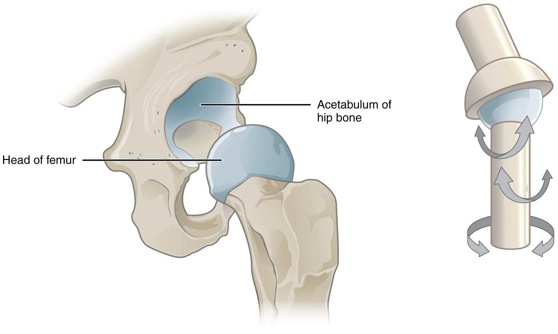
A synovial joint, also known as diarthrosis, joins bones or cartilage with a fibrous joint capsule that is continuous with the periosteum of the joined bones, constitutes the outer boundary of a synovial cavity, and surrounds the bones' articulating surfaces. This joint unites long bones and permits free bone movement and greater mobility. The synovial cavity/joint is filled with synovial fluid. The joint capsule is made up of an outer layer of fibrous membrane, which keeps the bones together structurally, and an inner layer, the synovial membrane, which seals in the synovial fluid. They are the most common and most movable type of joint in the body. As with most other joints, synovial joints achieve movement at the point of contact of the articulating bones. They originated 400 million years ago in the first jawed vertebrates. Structure Synovial joints contain the following structures: * Synovial cavity: all diarthroses have the characteristic space between the bones that is ... [...More Info...] [...Related Items...] OR: [Wikipedia] [Google] [Baidu] |
Joints
A joint or articulation (or articular surface) is the connection made between bones, ossicles, or other hard structures in the body which link an animal's skeletal system into a functional whole.Saladin, Ken. Anatomy & Physiology. 7th ed. McGraw-Hill Connect. Webp.274/ref> They are constructed to allow for different degrees and types of movement. Some joints, such as the knee, elbow, and shoulder, are self-lubricating, almost frictionless, and are able to withstand compression and maintain heavy loads while still executing smooth and precise movements. Other joints such as suture (joint), sutures between the bones of the skull permit very little movement (only during birth) in order to protect the brain and the sense organs. The connection between a tooth and the jawbone is also called a joint, and is described as a fibrous joint known as a gomphosis. Joints are classified both structurally and functionally. Joints play a vital role in the human body, contributing to movement, sta ... [...More Info...] [...Related Items...] OR: [Wikipedia] [Google] [Baidu] |


