|
Dendrodendritic Synapse
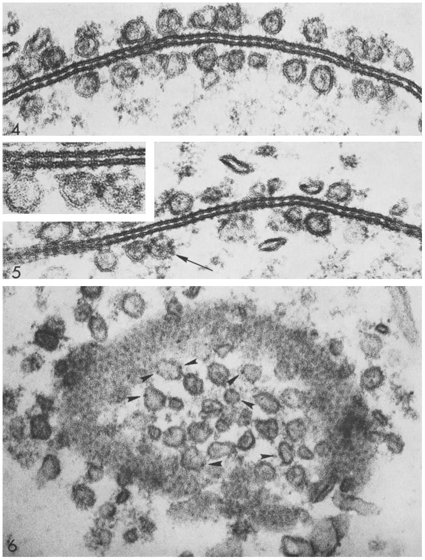
Dendrodendritic synapses are connections between the dendrites of two different neurons. This is in contrast to the more common axodendritic synapse (chemical synapse) where the axon sends signals and the dendrite receives them. Dendrodendritic synapses are activated in a similar fashion to axodendritic synapses in respects to using a chemical synapse. An incoming action potential permits the release of neurotransmitters to propagate the signal to the post synaptic cell. There is evidence that these synapses are bi-directional, in that either dendrite can signal at that synapse. Ordinarily, one of the dendrites will display inhibitory effects while the other will display excitatory effects. The actual signaling mechanism utilizes Na+ and Ca2+ pumps in a similar manner to those found in axodendritic synapses. History In 1966 Wilfrid Rall, Gordon Shepherd, Thomas Reese, and Milton Brightman found a novel pathway, dendrites that signaled to dendrites. While studying the mammalian ol ... [...More Info...] [...Related Items...] OR: [Wikipedia] [Google] [Baidu] |
Dendrite
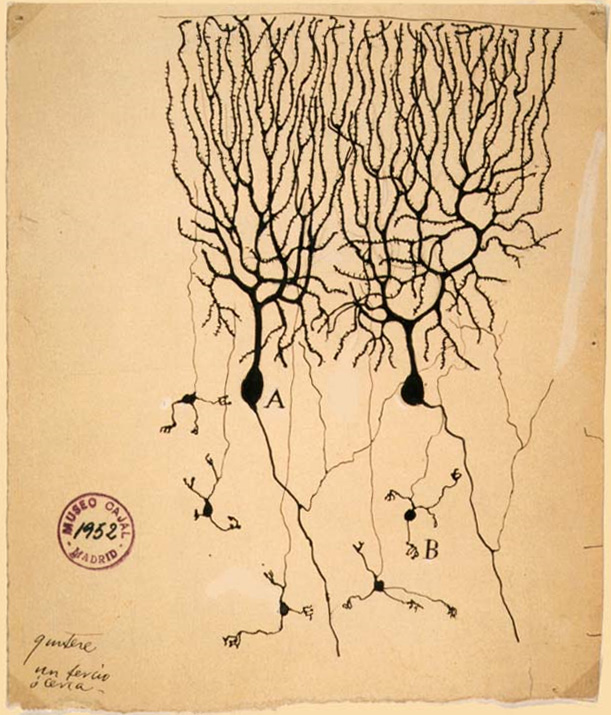
A dendrite (from Ancient Greek language, Greek δένδρον ''déndron'', "tree") or dendron is a branched cytoplasmic process that extends from a nerve cell that propagates the neurotransmission, electrochemical stimulation received from other neural cells to the cell body, or soma (biology), soma, of the neuron from which the dendrites project. Electrical stimulation is transmitted onto dendrites by upstream neurons (usually via their axons) via synapses which are located at various points throughout the dendritic tree. Dendrites play a critical role in integrating these synaptic inputs and in determining the extent to which action potentials are produced by the neuron. Structure and function Dendrites are one of two types of cytoplasmic processes that extrude from the cell body of a neuron, the other type being an axon. Axons can be distinguished from dendrites by several features including shape, length, and function. Dendrites often taper off in shape and are shorter, ... [...More Info...] [...Related Items...] OR: [Wikipedia] [Google] [Baidu] |
Granule Cell
The name granule cell has been used for a number of different types of neurons whose only common feature is that they all have very small cell bodies. Granule cells are found within the granular layer of the cerebellum, the dentate gyrus of the hippocampus, the superficial layer of the dorsal cochlear nucleus, the olfactory bulb, and the cerebral cortex. Cerebellar granule cells account for the majority of neurons in the human brain. These granule cells receive excitatory input from mossy fibers originating from pontine nuclei. Cerebellar granule cells project up through the Purkinje layer into the molecular layer where they branch out into parallel fibers that spread through Purkinje cell dendritic arbors. These parallel fibers form thousands of excitatory granule-cell–Purkinje-cell synapses onto the intermediate and distal dendrites of Purkinje cells using glutamate as a neurotransmitter. Layer 4 granule cells of the cerebral cortex receive inputs from the thala ... [...More Info...] [...Related Items...] OR: [Wikipedia] [Google] [Baidu] |
Neurodegeneration
A neurodegenerative disease is caused by the progressive loss of neurons, in the process known as neurodegeneration. Neuronal damage may also ultimately result in their cell death, death. Neurodegenerative diseases include amyotrophic lateral sclerosis, multiple sclerosis, Parkinson's disease, Alzheimer's disease, Huntington's disease, multiple system atrophy, Tauopathy, tauopathies, and prion diseases. Neurodegeneration can be found in the brain at many different levels of neuronal circuitry, ranging from molecular to systemic. Because there is no known way to reverse the progressive degeneration of neurons, these diseases are considered to be incurable; however research has shown that the two major contributing factors to neurodegeneration are oxidative stress and inflammation. Biomedical research has revealed many similarities between these diseases at the subcellular level, including atypical protein assemblies (like proteinopathy) and induced cell death. These similarities su ... [...More Info...] [...Related Items...] OR: [Wikipedia] [Google] [Baidu] |
Synaptogenesis
Synaptogenesis is the formation of synapses between neurons in the nervous system. Although it occurs throughout a healthy person's lifespan, an explosion of synapse formation occurs during early brain development, known as exuberant synaptogenesis. Synaptogenesis is particularly important during an individual's critical period, during which there is a certain degree of synaptic pruning due to competition for neural growth factors by neurons and synapses. Processes that are not used, or inhibited during their critical period will fail to develop normally later on in life. Exuberant synaptogenesis Brain growth and development begins during gestation and into the postnatal period. Brain development can be divided into stages including: neurogenesis, differentiation, proliferation, migration, synaptogenesis, gliogenesis and myelination, and apoptosis and synaptic pruning. Synaptogenesis occurs in the third trimester during gestation as well as the first two years postnatal. Dur ... [...More Info...] [...Related Items...] OR: [Wikipedia] [Google] [Baidu] |
Axotomy
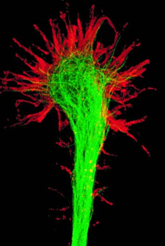
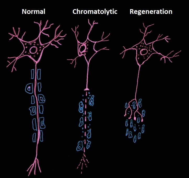
In cellular neuroscience, an axotomy () is the cutting or otherwise severing of an axon. This type of denervation is often used in experimental studies on neuronal physiology and neuronal death or survival as a method to better understand nervous system diseases. Axotomy may cause neuronal cell death, especially in embryonic or neonatal animals, as this is the period in which neurons are dependent on their targets for the supply of survival factors. In mature animals, where survival factors are derived locally or via autocrine loops, axotomy of peripheral neurons and motoneurons can lead to a robust regenerative response without any neuronal death. In both cases, autophagy is observed to markedly increase. Autophagy could either clear the way for neuronal degeneration or it could be a medium for cell destruction.Rubinsztein DC et al. (2005) Autophagy and Its Possible Roles in Nervous System Diseases, Damage and Repair. Autophagy 1(1):11-22. __TOC__ Axotomy response Peripher ... [...More Info...] [...Related Items...] OR: [Wikipedia] [Google] [Baidu] |
Neuroplasticity
Neuroplasticity, also known as neural plasticity or just plasticity, is the ability of neural networks in the brain to change through neurogenesis, growth and reorganization. Neuroplasticity refers to the brain's ability to reorganize and rewire its neural connections, enabling it to adapt and function in ways that differ from its prior state. This process can occur in response to learning new skills, experiencing environmental changes, recovering from injuries, or adapting to sensory or cognitive deficits. Such adaptability highlights the dynamic and ever-evolving nature of the brain, even into adulthood. These changes range from individual neuron pathways making new connections, to systematic adjustments like cortical remapping or neural oscillation. Other forms of neuroplasticity include homologous area adaptation, cross modal reassignment, map expansion, and compensatory masquerade. Examples of neuroplasticity include neural circuit, circuit and network changes that result fr ... [...More Info...] [...Related Items...] OR: [Wikipedia] [Google] [Baidu] |
Gap Junction
Gap junctions are membrane channels between adjacent cells that allow the direct exchange of cytoplasmic substances, such small molecules, substrates, and metabolites. Gap junctions were first described as ''close appositions'' alongside tight junctions, however, electron microscopy studies in 1967 led to gap junctions being named as such to be distinguished from tight junctions. They bridge a 2-4 nm gap between cell membranes. Gap junctions use protein complexes known as connexons, composed of connexin proteins to connect one cell to another. Gap junction proteins include the more than 26 types of connexin, as well as at least 12 non-connexin components that make up the gap junction complex or ''nexus,'' including the tight junction protein ZO-1—a protein that holds membrane content together and adds structural clarity to a cell, sodium channels, and aquaporin. More gap junction proteins have become known due to the development of next-generation sequencing. Connexins ... [...More Info...] [...Related Items...] OR: [Wikipedia] [Google] [Baidu] |
Antennal Lobe
The antennal lobe is the primary (first order) olfactory brain area in insects. The antennal lobe is a sphere-shaped deutocerebral neuropil in the brain that receives input from the olfactory sensory neurons in the antennae and mouthparts. Functionally, it shares some similarities with the olfactory bulb in vertebrates. The anatomy and physiology function of the insect brain can be studied by dissecting open the insect brain and imaging or carrying ou''in vivo'' electrophysiological recordingsfrom it. Structure In insects, the olfactory pathway starts at the antennae (though in some insects like ''Drosophila'' there are olfactory sensory neurons in other parts of the body) from where the sensory neurons carry the information about the odorant molecules impinging on the antenna to the antennal lobe. The antennal lobe is composed of densely packed neuropils, termed glomeruli, where the sensory neurons synapse with the two other kinds of neurons, the postsynaptic principal neuron ... [...More Info...] [...Related Items...] OR: [Wikipedia] [Google] [Baidu] |
Glomerulus (olfaction)
The glomerulus (: glomeruli) is a spherical structure located in the olfactory bulb of the brain where synapses form between the terminals of the olfactory nerve and the dendrites of mitral, periglomerular and tufted cells. Each glomerulus is surrounded by a heterogeneous population of juxtaglomerular neurons (that include periglomerular, short axon, and external tufted cells) and glial cells. All glomeruli are located near the surface of the olfactory bulb. The olfactory bulb also includes a portion of the anterior olfactory nucleus, the cells of which contribute fibers to the olfactory tract. They are the initial sites for synaptic processing of odor information coming from the nose. A glomerulus is made up of a globular tangle of axons from the olfactory receptor neurons, and dendrites from the mitral and tufted cells, as well as, from cells that surround the glomerulus such as the external tufted cells, periglomerular cells, short axon cells, and astrocytes. In mammals, ... [...More Info...] [...Related Items...] OR: [Wikipedia] [Google] [Baidu] |
Mitral Cell
Mitral cells are neurons that are part of the olfactory system. They are located in the olfactory bulb in the mammalian central nervous system. They receive information from the axons of olfactory receptor neurons, forming synapses in neuropils called glomeruli. Axons of the mitral cells transfer information to a number of areas in the brain, including the piriform cortex, entorhinal cortex, and amygdala. Mitral cells receive excitatory input from olfactory sensory neurons and external tufted cells on their primary dendrites, whereas inhibitory input arises either from granule cells onto their lateral dendrites and soma or from periglomerular cells onto their dendritic tuft. Mitral cells together with tufted cells form an obligatory relay for all olfactory information entering from the olfactory nerve. Mitral cell output is not a passive reflection of their input from the olfactory nerve. In mice, each mitral cell sends a single primary dendrite into a glomerulus receiving inp ... [...More Info...] [...Related Items...] OR: [Wikipedia] [Google] [Baidu] |
Locus Ceruleus
The locus coeruleus () (LC), also spelled locus caeruleus or locus ceruleus, is a nucleus in the pons of the brainstem involved with physiological responses to stress and panic. It is a part of the reticular activating system in the reticular formation. The locus coeruleus, which in Latin means "blue spot", is the principal site for brain synthesis of norepinephrine (noradrenaline). The locus coeruleus and the areas of the body affected by the norepinephrine it produces are described collectively as the locus coeruleus-noradrenergic system or LC-NA system. Norepinephrine may also be released directly into the blood from the adrenal medulla. Anatomy The locus coeruleus (LC) is located in the posterior area of the rostral pons in the lateral floor of the fourth ventricle. It is composed of mostly medium-size neurons. Melanin granules inside the neurons contribute to its blue colour. Thus, it is also known as the ''blue nucleus'', or the ''nucleus pigmentosus pontis'' (heavil ... [...More Info...] [...Related Items...] OR: [Wikipedia] [Google] [Baidu] |


