|
Dejerine–Klumpke Palsy
Klumpke's paralysis is a variety of partial palsy of the lower roots of the brachial plexus. p.1046 The brachial plexus is a network of spinal nerves that originates in the back of the neck, extends through the axilla (armpit), and gives rise to nerves to the upper limb.Warwick, R., & Williams, P.L. (1973). pp.1037-1047 pp.370-374 pp.76-77Shenaq S.M., & Spiegel A.J. Hand, Brachial Plexus Surgery. eMedicine.com. URLhttp://www.emedicine.com/plastic/topic450.htm Accessed on: April 13, 2007. The paralytic condition is named after Augusta Déjerine-Klumpke. Signs and symptoms Symptoms can range from minor to severe and can be obvious or subtle. The right arm and hand are more likely to be affected than the left. Symptoms include atrophy of the arm or hand, claw hand, constant crying (due to pain), intrinsic minus hand deformity, paralysis of intrinsic hand muscles, and C8/T1 Dermatome distribution numbness. Involvement of T1 may result in Horner's syndrome, with ptosis, and miosis. W ... [...More Info...] [...Related Items...] OR: [Wikipedia] [Google] [Baidu] |
Brachial Plexus
The brachial plexus is a network of nerves (nerve plexus) formed by the anterior rami of the lower four Spinal nerve#Cervical nerves, cervical nerves and first Spinal nerve#Thoracic nerves, thoracic nerve (cervical spinal nerve 5, C5, Cervical spinal nerve 6, C6, cervical spinal nerve 7, C7, cervical spinal nerve 8, C8, and thoracic spinal nerve 1, T1). This plexus extends from the spinal cord, through the cervicoaxillary canal in the neck, over the first rib, and into the axilla, armpit, it supplies Afferent nerve fiber, afferent and efferent nerve fibers to the chest, shoulder, arm, forearm, and hand. Structure The brachial plexus is divided into five ''roots'', three ''trunks'', six ''divisions'' (three anterior and three posterior), three ''cords'', and five ''branches''. There are five "terminal" branches and numerous other "pre-terminal" or "collateral" branches, such as the subscapular nerve, the thoracodorsal nerve, and the long thoracic nerve, that leave the plexus at vari ... [...More Info...] [...Related Items...] OR: [Wikipedia] [Google] [Baidu] |
Cervical Spinal Nerve 6
The cervical spinal nerve 6 (C6) is a spinal nerve of the cervical segment. It originates from the spinal column from above the cervical vertebra 6 (C6). The C6 nerve root shares a common branch from C5, and has a role in innervating many muscles of the rotator cuff and distal arm, including: * Subclavius *Supraspinatus * Infraspinatus *Biceps brachii * Brachialis *Deltoid * Teres minor *Brachioradialis *Serratus anterior *Subscapularis * Pectoralis major * Coracobrachialis * Teres major *Supinator * Extensor carpi radialis longus *Latissimus dorsi Damage to the C6 motor neuron, by way of impingement, ischemia, trauma, or degeneration of nerve tissue, can cause denervation Denervation is any loss of nerve supply regardless of the cause. If the nerves lost to denervation are part of neural communication to an organ system or for a specific tissue function, alterations to or compromise of physiological functioning ca ... of one or more of the associated muscles. Muscle atr ... [...More Info...] [...Related Items...] OR: [Wikipedia] [Google] [Baidu] |
Proprioception
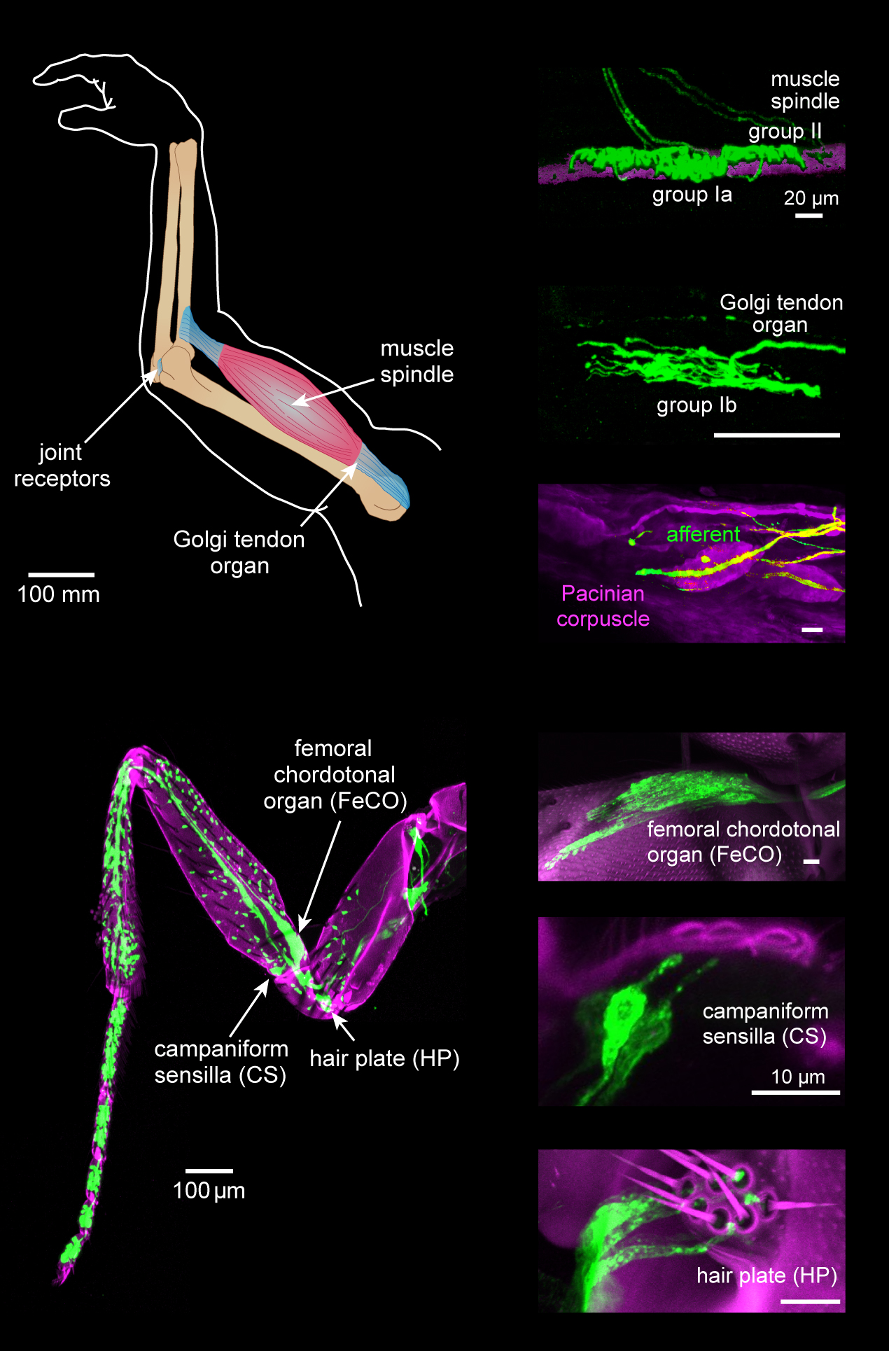
Proprioception ( ) is the sense of self-movement, force, and body position. Proprioception is mediated by proprioceptors, a type of sensory receptor, located within muscles, tendons, and joints. Most animals possess multiple subtypes of proprioceptors, which detect distinct kinesthetic parameters, such as joint position, movement, and load. Although all mobile animals possess proprioceptors, the structure of the sensory organs can vary across species. Proprioceptive signals are transmitted to the central nervous system, where they are integrated with information from other Sensory nervous system, sensory systems, such as Visual perception, the visual system and the vestibular system, to create an overall representation of body position, movement, and acceleration. In many animals, sensory feedback from proprioceptors is essential for stabilizing body posture and coordinating body movement. System overview In vertebrates, limb movement and velocity (muscle length and the rate ... [...More Info...] [...Related Items...] OR: [Wikipedia] [Google] [Baidu] |
Occupational Therapy
Occupational therapy (OT), also known as ergotherapy, is a healthcare profession. Ergotherapy is derived from the Greek wiktionary:ergon, ergon which is allied to work, to act and to be active. Occupational therapy is based on the assumption that engaging in meaningful activities, also referred to as occupations, is a basic human need and that purposeful activity has a health-promoting and therapeutic effect. Occupational science the study of humans as 'doers' or 'occupational beings' was developed by inter-disciplinary scholars, including occupational therapists, in the 1980s. The World Federation of Occupational Therapists (WFOT) defines occupational therapy as ‘a client-centred health profession concerned with promoting health and wellbeing through occupation. The primary goal of occupational therapy is to enable people to participate in the activities of everyday life. Occupational therapists achieve this outcome by working with people and communities to enhance their abi ... [...More Info...] [...Related Items...] OR: [Wikipedia] [Google] [Baidu] |
Electromyography
Electromyography (EMG) is a technique for evaluating and recording the electrical activity produced by skeletal muscles. EMG is performed using an instrument called an electromyograph to produce a record called an electromyogram. An electromyograph detects the electric potential generated by muscle cells when these cells are electrically or neurologically activated. The signals can be analyzed to detect abnormalities, activation level, or recruitment order, or to analyze the biomechanics of human or animal movement. Needle EMG is an electrodiagnostic medicine technique commonly used by neurologists. Surface EMG is a non-medical procedure used to assess muscle activation by several professionals, including physiotherapists, kinesiologists and biomedical engineers. In computer science, EMG is also used as middleware in gesture recognition towards allowing the input of physical action to a computer as a form of human-computer interaction. Clinical uses EMG testing has a varie ... [...More Info...] [...Related Items...] OR: [Wikipedia] [Google] [Baidu] |
Childbirth
Childbirth, also known as labour, parturition and delivery, is the completion of pregnancy, where one or more Fetus, fetuses exits the Womb, internal environment of the mother via vaginal delivery or caesarean section and becomes a newborn to the world. In 2019, there were about 140.11 million human births globally. In Developed country, developed countries, most deliveries occur in hospitals, while in Developing country, developing countries most are home births. The most common childbirth method worldwide is vaginal delivery. It involves four stages of labour: the cervical effacement, shortening and Cervical dilation, opening of the cervix during the first stage, descent and birth of the baby during the second, the delivery of the placenta during the third, and the recovery of the mother and infant during the fourth stage, which is referred to as the Postpartum period, postpartum. The first stage is characterised by abdominal cramping or also back pain in the case of B ... [...More Info...] [...Related Items...] OR: [Wikipedia] [Google] [Baidu] |
Flexor Digitorum Profundus
The flexor digitorum profundus or flexor digitorum communis profundus is a muscle in the forearm of humans that flexes the fingers (also known as digits). It is considered an Muscles of the hand#Extrinsic, extrinsic hand muscle because it acts on the hand while its muscle belly is located in the forearm. Together the Flexor pollicis longus muscle, flexor pollicis longus, Pronator quadratus muscle, pronator quadratus, and flexor digitorum profundus form the deep layer of ventral forearm muscles.Platzer 2004, p 162 The muscle is named . Structure Flexor digitorum profundus originates in the upper 3/4 of the anterior and medial surfaces of the ulna, interosseous membrane and deep fascia of the forearm. The muscle fans out into four tendons (one to each of the second to fifth fingers) to the palmar base of the distal phalanges, distal phalanx. Along with the flexor digitorum superficialis, it has long tendons that run down the arm and through the carpal tunnel and attach to the p ... [...More Info...] [...Related Items...] OR: [Wikipedia] [Google] [Baidu] |
Flexor Carpi Ulnaris
The flexor carpi ulnaris (FCU) is a skeletal muscle, muscle of the forearm that flexion, flexes and Adduction, adducts at the wrist joint. Structure Origin The flexor carpi ulnaris has two heads; a humeral head and ulnar head. The humeral head originates from the medial epicondyle of the humerus via the common flexor tendon. The ulnar head originates from the medial margin of the olecranon of the ulna and the upper two-thirds of the dorsal border of the ulna by an aponeurosis. Between the two heads passes the ulnar nerve and ulnar artery. Insertion The flexor carpi ulnaris inserts onto the pisiform bone, pisiform, hook of the hamate (via the pisohamate ligament) and the anterior surface of the base of the fifth metacarpal bone, fifth metacarpal (via the pisometacarpal ligament). Action The flexor carpi ulnaris flexes and adducts at the Wrist, wrist joint. Innervation The flexor carpi ulnaris is innervated by the ulnar nerve. The corresponding spinal nerves are Cervical spinal ... [...More Info...] [...Related Items...] OR: [Wikipedia] [Google] [Baidu] |
Hypothenar Muscles
The hypothenar muscles are a group of three muscles of the palm that control the motion of the little finger. The three muscles are: * Abductor digiti minimi * Flexor digiti minimi brevis * Opponens digiti minimi Structure The muscles of hypothenar eminence are from medial to lateral: * Opponens digiti minimi * Flexor digiti minimi brevis * Abductor digiti minimi The intrinsic muscles of hand can be remembered using the mnemonic, "A OF A OF A" for, Abductor pollicis brevis, Opponens pollicis, Flexor pollicis brevis (the three thenar muscles), Adductor pollicis, and the three hypothenar muscles, Opponens digiti minimi, Flexor digiti minimi brevis, Abductor digiti minimi. Clinical significance "Hypothenar atrophy" is associated with the lesion of the ulnar nerve, which supplies the three hypothenar muscles. Hypothenar hammer syndrome is a vascular occlusion of this region. See also * Thenar eminence * Palmaris brevis Palmaris brevis muscle is a thin, quadrilateral muscle, ... [...More Info...] [...Related Items...] OR: [Wikipedia] [Google] [Baidu] |
Thenar Muscles
The thenar eminence is the mound formed at the base of the thumb on the palm of the hand by the intrinsic group of muscles of the thumb. The skin overlying this region is the area stimulated when trying to elicit a palmomental reflex. The word thenar comes . Structure The following three muscles are considered part of the thenar eminence: * Abductor pollicis brevis abducts the thumb. This muscle is the most superficial of the thenar group. * Flexor pollicis brevis, which lies next to the abductor, will flex the thumb, curling it up in the palm. (The flexor pollicis longus, which is inserted into the distal phalanx of the thumb, is not considered part of the thenar eminence.) * Opponens pollicis lies deep to abductor pollicis brevis. As its name suggests it opposes the thumb, bringing it against the fingers. This is a very important movement, as most of human hand dexterity comes from this action. Another muscle that controls movement of the thumb is adductor pollicis. I ... [...More Info...] [...Related Items...] OR: [Wikipedia] [Google] [Baidu] |
Interossei
{{short description, Muscles between certain bones Interossei refer to muscles between certain bones. There are many interossei in a human body. Specific interossei include: On the hands * Dorsal interossei muscles of the hand * Palmar interossei muscles In human anatomy, the palmar or volar interossei (interossei volares in older literature) are four muscles, one on the thumb that is occasionally missing, and three small, unipennate, central muscles in the hand that lie between the Metacarpus, me ... File:Gray428.png, Dorsal interossei muscles of the hand File:Gray429.png, Palmar interossei muscles On the feet * Dorsal interossei muscles of the foot * Plantar interossei muscles File:Gray446.png, Dorsal interossei muscles of the foot File:Gray447.png, Plantar interossei muscles Muscular system ... [...More Info...] [...Related Items...] OR: [Wikipedia] [Google] [Baidu] |
Cervical Spinal Nerve 8
The cervical spinal nerve 8 (C8) is a spinal nerve of the cervical segment. It originates from the spinal column from below the cervical vertebra 7 (C7). Innervation The C8 nerve forms part of the radial and ulnar nerves via the brachial plexus, and therefore has motor and sensory function in the upper limb. Sensory The C8 nerve receives sensory afferents from the C8 dermatome. This consists of all the skin on the little finger, and continuing up slightly past the wrist on the palmar and dorsal aspects of the hand and forearm.Drake et al. Gray's Anatomy for Students. Second Edition (2010). Clinically, a test of the pad of the little finger is often used to assess C8 integrity.Aland et al. University of Queensland School of Medicine Clinical Skills Handbook 2010 Motor The C8 nerve contributes to the motor innervation of many of the muscles in the trunk and upper limb. Its primary function is the flexion of the fingers, and this is used as the clinical test for C8 inte ... [...More Info...] [...Related Items...] OR: [Wikipedia] [Google] [Baidu] |


