|
Wrist
In human anatomy, the wrist is variously defined as (1) the carpus or carpal bones, the complex of eight bones forming the proximal skeletal segment of the hand; "The wrist contains eight bones, roughly aligned in two rows, known as the carpal bones." (2) the wrist joint or radiocarpal joint, the joint between the radius and the carpus and; (3) the anatomical region surrounding the carpus including the distal parts of the bones of the forearm and the proximal parts of the metacarpus or five metacarpal bones and the series of joints between these bones, thus referred to as ''wrist joints''. "With the large number of bones composing the wrist (ulna, radius, eight carpas, and five metacarpals), it makes sense that there are many, many joints that make up the structure known as the wrist." This region also includes the carpal tunnel, the anatomical snuff box, bracelet lines, the flexor retinaculum, and the extensor retinaculum. As a consequence of these various definitions, f ... [...More Info...] [...Related Items...] OR: [Wikipedia] [Google] [Baidu] |
Distal Radius Fracture
A distal radius fracture, also known as wrist fracture, is a fracture (bone), break of the part of the radius (bone), radius bone which is close to the wrist. Symptoms include pain, bruising, and rapid-onset swelling. The ulna bone may also be broken. In younger people, these fractures typically occur during sports or a motor vehicle collision. In older people, the most common cause is falling on an outstretched hand. Specific types include Colles' fracture, Colles, Smith's fracture, Smith, Barton's fracture, Barton, and Chauffeur's fractures. The diagnosis is generally suspected based on symptoms and confirmed with radiography, X-rays. Treatment is with orthopedic cast, casting for six weeks or surgery. Surgery is generally indicated if the joint surface is broken and does not line up, the radius is overly short, or the joint surface of the radius is tilted more than 10% backwards. Among those who are cast, repeated X-rays are recommended within three weeks to verify that a go ... [...More Info...] [...Related Items...] OR: [Wikipedia] [Google] [Baidu] |
Carpal Bones
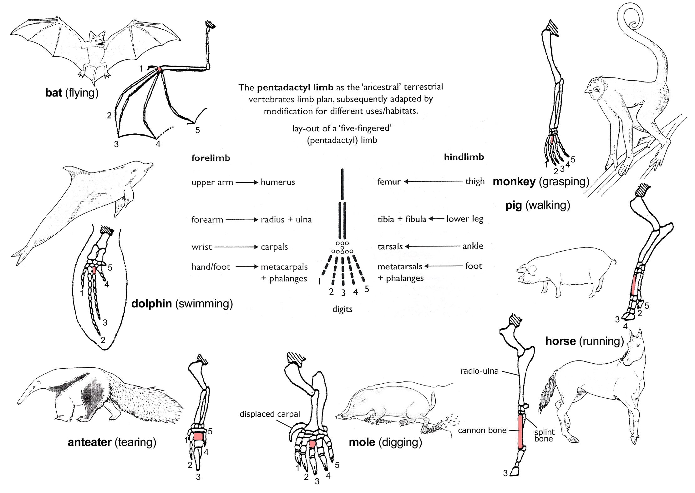
The carpal bones are the eight small bones that make up the wrist (carpus) that connects the hand to the forearm. The terms "carpus" and "carpal" are derived from the Latin wikt:carpus#Latin, carpus and the Greek language, Greek wikt:καρπός#Ancient Greek, καρπός (karpós), meaning "wrist". In human anatomy, the main role of the carpal bones is to joint, articulate with the radius (bone), radial and ulnar heads to form a highly mobile condyloid joint (i.e. wrist joint),Kingston 2000, pp 126-127 to provide attachments for thenar and hypothenar muscles, and to form part of the rigid carpal tunnel which allows the median nerve and tendons of the anterior compartment of the forearm, anterior forearm muscles to be transmitted to the hand and fingers. In tetrapods, the carpus is the sole cluster of bones in the wrist between the radius (bone), radius and ulna and the metacarpus. The bones of the carpus do not belong to individual fingers (or toes in quadrupeds), whereas those ... [...More Info...] [...Related Items...] OR: [Wikipedia] [Google] [Baidu] |
Head Of Ulna
The ulna or ulnar bone (: ulnae or ulnas) is a long bone in the forearm stretching from the elbow to the wrist. It is on the same side of the forearm as the little finger, running parallel to the radius, the forearm's other long bone. Longer and thinner than the radius, the ulna is considered to be the smaller long bone of the lower arm. The corresponding bone in the lower leg is the fibula. Structure The ulna is a long bone found in the forearm that stretches from the elbow to the wrist, and when in standard anatomical position, is found on the medial side of the forearm. It is broader close to the elbow, and narrows as it approaches the wrist. Close to the elbow, the ulna has a bony process, the olecranon process, a hook-like structure that fits into the olecranon fossa of the humerus. This prevents hyperextension and forms a hinge joint with the trochlea of the humerus. There is also a radial notch for the head of the radius, and the ulnar tuberosity to which muscle ... [...More Info...] [...Related Items...] OR: [Wikipedia] [Google] [Baidu] |
Ulna
The ulna or ulnar bone (: ulnae or ulnas) is a long bone in the forearm stretching from the elbow to the wrist. It is on the same side of the forearm as the little finger, running parallel to the Radius (bone), radius, the forearm's other long bone. Longer and thinner than the radius, the ulna is considered to be the smaller long bone of the lower arm. The corresponding bone in the Human leg#Structure, lower leg is the fibula. Structure The ulna is a long bone found in the forearm that stretches from the elbow to the wrist, and when in standard anatomical position, is found on the Medial (anatomy), medial side of the forearm. It is broader close to the elbow, and narrows as it approaches the wrist. Close to the elbow, the ulna has a bony Process (anatomy), process, the olecranon process, a hook-like structure that fits into the olecranon fossa of the humerus. This prevents hyperextension and forms a hinge joint with the trochlea of the humerus. There is also a radial notch for ... [...More Info...] [...Related Items...] OR: [Wikipedia] [Google] [Baidu] |
Hand
A hand is a prehensile, multi-fingered appendage located at the end of the forearm or forelimb of primates such as humans, chimpanzees, monkeys, and lemurs. A few other vertebrates such as the Koala#Characteristics, koala (which has two thumb#Opposition and apposition, opposable thumbs on each "hand" and fingerprints extremely similar to human fingerprints) are often described as having "hands" instead of paws on their front limbs. The raccoon is usually described as having "hands" though opposable thumbs are lacking. Some evolutionary anatomists use the term ''hand'' to refer to the appendage of digits on the forelimb more generally—for example, in the context of whether the three Digit (anatomy), digits of the bird hand involved the same Homology (biology), homologous loss of two digits as in the dinosaur hand. The human hand usually has five digits: Finger numbering#Four-finger system, four fingers plus one thumb; however, these are often referred to collectively as Finger ... [...More Info...] [...Related Items...] OR: [Wikipedia] [Google] [Baidu] |
Carpal Tunnel
In the human body, the carpal tunnel or carpal canal is a flattened body cavity on the flexor ( palmar/volar) side of the wrist, bounded by the carpal bones and flexor retinaculum. It forms the passageway that transmits the median nerve and the tendons of the extrinsic flexor muscles of the hand from the forearm to the hand. The median artery is an anatomical variant (increasingly found). When present it lies between the radial artery, and the ulnar artery and runs with the median nerve supplying the same structures innervated. When swelling or degeneration occurs in the tendons and sheaths of any of the nine flexor muscles ( flexor pollicis longus, four flexor digitorum profundus and four flexor digitorum superficialis) passing through the carpal tunnel, the canal can narrow and compress/entrap the median nerve, resulting in a compression neuropathy known as carpal tunnel syndrome (CTS). If untreated, neuropraxia, parasthesia and muscle atrophy (especially of the ... [...More Info...] [...Related Items...] OR: [Wikipedia] [Google] [Baidu] |
Radius (bone)
The radius or radial bone (: radii or radiuses) is one of the two large bones of the forearm, the other being the ulna. It extends from the Anatomical terms of location, lateral side of the Elbow-joint, elbow to the thumb side of the wrist and runs parallel to the ulna. The ulna is longer than the radius, but the radius is thicker. The radius is a long bone, Prism (geometry), prism-shaped and slightly curved longitudinally. The radius is part of two joint (anatomy), joints: the elbow and the wrist. At the elbow, it joins with the capitulum of the humerus, and in a separate region, with the ulna at the radial notch. At the wrist, the radius forms a joint with the ulna bone. The corresponding bone in the human leg, lower leg is the tibia. Structure The long narrow medullary cavity is enclosed in a strong wall of compact bone. It is thickest along the interosseous border and thinnest at the extremities, same over the cup-shaped articular surface (fovea) of the head. The tra ... [...More Info...] [...Related Items...] OR: [Wikipedia] [Google] [Baidu] |
Metacarpus
In human anatomy, the metacarpal bones or metacarpus, also known as the "palm bones", are the appendicular skeleton, appendicular bones that form the intermediate part of the hand between the phalanges (fingers) and the carpal bones (wrist, wrist bones), which joint, articulate with the forearm. The metacarpal bones are homologous to the metatarsal bones in the foot. Structure The metacarpals form a transverse arch to which the rigid row of distal carpal bones are fixed. The peripheral metacarpals (those of the thumb and little finger) form the sides of the cup of the palmar gutter and as they are brought together they deepen this concavity. The index metacarpal is the most firmly fixed, while the thumb metacarpal articulates with the trapezium and acts independently from the others. The middle metacarpals are tightly united to the carpus by intrinsic interlocking bone elements at their bases. The ring metacarpal is somewhat more mobile while the fifth metacarpal is semi-indepen ... [...More Info...] [...Related Items...] OR: [Wikipedia] [Google] [Baidu] |
Pronation
Motion, the process of movement, is described using specific anatomical terminology, anatomical terms. Motion includes movement of Organ (anatomy), organs, joints, Limb (anatomy), limbs, and specific sections of the body. The terminology used describes this motion according to its direction relative to the anatomical position of the body parts involved. Anatomy, Anatomists and others use a unified set of terms to describe most of the movements, although other, more specialized terms are necessary for describing unique movements such as those of the hands, feet, and eyes. In general, motion is classified according to the anatomical plane it occurs in. ''Flexion'' and ''extension'' are examples of ''angular'' motions, in which two axes of a joint are brought closer together or moved further apart. ''Rotational'' motion may occur at other joints, for example the shoulder, and are described as ''internal'' or ''external''. Other terms, such as ''elevation'' and ''depression'', descri ... [...More Info...] [...Related Items...] OR: [Wikipedia] [Google] [Baidu] |
Supination
Motion, the process of movement, is described using specific anatomical terms. Motion includes movement of organs, joints, limbs, and specific sections of the body. The terminology used describes this motion according to its direction relative to the anatomical position of the body parts involved. Anatomists and others use a unified set of terms to describe most of the movements, although other, more specialized terms are necessary for describing unique movements such as those of the hands, feet, and eyes. In general, motion is classified according to the anatomical plane it occurs in. ''Flexion'' and ''extension'' are examples of ''angular'' motions, in which two axes of a joint are brought closer together or moved further apart. ''Rotational'' motion may occur at other joints, for example the shoulder, and are described as ''internal'' or ''external''. Other terms, such as ''elevation'' and ''depression'', describe movement above or below the horizontal plane. Many anatomic ... [...More Info...] [...Related Items...] OR: [Wikipedia] [Google] [Baidu] |


