|
Testicle
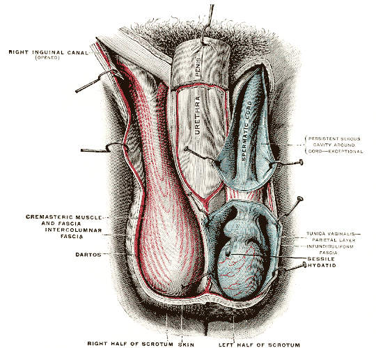
A testicle or testis ( testes) is the gonad in all male bilaterians, including humans, and is Homology (biology), homologous to the ovary in females. Its primary functions are the production of sperm and the secretion of Androgen, androgens, primarily testosterone. The release of testosterone is regulated by luteinizing hormone (LH) from the anterior pituitary gland. Sperm production is controlled by follicle-stimulating hormone (FSH) from the anterior pituitary gland and by testosterone produced within the gonads. Structure Appearance Males have two testicles of similar size contained within the scrotum, which is an extension of the abdominal wall. Scrotal asymmetry, in which one testicle extends farther down into the scrotum than the other, is common. This is because of the differences in the vasculature's anatomy. For 85% of men, the right testis hangs lower than the left one. Measurement and volume The volume of the testicle can be estimated by palpating it and compari ... [...More Info...] [...Related Items...] OR: [Wikipedia] [Google] [Baidu] |
Testosterone
Testosterone is the primary male sex hormone and androgen in Male, males. In humans, testosterone plays a key role in the development of Male reproductive system, male reproductive tissues such as testicles and prostate, as well as promoting secondary sexual characteristics such as increased muscle and bone mass, and the growth of androgenic hair, body hair. It is associated with increased aggression, sex drive, Dominance hierarchy, dominance, courtship display, and a wide range of behavioral characteristics. In addition, testosterone in both sexes is involved in health and well-being, where it has a significant effect on overall mood, cognition, social and sexual behavior, metabolism and energy output, the cardiovascular system, and in the prevention of osteoporosis. Insufficient levels of testosterone in men may lead to abnormalities including frailty, accumulation of adipose fat tissue within the body, anxiety and depression, sexual performance issues, and bone loss. Excessiv ... [...More Info...] [...Related Items...] OR: [Wikipedia] [Google] [Baidu] |
Scrotum
In most terrestrial mammals, the scrotum (: scrotums or scrota; possibly from Latin ''scortum'', meaning "hide" or "skin") or scrotal sac is a part of the external male genitalia located at the base of the penis. It consists of a sac of skin containing the external spermatic fascia, testicles, epididymides, and vasa deferentia. The scrotum will usually tighten when exposed to cold temperatures. The scrotum is homologous to the labia majora in females. Structure In regards to humans, the scrotum is a suspended two-chambered sac of skin and muscular tissue containing the testicles and the lower part of the spermatic cords. It is located behind the penis and above the perineum. The perineal raphe is a small, vertical ridge of skin that expands from the anus and runs through the middle of the scrotum front to back. The scrotum is also a distention of the perineum and carries some abdominal tissues into its cavity including the testicular artery, testicular vein, and ... [...More Info...] [...Related Items...] OR: [Wikipedia] [Google] [Baidu] |
Puberty
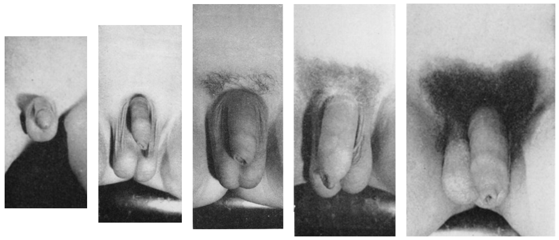
Puberty is the process of physical changes through which a child's body matures into an adult body capable of sexual reproduction. It is initiated by hormonal signals from the brain to the gonads: the ovaries in a female, the testicles in a male. In response to the signals, the gonads produce hormones that stimulate libido and the growth, function, and transformation of the brain, bones, muscle, blood, skin, hair, breasts, and sex organs. Physical growth—height and weight—accelerates in the first half of puberty and is completed when an adult body has been developed. Before puberty, the external sex organs, known as primary sexual characteristics, are sex characteristics that distinguish males and females. Puberty leads to sexual dimorphism through the development of the secondary sex characteristics, which further distinguish the sexes. On average, females begin puberty at age 10½ and complete puberty at ages 15-17; males begin at ages 11½-12 and complete pube ... [...More Info...] [...Related Items...] OR: [Wikipedia] [Google] [Baidu] |
Tunica Albuginea Of Testis
The tunica albuginea is a dense, blue-white layer of fibrous tissue surrounding the testis. It is the middle of three envelopes forming the capsule of the testis; it is deep to the visceral layer of tunica vaginalis, and superficial to the tunica vasculosa testis (vascular layer of testis). The connective tissue of the tunica albuginea testis extends into the substance of the testis to form fibrous partitions - the septa testis. At the posterior aspect of the testis (where the serosa of testis is deficient to allow for the attachment of the epididymis), the tunica albuginea extends into the testis to form the mediastinum testis. Anatomy It is thicker than the tunica albuginea of the ovary. Histology It is composed of bundles of white fibrous connective tissue (from which it derives its name '' albuginea'') which interlace in every direction. Additional images File:Gray1145.png, Transverse section through the left side of the scrotum and the left testis. File:Gray111 ... [...More Info...] [...Related Items...] OR: [Wikipedia] [Google] [Baidu] |
Testicular Artery
The testicular artery (the male gonadal artery, also called the internal spermatic arteries in older texts) is a branch of the abdominal aorta that supplies blood to the testicle. It is a paired artery, with one for each of the testicles. It is the male equivalent of the ovarian artery. Because the testis is found in a different location than that of its female equivalent, it has a different course than the ovarian artery. They are two slender vessels of considerable length, and arise from the front of the aorta a little below the renal arteries. Each passes obliquely downward and lateralward behind the peritoneum, resting on the psoas major, the right lying in front of the inferior vena cava and behind the middle colic and ileocolic arteries and the terminal part of the ileum, the left behind the left colic and sigmoid arteries and the iliac colon. Each crosses obliquely over the ureter and the lower part of the external iliac artery to reach the abdominal inguinal rin ... [...More Info...] [...Related Items...] OR: [Wikipedia] [Google] [Baidu] |
Sertoli Cells
Sertoli cells are a type of sustentacular "nurse" cell found in human testes which contribute to the process of spermatogenesis (the production of sperm) as a structural component of the seminiferous tubules. They are activated by follicle-stimulating hormone (FSH) secreted by the adenohypophysis and express FSH receptor on their membranes. History Sertoli cells are named after Enrico Sertoli, an Italian physiologist who discovered them while studying medicine at the University of Pavia, Italy. He published a description of his eponymous cell in 1865. The cell was discovered by Sertoli with a Belthle microscope which had been purchased in 1862. In the 1865 publication, his first description used the terms "tree-like cell" or "stringy cell"; most importantly, he referred to these as "mother cells". Other scientists later used Enrico's family name to label these cells in publications, beginning in 1888. As of 2006, two textbooks that are devoted specifically to the Sertoli cell ... [...More Info...] [...Related Items...] OR: [Wikipedia] [Google] [Baidu] |
Orchidometer
An orchidometer (or orchiometer) is a medical instrument used to measure the volume of the testicles. Instrument The orchidometer was introduced in 1966 by Swiss pediatric endocrinologist Andrea Prader of the University of Zurich.[ Page 16] in: It consists of a string of twelve numbered wooden or plastic beads of increasing size from about 1 to 25 millilitres. Physician, Doctors sometimes informally refer to them as "Prader's balls", "the medical worry beads", or the "endocrine rosary".[ Page 86] in: The beads are compared with the testicles of the patient, and the volume is read off the bead which matches most closely in size. Prepubertal sizes are 1–3 ml, pubertal sizes are considered 4 ml and up and adult sizes are 15-25 ml. The orchidometer can be used to accurately determine size of testes. Discrepancy of testicular size with other parameters of maturation can be an important clue to various diseases. Small testes can indicate either primary or secondary hypogonadism. ... [...More Info...] [...Related Items...] OR: [Wikipedia] [Google] [Baidu] |
Androgen
An androgen (from Greek ''andr-'', the stem of the word meaning ) is any natural or synthetic steroid hormone that regulates the development and maintenance of male characteristics in vertebrates by binding to androgen receptors. This includes the embryological development of the primary male sex organs, and the development of male secondary sex characteristics at puberty. Androgens are synthesized in the testes, the ovaries, and the adrenal glands. Androgens increase in both males and females during puberty. The major androgen in males is testosterone. Dihydrotestosterone (DHT) and androstenedione are of equal importance in male development. DHT ''in utero'' causes differentiation of the penis, scrotum and prostate. In adulthood, DHT contributes to balding, prostate growth, and sebaceous gland activity. Although androgens are commonly thought of only as male Sex steroids, sex hormones, females also have them, but at lower levels: they function in libido and sexual arousal. An ... [...More Info...] [...Related Items...] OR: [Wikipedia] [Google] [Baidu] |
Testicular Vein
The testicular vein (or spermatic vein), the male gonadal vein, carries deoxygenated blood from its corresponding testis to the inferior vena cava or one of its tributaries. It is the male equivalent of the ovarian vein, and is the venous counterpart of the testicular artery. Structure It is a paired vein, with one supplying each testis: * the right testicular vein generally joins the inferior vena cava; * the left testicular vein, unlike the right one, joins the left renal vein instead of the inferior vena cava. The veins emerge from the back of the testis, and receive tributaries from the epididymis. They unite and form a convoluted plexus, called the pampiniform plexus, which constitutes the greater mass of the spermatic cord; the vessels composing this plexus are very numerous, and ascend along the cord, in front of the ductus deferens. Below the subcutaneous inguinal ring, they unite to form three or four veins, which pass along the inguinal canal, and, entering the abd ... [...More Info...] [...Related Items...] OR: [Wikipedia] [Google] [Baidu] |
Seminiferous Tubule
Seminiferous tubules are located within the testicles, and are the specific location of meiosis, and the subsequent creation of male gametes, namely spermatozoa. Structure The epithelium of the tubule consists of a type of sustentacular cells known as Sertoli cells, which are tall, columnar type cells that line the tubule. In between the Sertoli cells are spermatogenic cells, which differentiate through meiosis to sperm cells. Sertoli cells function to nourish the developing sperm cells. They secrete androgen-binding protein, a binding protein which increases the concentration of testosterone. There are two types: convoluted and straight, convoluted toward the lateral side, and straight as the tubule comes medially to form ducts that will exit the testis. The seminiferous tubules are formed from the testis cords that develop from the primitive gonadal cords, formed from the gonadal ridge. Function Spermatogenesis, the process for producing spermatozoa, takes place in ... [...More Info...] [...Related Items...] OR: [Wikipedia] [Google] [Baidu] |
Homology (biology)
In biology, homology is similarity in anatomical structures or genes between organisms of different taxa due to shared ancestry, ''regardless'' of current functional differences. Evolutionary biology explains homologous structures as retained heredity from a common descent, common ancestor after having been subjected to adaptation (biology), adaptive modifications for different purposes as the result of natural selection. The term was first applied to biology in a non-evolutionary context by the anatomist Richard Owen in 1843. Homology was later explained by Charles Darwin's theory of evolution in 1859, but had been observed before this from Aristotle's biology onwards, and it was explicitly analysed by Pierre Belon in 1555. A common example of homologous structures is the forelimbs of vertebrates, where the bat wing development, wings of bats and origin of avian flight, birds, the arms of primates, the front flipper (anatomy), flippers of whales, and the forelegs of quadrupedalis ... [...More Info...] [...Related Items...] OR: [Wikipedia] [Google] [Baidu] |


