|
Rhizodonts
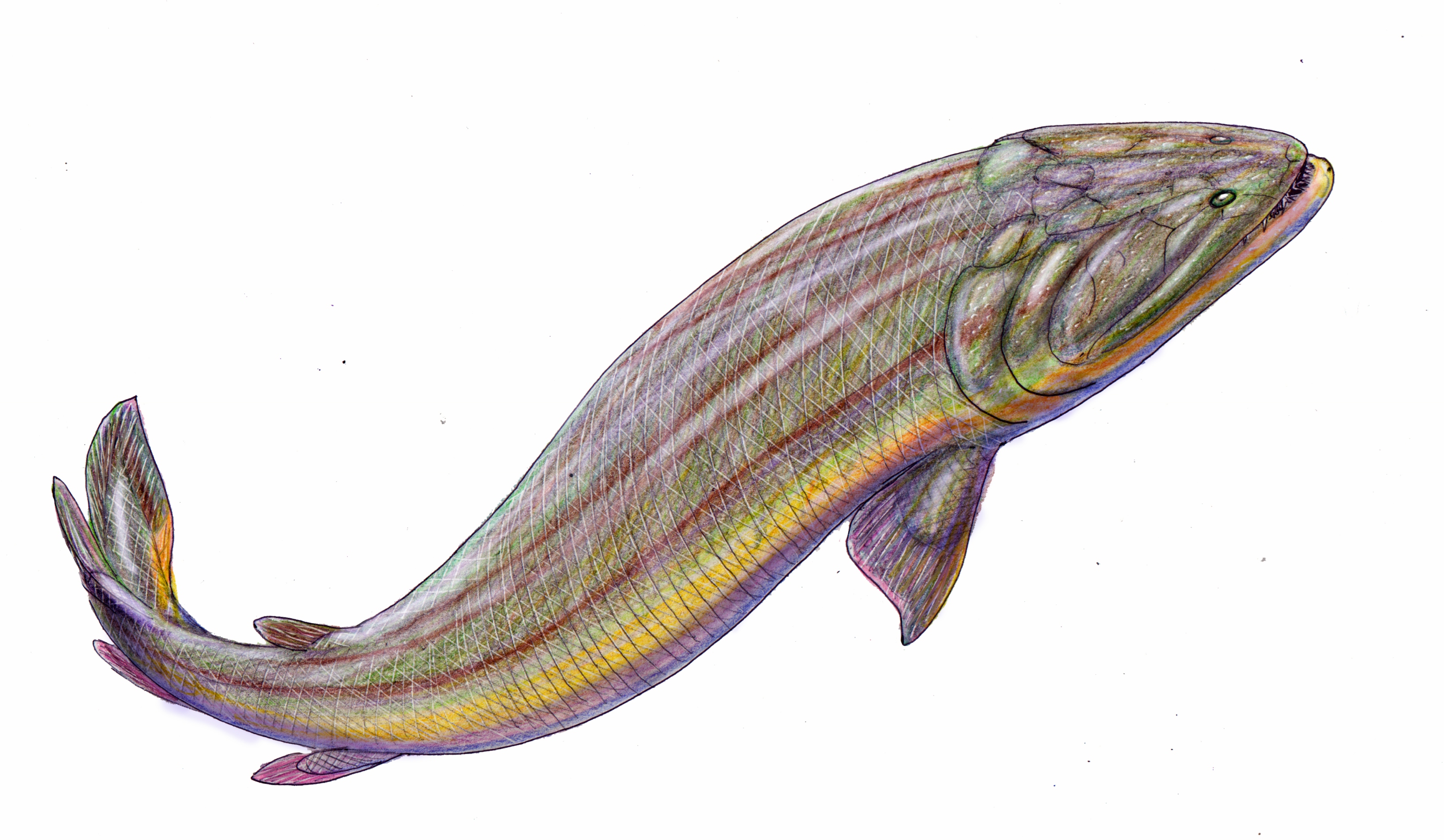
Rhizodontida is an extinct group of predatory tetrapodomorphs known from many areas of the world from the Givetian through to the Pennsylvanian (geology), Pennsylvanian - the earliest known species is about 377 million years ago (Mya), the latest around 310 Mya. Rhizodonts lived in tropical rivers and freshwater lakes and were the dominant predators of their age. They reached huge sizes - the largest known species, ''Rhizodus, Rhizodus hibberti'' from Europe and North America, was an estimated 7 m in length, making it the largest freshwater fish known. Description The upper jaw had a marginal row of small teeth on the maxilla and premaxilla, medium-sized fangs on the ectopterygoid and dermopalatine bones, and large tusks on the vomers and premaxillae. On the lower jaw were marginal teeth on the dentary, with fangs on the three coronoid process of the mandible, coronoids and a huge tusk at the symphysis, symphysial tip of the dentary. Apparently, the left and right mandibles rota ... [...More Info...] [...Related Items...] OR: [Wikipedia] [Google] [Baidu] |
Givetian
The Givetian is one of two faunal stages in the Middle Devonian Period. It lasted from million years ago to million years ago. It was preceded by the Eifelian Stage and followed by the Frasnian Stage. It is named after the town of Givet in France. The oldest forests occurred during the late Givetian. The lower GSSP A Global Boundary Stratotype Section and Point (GSSP), sometimes referred to as a golden spike, is an internationally agreed upon reference point on a stratigraphic section which defines the lower boundary of a stage on the geologic time scale. ... is located at Jebel Mech Irdane, Tafilalt, Morocco. Name and definition The Givetian Stage was proposed in 1879 by French geologist Jules Gosselet and was accepted for the higher stage of the Middle Devonian by the Subcommission on Devonian Stratigraphy in 1981. References Further reading * {{Geological history, p, p Middle Devonian Devonian geochronology . Devonian North America ... [...More Info...] [...Related Items...] OR: [Wikipedia] [Google] [Baidu] |
Dorsal Fin
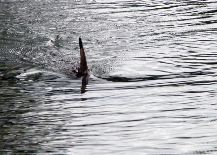
A dorsal fin is a fin on the back of most marine and freshwater vertebrates. Dorsal fins have evolved independently several times through convergent evolution adapting to marine environments, so the fins are not all homologous. They are found in most fish, in mammals such as whales, and in extinct ancient marine reptiles such as ichthyosaurs. Most have only one dorsal fin, but some have two or three. Wildlife biologists often use the distinctive nicks and wear patterns which develop on the dorsal fins of whales to identify individuals in the field. The bones or cartilages that support the dorsal fin in fish are called pterygiophores. Functions The main purpose of the dorsal fin is usually to stabilize the animal against rolling and to assist in sudden turns. Some species have further adapted their dorsal fins to other uses. The sunfish uses the dorsal fin (and the anal fin Fins are moving appendages protruding from the body of fish that interact with water to ge ... [...More Info...] [...Related Items...] OR: [Wikipedia] [Google] [Baidu] |
Pelvic
The pelvis (: pelves or pelvises) is the lower part of an anatomical trunk, between the abdomen and the thighs (sometimes also called pelvic region), together with its embedded skeleton (sometimes also called bony pelvis or pelvic skeleton). The pelvic region of the trunk includes the bony pelvis, the pelvic cavity (the space enclosed by the bony pelvis), the pelvic floor, below the pelvic cavity, and the perineum, below the pelvic floor. The pelvic skeleton is formed in the area of the back, by the sacrum and the coccyx and anteriorly and to the left and right sides, by a pair of hip bones. The two hip bones connect the spine with the lower limbs. They are attached to the sacrum posteriorly, connected to each other anteriorly, and joined with the two femurs at the hip joints. The gap enclosed by the bony pelvis, called the pelvic cavity, is the section of the body underneath the abdomen and mainly consists of the reproductive organs and the rectum, while the pelvic floor at ... [...More Info...] [...Related Items...] OR: [Wikipedia] [Google] [Baidu] |
Mandible
In jawed vertebrates, the mandible (from the Latin ''mandibula'', 'for chewing'), lower jaw, or jawbone is a bone that makes up the lowerand typically more mobilecomponent of the mouth (the upper jaw being known as the maxilla). The jawbone is the skull's only movable, posable bone, sharing Temporomandibular joint, joints with the cranium's temporal bones. The mandible hosts the lower Human tooth, teeth (their depth delineated by the alveolar process). Many muscles attach to the bone, which also hosts nerves (some connecting to the teeth) and blood vessels. Amongst other functions, the jawbone is essential for chewing food. Owing to the Neolithic Revolution, Neolithic advent of agriculture (), human jaws evolved to be Human jaw shrinkage, smaller. Although it is the strongest bone of the facial skeleton, the mandible tends to deform in old age; it is also subject to Mandibular fracture, fracturing. Surgery allows for the removal of jawbone fragments (or its entirety) as well a ... [...More Info...] [...Related Items...] OR: [Wikipedia] [Google] [Baidu] |
Symphysis
A symphysis (, : symphyses) is a fibrocartilaginous fusion between two bones. It is a type of cartilaginous joint, specifically a secondary cartilaginous joint. # A symphysis is an amphiarthrosis, a slightly movable joint. # A growing together of parts or structures. Unlike synchondroses, symphyses are permanent. Examples The more prominent symphyses are: * the pubic symphysis * sacrococcygeal symphysis * intervertebral disc between two vertebrae * in the sternum, between the manubrium and body * mandibular symphysis In human anatomy, the facial skeleton of the skull the external surface of the mandible is marked in the median line by a faint ridge, indicating the mandibular symphysis (Latin: ''symphysis menti'') or line of junction where the two lateral ha ..., in the jaw Symphysis disorders Pubic symphysis diastasis Pubic symphysis diastasis is an extremely rare complication that occurs in women who are giving birth. Separation of the two pubic bones during deli ... [...More Info...] [...Related Items...] OR: [Wikipedia] [Google] [Baidu] |
Coronoid Process Of The Mandible
In human anatomy, the mandible's coronoid process () is a thin, triangular eminence, which is flattened from side to side and varies in shape and size. Its anterior border is convex and is continuous below with the anterior border of the ramus. Its ''posterior border'' is concave and forms the anterior boundary of the mandibular notch. The ''lateral surface'' is smooth, and affords insertion to the temporalis and masseter muscles. Its ''medial surface'' gives insertion to the temporalis, and presents a ridge which begins near the apex of the process and runs downward and forward to the inner side of the last molar tooth. Between this ridge and the anterior border is a grooved triangular area, the upper part of which gives attachment to the temporalis, the lower part to some fibers of the buccinator. Clinical significance Fractures of the mandible are common. However, coronoid process fractures are very rare. Isolated fractures of the coronoid process caused by direct trauma ... [...More Info...] [...Related Items...] OR: [Wikipedia] [Google] [Baidu] |
Dentary
In jawed vertebrates, the mandible (from the Latin ''mandibula'', 'for chewing'), lower jaw, or jawbone is a bone that makes up the lowerand typically more mobilecomponent of the mouth (the upper jaw being known as the maxilla). The jawbone is the skull's only movable, posable bone, sharing joints with the cranium's temporal bones. The mandible hosts the lower teeth (their depth delineated by the alveolar process). Many muscles attach to the bone, which also hosts nerves (some connecting to the teeth) and blood vessels. Amongst other functions, the jawbone is essential for chewing food. Owing to the Neolithic advent of agriculture (), human jaws evolved to be smaller. Although it is the strongest bone of the facial skeleton, the mandible tends to deform in old age; it is also subject to fracturing. Surgery allows for the removal of jawbone fragments (or its entirety) as well as regenerative methods. Additionally, the bone is of great forensic significance. Structure ... [...More Info...] [...Related Items...] OR: [Wikipedia] [Google] [Baidu] |
Vomer
The vomer (; ) is one of the unpaired facial bones of the skull. It is located in the midsagittal line, and articulates with the sphenoid, the ethmoid, the left and right palatine bones, and the left and right maxillary bones. The vomer forms the inferior part of the nasal septum in humans, with the superior part formed by the perpendicular plate of the ethmoid bone. The name is derived from the Latin word for a ploughshare and the shape of the bone. In humans The vomer is situated in the median plane, but its anterior portion is frequently bent to one side. It is thin, somewhat quadrilateral in shape, and forms the hinder and lower part of the nasal septum; it has two surfaces and four borders. The surfaces are marked by small furrows for blood vessels, and on each is the nasopalatine groove, which runs obliquely downward and forward, and lodges the nasopalatine nerve and vessels. Borders The ''superior border'', the thickest, presents a deep furrow, bounded on eith ... [...More Info...] [...Related Items...] OR: [Wikipedia] [Google] [Baidu] |
Dermopalatine
In anatomy, the palatine bones (; derived from the Latin ''palatum'') are two irregular bones of the facial skeleton in many animal species, located above the uvula in the throat. Together with the maxilla, they comprise the hard palate. Structure The palatine bones are situated at the back of the nasal cavity between the maxilla and the pterygoid process of the sphenoid bone. They contribute to the walls of three cavities: the floor and lateral walls of the nasal cavity, the roof of the mouth, and the floor of the orbits. They help to form the pterygopalatine and pterygoid fossae, and the inferior orbital fissures. Each palatine bone somewhat resembles the letter L, and consists of a horizontal plate, a perpendicular plate, and three projecting processes—the pyramidal process, which is directed backward and lateral from the junction of the two parts, and the orbital and sphenoidal processes, which surmount the vertical part, and are separated by a deep notch, the sphe ... [...More Info...] [...Related Items...] OR: [Wikipedia] [Google] [Baidu] |
Premaxilla
The premaxilla (or praemaxilla) is one of a pair of small cranial bones at the very tip of the upper jaw of many animals, usually, but not always, bearing teeth. In humans, they are fused with the maxilla. The "premaxilla" of therian mammals has been usually termed as the incisive bone. Other terms used for this structure include premaxillary bone or ''os premaxillare'', intermaxillary bone or ''os intermaxillare'', and Goethe's bone. Human anatomy In human anatomy, the premaxilla is referred to as the incisive bone (') and is the part of the maxilla which bears the incisor teeth, and encompasses the anterior nasal spine and alar region. In the nasal cavity, the premaxillary element projects higher than the maxillary element behind. The palatal portion of the premaxilla is a bony plate with a generally transverse orientation. The incisive foramen is bound anteriorly and laterally by the premaxilla and posteriorly by the palatine process of the maxilla. It is formed from ... [...More Info...] [...Related Items...] OR: [Wikipedia] [Google] [Baidu] |


