|
Myoblasts
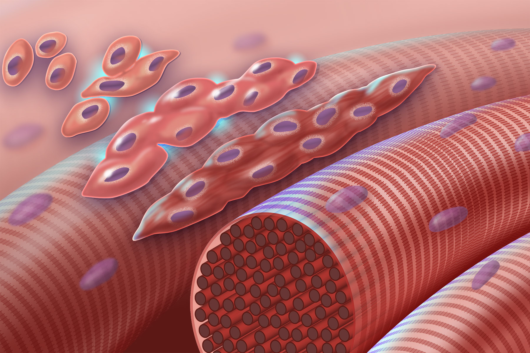
Myogenesis is the formation of skeletal muscular tissue, particularly during embryonic development. Muscle fibers generally form through the fusion of precursor myoblasts into multinucleated fibers called myotubes. In the early development of an embryo, myoblasts can either proliferate, or differentiate into a myotube. What controls this choice in vivo is generally unclear. If placed in cell culture, most myoblasts will proliferate if enough fibroblast growth factor (FGF) or another growth factor is present in the medium surrounding the cells. When the growth factor runs out, the myoblasts cease division and undergo terminal differentiation into myotubes. Myoblast differentiation proceeds in stages. The first stage involves cell cycle exit and the commencement of expression of certain genes. The second stage of differentiation involves the alignment of the myoblasts with one another. Studies have shown that even rat and chick myoblasts can recognise and align with one an ... [...More Info...] [...Related Items...] OR: [Wikipedia] [Google] [Baidu] |
Myoblast Fusion - Myogenesis
Myogenesis is the formation of skeletal muscle, skeletal muscular tissue, particularly during embryonic development. Skeletal muscle#Skeletal muscle cells, Muscle fibers generally form through the fusion of precursor cell, precursor myoblasts into Multinucleate, multinucleated fibers called myotubes. In the early development of an embryo, myoblasts can either cell proliferation, proliferate, or Cellular differentiation, differentiate into a myotube. What controls this choice in vivo is generally unclear. If placed in cell culture, most myoblasts will proliferate if enough fibroblast growth factor (FGF) or another growth factor is present in the medium surrounding the cells. When the growth factor runs out, the myoblasts cease division and undergo terminal differentiation into myotubes. Myoblast differentiation proceeds in stages. The first stage involves cell cycle exit and the commencement of expression of certain genes. The second stage of differentiation involves the align ... [...More Info...] [...Related Items...] OR: [Wikipedia] [Google] [Baidu] |
Skeletal Muscle
Skeletal muscle (commonly referred to as muscle) is one of the three types of vertebrate muscle tissue, the others being cardiac muscle and smooth muscle. They are part of the somatic nervous system, voluntary muscular system and typically are attached by tendons to bones of a skeleton. The skeletal muscle cells are much longer than in the other types of muscle tissue, and are also known as ''muscle fibers''. The tissue of a skeletal muscle is striated muscle tissue, striated – having a striped appearance due to the arrangement of the sarcomeres. A skeletal muscle contains multiple muscle fascicle, fascicles – bundles of muscle fibers. Each individual fiber and each muscle is surrounded by a type of connective tissue layer of fascia. Muscle fibers are formed from the cell fusion, fusion of developmental myoblasts in a process known as myogenesis resulting in long multinucleated cells. In these cells, the cell nucleus, nuclei, termed ''myonuclei'', are located along the inside ... [...More Info...] [...Related Items...] OR: [Wikipedia] [Google] [Baidu] |
Myogenin
Myogenin, is a transcriptional activator encoded by the ''MYOG'' gene. Myogenin is a muscle-specific basic-helix-loop-helix (bHLH) transcription factor involved in the coordination of skeletal muscle development or myogenesis and repair. Myogenin is a member of the MyoD family of transcription factors, which also includes MyoD, Myf5, and MRF4. In mice, myogenin is essential for the development of functional skeletal muscle. Myogenin is required for the proper differentiation of most myogenic precursor cells during the process of myogenesis. When the DNA coding for myogenin was knocked out of the mouse genome, severe skeletal muscle defects were observed. Mice lacking both copies of myogenin (homozygous Zygosity (the noun, zygote, is from the Greek "yoked," from "yoke") () is the degree to which both copies of a chromosome or gene have the same genetic sequence. In other words, it is the degree of similarity of the alleles in an organism. Mos ...-null) suffer from perin ... [...More Info...] [...Related Items...] OR: [Wikipedia] [Google] [Baidu] |
MyoD
MyoD, also known as myoblast determination protein 1, is a protein in animals that plays a major role in regulating muscle differentiation. MyoD, which was discovered in the laboratory of Harold M. Weintraub, belongs to a family of proteins known as myogenic regulatory factors (MRFs). These bHLH (basic helix loop helix) transcription factors act sequentially in myogenic differentiation. Vertebrate MRF family members include MyoD1, Myf5, myogenin, and MRF4 (Myf6). In non-vertebrate animals, a single MyoD protein is typically found. MyoD is one of the earliest markers of myogenic commitment. MyoD is expressed at extremely low and essentially undetectable levels in quiescent satellite cells, but expression of MyoD is activated in response to exercise or muscle tissue damage. The effect of MyoD on satellite cells is dose-dependent; high MyoD expression represses cell renewal, promotes terminal differentiation and can induce apoptosis. Although MyoD marks myoblast commitment, ... [...More Info...] [...Related Items...] OR: [Wikipedia] [Google] [Baidu] |
MSX1
Homeobox protein MSX-1, is a protein that in humans is encoded by the ''MSX1'' gene. MSX1 transcripts are not only found in thyrotrope-derived TSH cells, but also in the TtT97 thyrotropic tumor, which is a well differentiated hyperplastic tissue that produces both TSHß- and a-subunits and is responsive to thyroid hormone. MSX1 is also expressed in highly differentiated pituitary cells which until recently was thought to be expressed exclusively during embryogenesis. There is a highly conserved structural organization of the members of the MSX family of genes and their abundant expression at sites of inductive cell–cell interactions in the embryo suggest that they have a pivotal role during early development. Function This gene encodes a member of the muscle segment homeobox gene family. The encoded protein functions as a transcriptional repressor during embryogenesis through interactions with components of the core transcription complex and other homeoproteins. It may also ha ... [...More Info...] [...Related Items...] OR: [Wikipedia] [Google] [Baidu] |
LBX1
Transcription factor LBX1 is a protein that in humans is encoded by the ''LBX1'' gene. This gene and the orthologous mouse gene were found by their homology to the Drosophila lady bird early and late homeobox genes. In the mouse, this gene is a key regulator of muscle precursor cell migration Cell migration is a central process in the development and maintenance of multicellular organisms. Tissue formation during embryogenesis, embryonic development, wound healing and immune system, immune responses all require the orchestrated movemen ... and is required for the acquisition of dorsal identities of forelimb muscles. References Further reading * * * * * * {{gene-10-stub ... [...More Info...] [...Related Items...] OR: [Wikipedia] [Google] [Baidu] |
Hepatocyte Growth Factor
Hepatocyte growth factor (HGF) or scatter factor (SF) is a paracrine cellular growth, motility and morphogenic factor. It is secreted by mesenchymal cells and targets and acts primarily upon epithelial cells and endothelial cells, but also acts on haemopoietic progenitor cells and T cells. It has been shown to have a major role in embryonic organ development, specifically in myogenesis, in adult organ regeneration, and in wound healing. Function Hepatocyte growth factor regulates cell growth, cell motility, and morphogenesis by activating a tyrosine kinase signaling cascade after binding to the proto-oncogenic c-Met receptor. Hepatocyte growth factor is secreted by platelets, and mesenchymal cells and acts as a multi-functional cytokine on cells of mainly epithelial origin. Its ability to stimulate mitogenesis, cell motility, and matrix invasion gives it a central role in angiogenesis, tumorogenesis, and tissue regeneration. Structure It is secreted as a single inactive ... [...More Info...] [...Related Items...] OR: [Wikipedia] [Google] [Baidu] |
C-Met
Hepatocyte growth factor receptor (HGF receptor) is a protein that in humans is encoded by the ''MET'' gene. The protein possesses tyrosine kinase activity. The primary single chain precursor protein is post-translationally cleaved to produce the alpha and beta subunits, which are disulfide linked to form the mature receptor. HGF receptor is a single pass tyrosine kinase receptor essential for embryonic development, organogenesis and wound healing. Hepatocyte growth factor/Scatter Factor (HGF/SF) and its splicing isoform (NK1, NK2) are the only known ligands of the HGF receptor. MET is normally expressed by cells of epithelial origin, while expression of HGF/SF is restricted to cells of mesenchymal origin. When HGF/SF binds its cognate receptor MET it induces its dimerization through a not yet completely understood mechanism leading to its activation. Sometimes MET is misunderstood as of an abbreviation of Mesenchymal-Epithelial Transition. It is incorrect. The three letters ... [...More Info...] [...Related Items...] OR: [Wikipedia] [Google] [Baidu] |
PAX3
The PAX3 (paired box gene 3) gene encodes a member of the paired box or Pax genes, PAX family of transcription factors. The PAX family consists of nine human (PAX1-PAX9) and nine mouse (Pax1-Pax9) members arranged into four subfamilies. Human PAX3 and mouse Pax3 are present in a subfamily along with the highly homologous human PAX7 and mouse Pax7 genes. The human PAX3 gene is located in the 2q36.1 chromosomal region, and contains 10 exons within a 100 kb region. Transcript splicing Alternative splicing and processing generates multiple PAX3 isoforms that have been detected at the mRNA level. PAX3e is the longest isoform and consists of 10 exons that encode a 505 amino acid protein. In other mammalian species, including mouse, the longest mRNAs correspond to the human PAX3c and PAX3d isoforms, which consist of the first 8 or 9 exons of the PAX3 gene, respectively. Shorter PAX3 isoforms include mRNAs that skip exon 8 (PAX3g and PAX3h) and mRNAs containing 4 or 5 exons (PAX3a and P ... [...More Info...] [...Related Items...] OR: [Wikipedia] [Google] [Baidu] |
Androgen Receptor
The androgen receptor (AR), also known as NR3C4 (nuclear receptor subfamily 3, group C, member 4), is a type of nuclear receptor that is activated by binding any of the androgenic hormones, including testosterone and dihydrotestosterone, in the cytoplasm and then translocating into the Cell nucleus, nucleus. The androgen receptor is most closely related to the progesterone receptor, and progestins in higher dosages can block the androgen receptor. The main function of the androgen receptor is as a DNA-binding protein, DNA-binding transcription factor that Gene expression regulation, regulates gene expression; however, the androgen receptor has other functions as well. Androgen-regulated genes are critical for the development and maintenance of the male sexual phenotype. Function Effect on development In some cell types, testosterone interacts directly with androgen receptors, whereas, in others, testosterone is converted by 5-alpha reductase, 5-alpha-reductase to dihydrot ... [...More Info...] [...Related Items...] OR: [Wikipedia] [Google] [Baidu] |


