|
Horse Anatomy
Equine anatomy encompasses the gross and microscopic anatomy of horses, ponies and other equids, including donkeys, mules and zebras. While all anatomical features of equids are described in the same terms as for other animals by the International Committee on Veterinary Gross Anatomical Nomenclature in the book '' Nomina Anatomica Veterinaria'', there are many horse-specific colloquial terms used by equestrians. External anatomy * Back: the area where the saddle sits, beginning at the end of the withers, extending to the last thoracic vertebrae (colloquially includes the loin or "coupling", though technically incorrect usage) * Barrel: the body of the horse, enclosing the rib cage and the major internal organs * Buttock: the part of the hindquarters behind the thighs and below the root of the tail * Cannon or cannon bone: the area between the knee or hock and the fetlock joint, sometimes called the "shin" of the horse, though technically it is the third metacarpal * Chestn ... [...More Info...] [...Related Items...] OR: [Wikipedia] [Google] [Baidu] |
Root Of The Tail
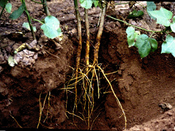
In vascular plants, the roots are the plant organ, organs of a plant that are modified to provide anchorage for the plant and take in water and nutrients into the plant body, which allows plants to grow taller and faster. They are most often below the surface of the soil, but roots can also be aerial root, aerial or aerating, that is, growing up above the ground or especially above water. Function The major functions of roots are absorption of water, plant nutrition and anchoring of the plant body to the ground. Types of Roots (major rooting system) Plants exhibit two main root system types: ''taproot'' and ''fibrous'', with variations like adventitious, aerial, and buttress roots, each serving specific functions. Taproot System Characterized by a single, main root growing vertically downward, with smaller lateral roots branching off. Examples. Dandelions, carrots, and many dicot plants. Fibrous RootSystem Consists of a network of thin, branching roots that spread out from ... [...More Info...] [...Related Items...] OR: [Wikipedia] [Google] [Baidu] |
Ergot (horse Anatomy)
The ergot is a small callosity (''Calcar metacarpeum and Calcar metatarseum)'' on the underside of the fetlock of a horse or other equine. Some equines have them on all four fetlocks; others have few or no detectable ergots. In horses, the ergot varies from very small to the size of a pea or bean, larger ergots occurring in horses with "Feathering (horse), feather" – long hairs on the lower legs. In some other equines, the ergot can be as much as in diameter. The word ''ergot'' comes from French ''ergot'' 'rooster's spur'.Clothier, Jane, ''The Ergot, p.15, ''Equine News'' Autumn 2010 Evolution Like the chestnut (horse anatomy), chestnut, the ergot is thought to be a Vestigiality, vestige of some part of the ancestral foot of the multi-toed Equidae, the ergot corresponding to the sole pad of other extant taxon, extant members of Perissodactyla, such as the tapir and rhinoceros. Unlike the chestnut, which in the same individual may be large on the forelegs and smaller o ... [...More Info...] [...Related Items...] OR: [Wikipedia] [Google] [Baidu] |
Homology (biology)
In biology, homology is similarity in anatomical structures or genes between organisms of different taxa due to shared ancestry, ''regardless'' of current functional differences. Evolutionary biology explains homologous structures as retained heredity from a common descent, common ancestor after having been subjected to adaptation (biology), adaptive modifications for different purposes as the result of natural selection. The term was first applied to biology in a non-evolutionary context by the anatomist Richard Owen in 1843. Homology was later explained by Charles Darwin's theory of evolution in 1859, but had been observed before this from Aristotle's biology onwards, and it was explicitly analysed by Pierre Belon in 1555. A common example of homologous structures is the forelimbs of vertebrates, where the bat wing development, wings of bats and origin of avian flight, birds, the arms of primates, the front flipper (anatomy), flippers of whales, and the forelegs of quadrupedalis ... [...More Info...] [...Related Items...] OR: [Wikipedia] [Google] [Baidu] |
Ligament
A ligament is a type of fibrous connective tissue in the body that connects bones to other bones. It also connects flight feathers to bones, in dinosaurs and birds. All 30,000 species of amniotes (land animals with internal bones) have ligaments. It is also known as ''articular ligament'', ''articular larua'', ''fibrous ligament'', or ''true ligament''. Comparative anatomy Ligaments are similar to tendons and fasciae as they are all made of connective tissue. The differences among them are in the connections that they make: ligaments connect one bone to another bone, tendons connect muscle to bone, and fasciae connect muscles to other muscles. These are all found in the skeletal system of the human body. Ligaments cannot usually be regenerated naturally; however, there are periodontal ligament stem cells located near the periodontal ligament which are involved in the adult regeneration of periodontist ligament. The study of ligaments is known as . Humans Other ligame ... [...More Info...] [...Related Items...] OR: [Wikipedia] [Google] [Baidu] |
Muscles
Muscle is a soft tissue, one of the four basic types of animal tissue. There are three types of muscle tissue in vertebrates: skeletal muscle, cardiac muscle, and smooth muscle. Muscle tissue gives skeletal muscles the ability to muscle contraction, contract. Muscle tissue contains special Muscle contraction, contractile proteins called actin and myosin which interact to cause movement. Among many other muscle proteins, present are two regulatory proteins, troponin and tropomyosin. Muscle is formed during embryonic development, in a process known as myogenesis. Skeletal muscle tissue is striated consisting of elongated, multinucleate muscle cells called muscle fibers, and is responsible for movements of the body. Other tissues in skeletal muscle include tendons and perimysium. Smooth and cardiac muscle contract involuntarily, without conscious intervention. These muscle types may be activated both through the interaction of the central nervous system as well as by innervation ... [...More Info...] [...Related Items...] OR: [Wikipedia] [Google] [Baidu] |
Coccygeal Vertebrae
The coccyx (: coccyges or coccyxes), commonly referred to as the tailbone, is the final segment of the vertebral column in all apes, and analogous structures in certain other mammals such as horses. In tailless primates (e.g. humans and other great apes) since '' Nacholapithecus'' (a Miocene hominoid),Nakatsukasa 2004, ''Acquisition of bipedalism'' (SeFig. 5entitled ''First coccygeal/caudal vertebra in short-tailed or tailless primates.''.) the coccyx is the remnant of a vestigial tail. In animals with bony tails, it is known as ''tailhead'' or ''dock'', in bird anatomy as ''tailfan''. It comprises three to five separate or fused coccygeal vertebrae below the sacrum, attached to the sacrum by a fibrocartilaginous joint, the sacrococcygeal symphysis, which permits limited movement between the sacrum and the coccyx. Structure The coccyx is formed of three, four or five rudimentary vertebrae. It articulates superiorly with the sacrum. In each of the first three segments may ... [...More Info...] [...Related Items...] OR: [Wikipedia] [Google] [Baidu] |
Sacral Vertebrae
The sacrum (: sacra or sacrums), in human anatomy, is a triangular bone at the base of the spine that forms by the fusing of the sacral vertebrae (S1S5) between ages 18 and 30. The sacrum situates at the upper, back part of the pelvic cavity, between the two wings of the pelvis. It forms joints with four other bones. The two projections at the sides of the sacrum are called the alae (wings), and articulate with the ilium at the L-shaped sacroiliac joints. The upper part of the sacrum connects with the last lumbar vertebra (L5), and its lower part with the coccyx (tailbone) via the sacral and coccygeal cornua. The sacrum has three different surfaces which are shaped to accommodate surrounding pelvic structures. Overall, it is concave (curved upon itself). The base of the sacrum, the broadest and uppermost part, is tilted forward as the sacral promontory internally. The central part is curved outward toward the posterior, allowing greater room for the pelvic cavity. In all o ... [...More Info...] [...Related Items...] OR: [Wikipedia] [Google] [Baidu] |
Croup (horse)
The rump or croup, in the external morphology of an animal, is the portion of the posterior dorsum – that is, posterior to the loins and anterior to the tail. Anatomically, the rump corresponds to the sacrum. The tailhead or dock is the beginning of the tail, where the tail joins the rump. It is known also as the base or root of the tail, and corresponds to the human sacrococcygeal symphysis. In some mammals the tail may be said to consist of the tailbone (meaning the bony column, muscles, and skin) and the skirt (meaning the long hairs growing from the tailbone). In birds, similarly, the tail consists of tailbone and tailfan (tail fan). Some animals are subjected to docking, the amputation of the tailbone at or near the dock. These include dogs, cats, sheep, pigs, and horses. Humans have a remnant tail, the coccyx, and the human equivalent of docking is coccygectomy. Usage varies from animal to animal. Birds and cattle are said to have a rump and tailhead. Dogs ... [...More Info...] [...Related Items...] OR: [Wikipedia] [Google] [Baidu] |
Horse Hoof
A horse hoof is the lower extremity of each leg of a horse, the part that makes contact with the ground and carries the weight of the animal. It is both hard and flexible. It is a complex structure surrounding the distal Phalanx bones, phalanx of the 3rd digit (digit III of the basic pentadactyl limb of vertebrates, evolved into a single weight-bearing digit in horses) of each of the four limbs, which is covered by soft tissue and keratinised (cornified) matter. Anatomy The hoof is made up of two parts. The outer part, called the hoof capsule, is composed of various cornified specialized structures. The inner, living part of the hoof, is made up of soft tissues and bone. The cornified materials of the hoof capsule differ in structure and properties. Dorsally, it covers, protects, and supports P3 (also known as the coffin bone, pedal bone, or PIII). Palmarly/plantarly, it covers and protects specialised soft tissues, such as tendons, ligaments, fibro-fatty and/or fibrocartilagin ... [...More Info...] [...Related Items...] OR: [Wikipedia] [Google] [Baidu] |
Curb Chain
A curb chain, or curb strap, is a piece of horse tack required for proper use on any type of curb bit. It is a flat linked chain or flat strap that runs under the chin groove of the horse, between the bit shank's purchase arms. It has a buckle or hook attachment and English designs have a "fly link" in the middle to hold a lip strap. On English bridles the horse is bridled with the curb chain undone on one side, then connected once on the horse. On western bridles, the curb chain is kept buckled to both sides of the bit. Action The main use of the curb chain is to enhance and control the lever A lever is a simple machine consisting of a beam (structure), beam or rigid rod pivoted at a fixed hinge, or '':wikt:fulcrum, fulcrum''. A lever is a rigid body capable of rotating on a point on itself. On the basis of the locations of fulcrum, l ... action of a curb bit. Additionally, it helps to keep the bit steady and in place within the mouth. On English pelham and double b ... [...More Info...] [...Related Items...] OR: [Wikipedia] [Google] [Baidu] |
Callosity
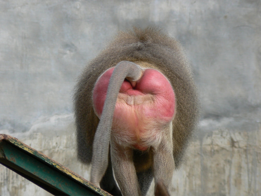
A callosity is a type of callus, a piece of skin that has become thickened as a result of repeated contact and friction. Primates All Old World monkeys, gibbons, and some chimpanzees have pads on their rears known as ''ischium, ischial callosities''. The pads enable the monkeys to sleep sitting upright on thin branches, beyond reach of predators, without falling. Humans do not possess ischial callosities due to the gluteal muscles being large enough to provide the same cushioning. The ischial callosities are one of the most distinctive pelvic features which separates Old World monkeys from New World monkeys. Right whales In whales, callosities are rough, calcified skin patches found on the heads of the three species of right whales. Callosities are a characteristic feature of the whale genus ''Eubalaena''. Because they are found on the head of the whale and appear white against the dark background of the whale's skin, they allow the reliable identification of individuals ... [...More Info...] [...Related Items...] OR: [Wikipedia] [Google] [Baidu] |


