|
Dental Anatomy
Dental anatomy is a field of anatomy dedicated to the study of Tooth (human), human tooth structures. The development, appearance, and classification of teeth fall within its purview. (The function of teeth as they contact one another falls elsewhere, under dental occlusion.) Tooth formation begins before birth, and the teeth's eventual Morphology (biology), morphology is dictated during this time. Dental anatomy is also a Taxonomy (general), taxonomical science: it is concerned with the naming of teeth and the structures of which they are made, this information serving a practical purpose in dental treatment. Usually, there are 20 primary ("baby") teeth and 32 permanent teeth, the last four being third molars or "wisdom teeth", each of which may or may not grow in. Among primary teeth, 10 usually are found in the maxilla (upper jaw) and the other 10 in the human mandible, mandible (lower jaw). Among permanent teeth, 16 are found in the maxilla and the other 16 in the mandible. Ea ... [...More Info...] [...Related Items...] OR: [Wikipedia] [Google] [Baidu] |
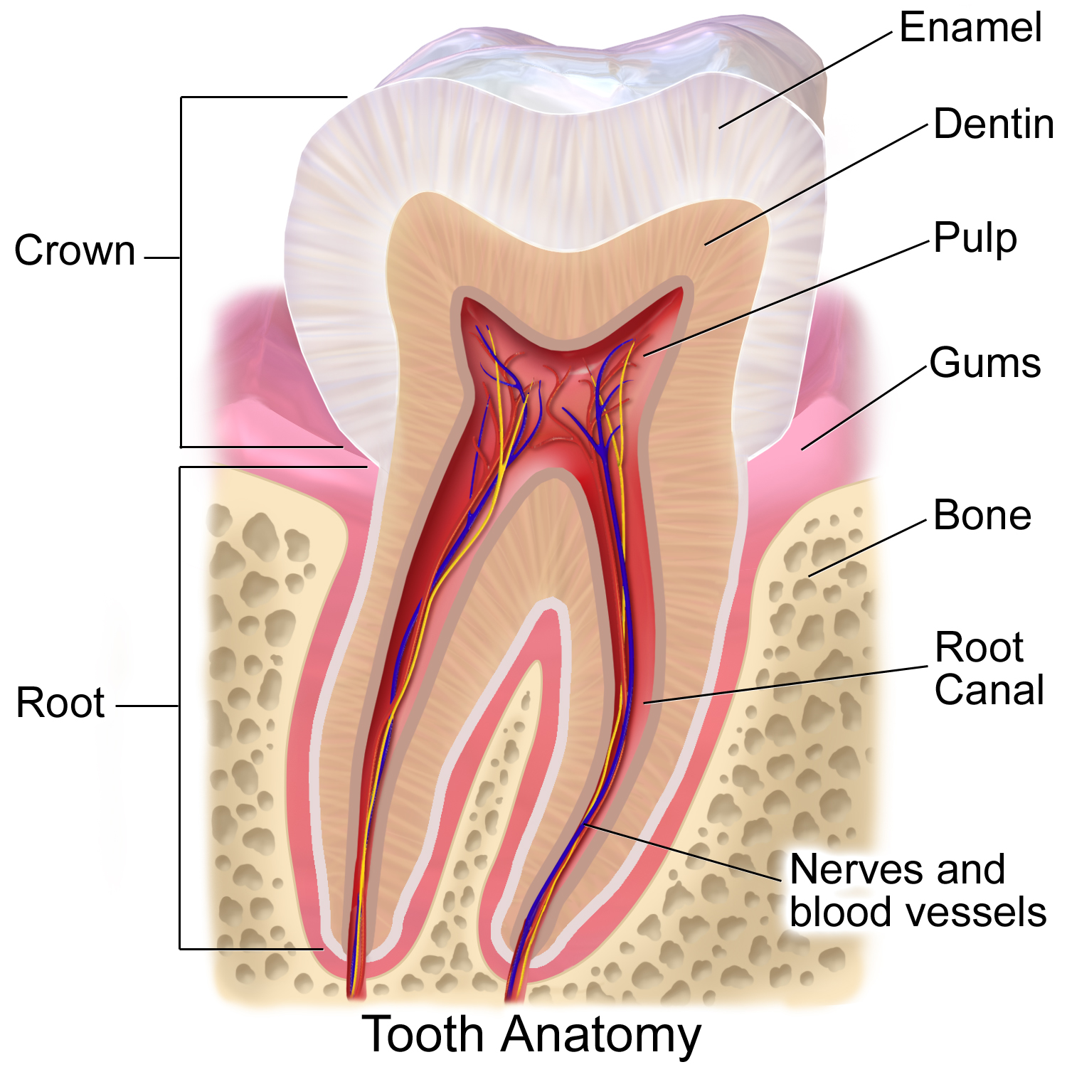
Blausen 0863 ToothAnatomy 02
Blausen Medical Communications, Inc. (BMC) is the creator and owner of a library of two- and three-dimensional medical and scientific images and animations, a developer of information technology allowing access to that content, and a business focused on licensing and distributing the content. It was founded by Bruce Blausen in Houston, Texas, in 1991, and is privately held. Background Blausen Medical Communications is a privately held company founded by Bruce Blausen in Houston, Texas in 1991. BMC created and owns a library of medical and scientific images and animations, and has developed information technology Information technology (IT) is a set of related fields within information and communications technology (ICT), that encompass computer systems, software, programming languages, data processing, data and information processing, and storage. Inf ... tools allowing access to the library; as well, it licenses and otherwise works to distribute the content. As of this da ... [...More Info...] [...Related Items...] OR: [Wikipedia] [Google] [Baidu] |
Dentin
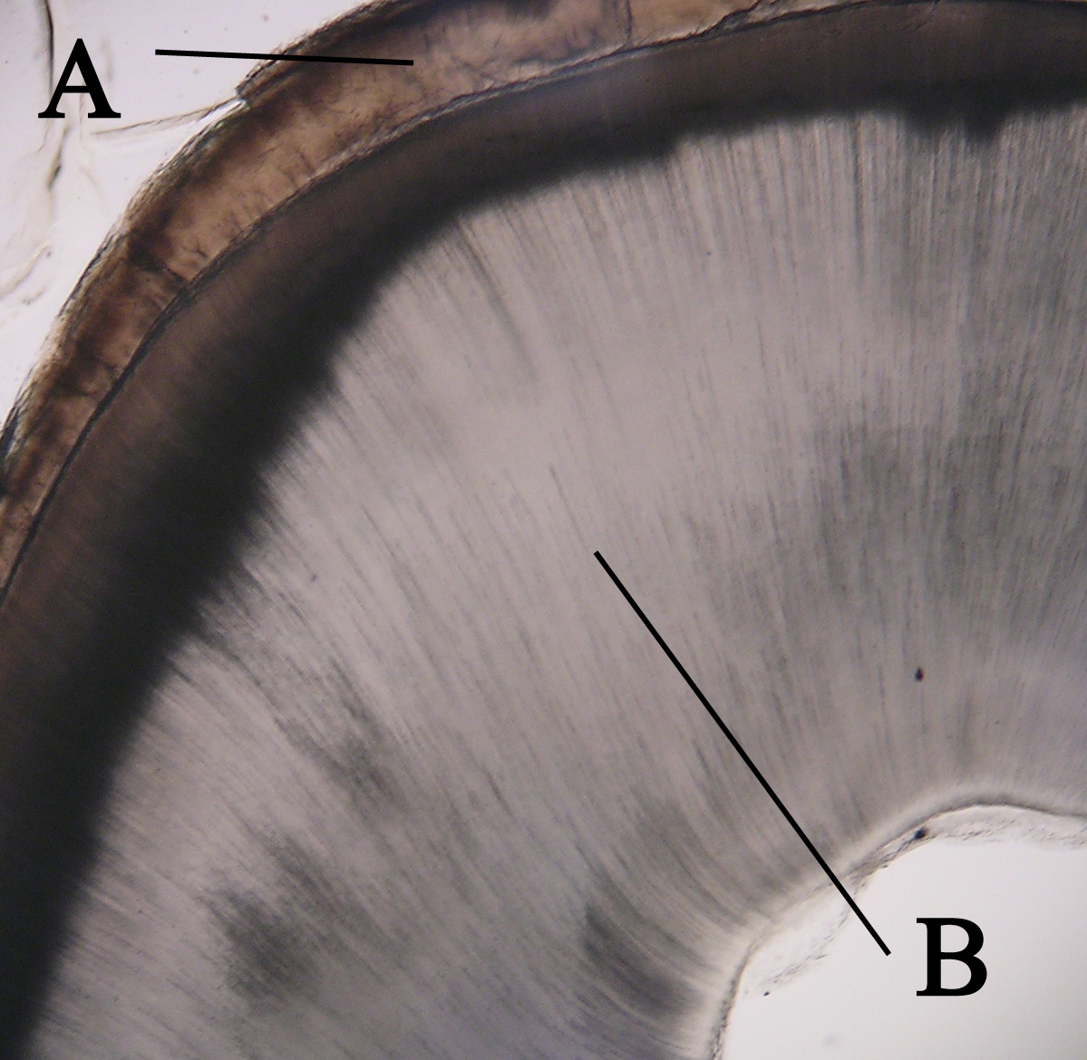
Dentin ( ) (American English) or dentine ( or ) (British English) () is a calcified tissue (biology), tissue of the body and, along with tooth enamel, enamel, cementum, and pulp (tooth), pulp, is one of the four major components of teeth. It is usually covered by enamel on the crown and cementum on the root and surrounds the entire pulp. By volume, 45% of dentin consists of the mineral hydroxyapatite, 33% is organic material, and 22% is water. Yellow in appearance, it greatly affects the color of a tooth due to the translucency of enamel. Dentin, which is less mineralized and less brittle than enamel, is necessary for the support of enamel. Dentin rates approximately 3 on the Mohs scale of mineral hardness. There are two main characteristics which distinguish dentin from enamel: firstly, dentin forms throughout life; secondly, dentin is sensitive and can become hypersensitive to changes in temperature due to the sensory function of odontoblasts, especially when enamel recedes an ... [...More Info...] [...Related Items...] OR: [Wikipedia] [Google] [Baidu] |
Stellate Reticulum
In animal tooth development, the stellate reticulum is a group of cells located in the center of the enamel organ of a developing tooth. These cells are star-shaped (hence ''stellate'') and synthesize glycosaminoglycans. As glycosaminoglycans are produced, water is drawn in between the cells, stretching them apart. As they are moved further away from one another, the stellate reticular cells maintain contact with one another through desmosome A desmosome (; "binding body"), also known as a macula adherens (plural: maculae adherentes) (Latin for ''adhering spot''), is a cell structure specialized for cell-to-cell adhesion. A type of junctional complex, they are localized spot-like ad ...s, resulting in their unique appearance. The stellate reticulum is lost after the first layer of enamel is laid down. This brings cells in the inner enamel epithelium closer to blood vessels at the periphery. References * Orbans Oral histology and embryology – 10th ed. * Cate, A.R. Ten. O ... [...More Info...] [...Related Items...] OR: [Wikipedia] [Google] [Baidu] |
Inner Enamel Epithelium
Interior may refer to: Arts and media * ''Interior'' (Degas) (also known as ''The Rape''), painting by Edgar Degas * ''Interior'' (play), 1895 play by Belgian playwright Maurice Maeterlinck * ''The Interior'' (novel), by Lisa See * Interior design, the trade of designing an architectural interior * ''The Interior'' (Presbyterian periodical), an American Presbyterian periodical * Interior architecture, process of designing building interiors or renovating existing home interiors Places * Interior, South Dakota * Interior, Washington * Interior Township, Michigan * British Columbia Interior, commonly known as "The Interior" Government agencies * Interior ministry, sometimes called the ministry of home affairs * United States Department of the Interior Other uses * Interior (topology), mathematical concept that includes, for example, the inside of a shape * Interior FC, a football team in Gambia See also * * * List of geographic interiors * Interiors (other) ... [...More Info...] [...Related Items...] OR: [Wikipedia] [Google] [Baidu] |
Outer Enamel Epithelium
The outer enamel epithelium, also known as the external enamel epithelium, is a layer of cuboidal cells located on the periphery of the enamel organ in a developing tooth. This layer is first seen during the bell stage. The rim of the enamel organ, where the outer and inner enamel epithelium Interior may refer to: Arts and media * ''Interior'' (Degas) (also known as ''The Rape''), painting by Edgar Degas * ''Interior'' (play), 1895 play by Belgian playwright Maurice Maeterlinck * ''The Interior'' (novel), by Lisa See * Interior de ... join is called the cervical loop. References *Cate, A.R. Ten. Oral Histology: development, structure, and function. 5th ed. 1998. . *Ross, Michael H., Gordon I. Kaye, and Wojciech Pawlina. Histology: a text and atlas. 4th edition. 2003. . Tooth development {{dentistry-stub ... [...More Info...] [...Related Items...] OR: [Wikipedia] [Google] [Baidu] |
Dental Follicle
{{Disambig ...
Dental may refer to: * Dental consonant, in phonetics * Dental Records, an independent UK record label * Dentistry, oral medicine * Teeth See also * * Dental care (other) * Dentist (other) * Tooth (other) A tooth (: teeth) is a small, calcified, whitish structure found in the jaws (or mouths) of many vertebrates. Tooth or Teeth may also refer to: Music *Teeth (Filipino band), a Filipino rock band *Teeth (electronic band), UK electronic pop punk b ... [...More Info...] [...Related Items...] OR: [Wikipedia] [Google] [Baidu] |
Dental Papilla
In embryology and prenatal development, the dental papilla is a condensation of ectomesenchymal cells called odontoblasts, seen in histologic sections of a developing tooth. It lies below a cellular aggregation known as the enamel organ. The dental papilla appears after 8–10 weeks of intra uteral life. The dental papilla gives rise to the dentin and pulp of a tooth. The enamel organ, dental papilla, and dental follicle together forms one unit, called the tooth germ. This is of importance because all the tissues of a tooth and its supporting structures form from these distinct cellular aggregations. Similar to dental follicle, the dental papilla has a very rich blood supply and provides nutrition to the enamel organ. Embryology Formation of dental papilla occurs in the cap stage of odontogenesis. Cap stage The cap stage is the second stage of tooth development and occurs during the ninth or tenth week of prenatal development. Unequal proliferation of the tooth bu ... [...More Info...] [...Related Items...] OR: [Wikipedia] [Google] [Baidu] |
Enamel Organ
The enamel organ, also known as the dental organ, is a cellular aggregation seen in a developing tooth and it lies above the dental papilla. The enamel organ which is differentiated from the primitive oral epithelium lining the stomodeum. The enamel organ is responsible for the formation of enamel, initiation of dentine formation, establishment of the shape of a tooth's crown, and establishment of the dentoenamel junction. The enamel organ has four layers; the inner enamel epithelium, outer enamel epithelium, stratum intermedium, and the stellate reticulum. The dental papilla, the differentiated ectomesenchyme deep to the enamel organ, will produce dentin and the dental pulp. The surrounding ectomesenchyme tissue, the dental follicle, is the primitive cementum, periodontal ligament and alveolar bone beneath the tooth root. The site where the internal enamel epithelium and external enamel epithelium coalesce is the cervical root, important in proliferation of the dental root ... [...More Info...] [...Related Items...] OR: [Wikipedia] [Google] [Baidu] |
Branchial Arch
Branchial arches or gill arches are a series of paired bony/ cartilaginous "loops" behind the throat ( pharyngeal cavity) of fish, which support the fish gills. As chordates, all vertebrate embryos develop pharyngeal arches, though the eventual fate of these arches varies between taxa. In all jawed fish (gnathostomes), the first arch pair (mandibular arches) develops into the jaw, the second gill arches (the hyoid arches) develop into the hyomandibular complex (which supports the back of the jaw and the front of the gill series), and the remaining posterior arches (simply called branchial arches) support the gills. In tetrapods, a mostly terrestrial clade evolved from lobe-finned fish, many pharyngeal arch elements are lost, including the gill arches. In amphibians and reptiles, only the oral jaws and a hyoid apparatus remains, and in mammals and birds the hyoid is simplified further to support the tongue and floor of the mouth. In mammals, the first and second pharyng ... [...More Info...] [...Related Items...] OR: [Wikipedia] [Google] [Baidu] |
Permanent Teeth
Permanent teeth or adult teeth are the second set of teeth formed in diphyodont mammals. In humans and old world simians, there are thirty-two permanent teeth, consisting of six maxillary and six mandibular molars, four maxillary and four mandibular premolars, two maxillary and two mandibular canines, four maxillary and four mandibular incisors. Timeline The first permanent tooth usually appears in the mouth at around 5-6 years of age, and the mouth will then be in a transition time with both primary (or deciduous dentition) teeth and permanent teeth during the mixed dentition period until the last primary tooth is lost or shed. The first of the permanent teeth to erupt are the permanent first molars, right behind the last 'milk' molars of the primary dentition. These first permanent molars are important for the correct development of a permanent dentition. Up to thirteen years of age, 28 of the 32 permanent teeth will appear. The full permanent dentition is completed much l ... [...More Info...] [...Related Items...] OR: [Wikipedia] [Google] [Baidu] |
Uterus
The uterus (from Latin ''uterus'', : uteri or uteruses) or womb () is the hollow organ, organ in the reproductive system of most female mammals, including humans, that accommodates the embryonic development, embryonic and prenatal development, fetal development of one or more Fertilized egg, fertilized eggs until birth. The uterus is a hormone-responsive sex organ that contains uterine gland, glands in its endometrium, lining that secrete uterine milk for embryonic nourishment. (The term ''uterus'' is also applied to analogous structures in some non-mammalian animals.) In humans, the lower end of the uterus is a narrow part known as the Uterine isthmus, isthmus that connects to the cervix, the anterior gateway leading to the vagina. The upper end, the body of the uterus, is connected to the fallopian tubes at the uterine horns; the rounded part, the fundus, is above the openings to the fallopian tubes. The connection of the uterine cavity with a fallopian tube is called the utero ... [...More Info...] [...Related Items...] OR: [Wikipedia] [Google] [Baidu] |
Deciduous Teeth
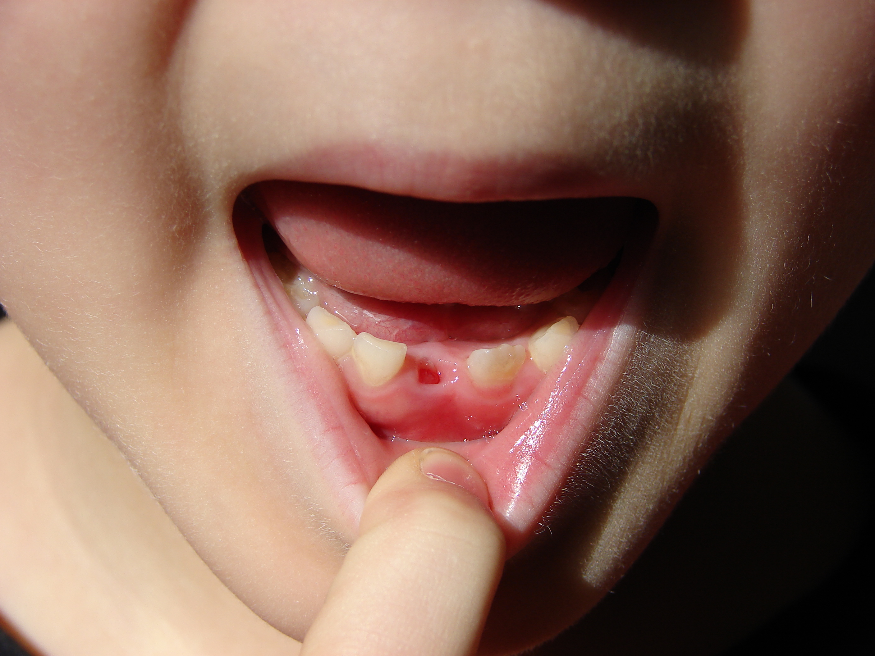
Deciduous teeth or primary teeth, also informally known as baby teeth, milk teeth, or temporary teeth,Fehrenbach, MJ and Popowics, T. (2026). ''Illustrated Dental Embryology, Histology, and Anatomy'', 6th edition, Elsevier, page 287–296. are the first set of teeth in the growth and development of humans and other diphyodonts, which include most mammals but not elephants, kangaroos, or manatees, which are polyphyodonts. Deciduous teeth Animal tooth development, develop during the embryonic stage of development and tooth eruption, erupt (break through the gums and become visible in the mouth) during infancy. They are usually lost and replaced by permanent teeth, but in the absence of their permanent replacements, they can remain functional for many years into adulthood. Development Formation Primary teeth start to form during the embryonic phase of human development (biology), human life. The development of primary teeth starts at the sixth week of tooth development as the d ... [...More Info...] [...Related Items...] OR: [Wikipedia] [Google] [Baidu] |


