|
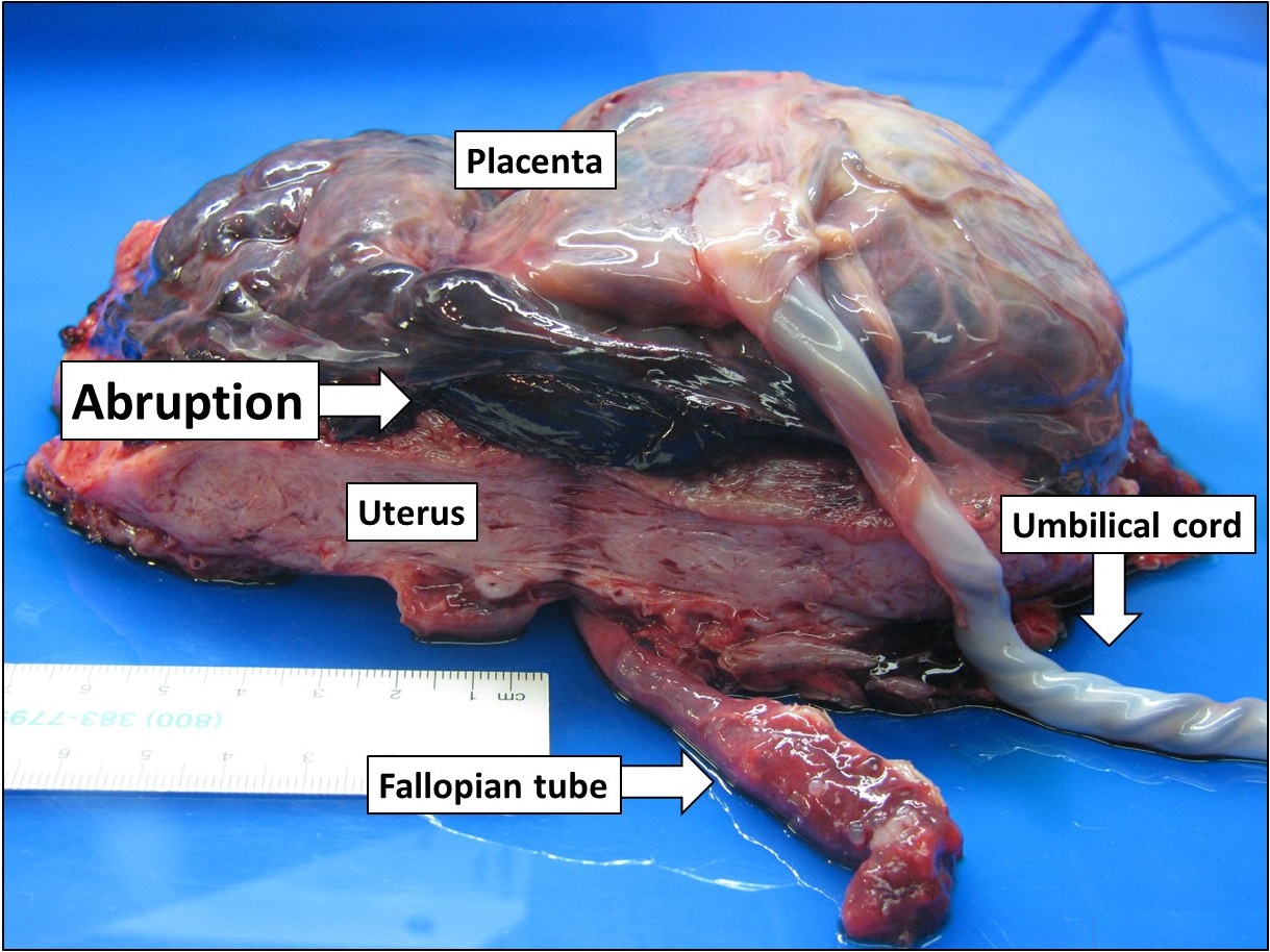
Couvelaire Uterus
Couvelaire uterus (also known as uteroplacental apoplexy) is a rare but not a life-threatening condition in which loosening of the placenta (abruptio placentae) causes bleeding that penetrates into the uterine myometrium forcing its way into the peritoneal cavity. This condition makes the uterus very tense and rigid. Symptoms and signs Patients can have pain secondary to uterine contractions, uterine tetany or localized uterine tenderness. Signs can also be due to abruptio placentae including uterine hypertonus, fetal distress, fetal death, and rarely, hypovolemic shock (shock secondary to severe blood loss). The uterus may adopt a bluish/purplish, mottled appearance due to extravasation of blood into uterine muscle. Pathophysiology Couvelaire uterus is a phenomenon where the retroplacental blood may penetrate through the thickness of the wall of the uterus into the peritoneal cavity. This may occur after abruptio placentae. The hemorrhage that gets into the decidua basalis ultim ... [...More Info...] [...Related Items...] OR: [Wikipedia] [Google] [Baidu] |
Abruptio Placentae
Placental abruption is when the placenta separates early from the uterus, in other words separates before childbirth. It occurs most commonly around 25 weeks of pregnancy. Symptoms may include vaginal bleeding, lower abdominal pain, and dangerously low blood pressure. Complications for the mother can include disseminated intravascular coagulopathy and kidney failure. Complications for the baby can include fetal distress, low birthweight, preterm delivery, and stillbirth. The cause of placental abruption is not entirely clear. Risk factors include smoking, pre-eclampsia, prior abruption (most important and predictive risk factor), trauma during pregnancy, cocaine use, and previous cesarean section. Diagnosis is based on symptoms and supported by ultrasound. It is classified as a complication of pregnancy. For small abruption, bed rest may be recommended, while for more significant abruptions or those that occur near term, delivery may be recommended. If everything is stable, v ... [...More Info...] [...Related Items...] OR: [Wikipedia] [Google] [Baidu] |
Myometrium
The myometrium is the middle layer of the uterine wall, consisting mainly of uterine smooth muscle cells (also called uterine myocytes) but also of supporting stromal and vascular tissue. Its main function is to induce uterine contractions. Structure The myometrium is located between the endometrium (the inner layer of the uterine wall) and the serosa or perimetrium (the outer uterine layer). The inner one-third of the myometrium (termed the ''junctional'' or ''sub-endometrial'' layer) appears to be derived from the Müllerian duct, while the outer, more predominant layer of the myometrium appears to originate from non-Müllerian tissue and is the major contractile tissue during parturition and abortion. The junctional layer appears to function like a circular muscle layer, capable of peristaltic and anti-peristaltic activity, equivalent to the muscular layer of the intestines. Muscular structure The molecular structure of the smooth muscle of myometrium is very similar to ... [...More Info...] [...Related Items...] OR: [Wikipedia] [Google] [Baidu] |
Peritoneal Cavity
The peritoneal cavity is a potential space between the parietal peritoneum (the peritoneum that surrounds the abdominal wall) and visceral peritoneum (the peritoneum that surrounds the internal organs). The parietal and visceral peritonea are layers of the peritoneum named depending on their function/location. It is one of the spaces derived from the coelomic cavity of the embryo, the others being the pleural cavities around the lungs and the pericardial cavity around the heart. It is the largest serosal sac, and the largest fluid-filled cavity, in the body and secretes approximately 50 ml of fluid per day. This fluid acts as a lubricant and has anti-inflammatory properties. The peritoneal cavity is divided into two compartments – one above, and one below the transverse colon. Compartments The peritoneal cavity is divided by the transverse colon (and its mesocolon) into an upper supracolic compartment, and a lower infracolic compartment. The liver, spleen, stomach, and le ... [...More Info...] [...Related Items...] OR: [Wikipedia] [Google] [Baidu] |
Hypovolemic Shock
Hypovolemic shock is a form of shock caused by severe hypovolemia (insufficient blood volume or extracellular fluid in the body). It could be the result of severe dehydration through a variety of mechanisms or blood loss. Hypovolemic shock is a medical emergency; if left untreated, the insufficient blood flow can cause damage to organs, leading to multiple organ failure. In treating hypovolemic shock, it is important to determine the cause of the underlying hypovolemia, which may be the result of bleeding or other fluid losses. To minimize ischemic damage to tissues, treatment involves quickly replacing lost blood or fluids, with consideration of both rate and the type of fluids used. Tachycardia, a fast heart rate, is typically the first abnormal vital sign. When resulting from blood loss, trauma is the most common root cause, but severe blood loss can also happen in various body systems without clear traumatic injury. The body in hypovolemic shock prioritizes getting o ... [...More Info...] [...Related Items...] OR: [Wikipedia] [Google] [Baidu] |
