|
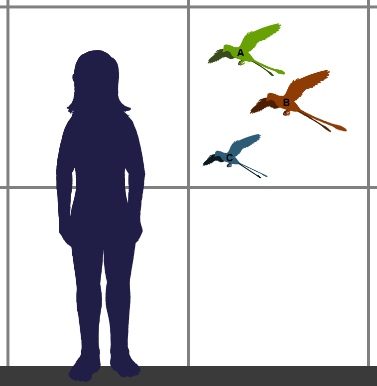
Confuciusornithidae Sizes
Confuciusornithidae is an extinct family of pygostylian avialans known from the Early Cretaceous, found in northern China. They are commonly placed as a sister group to Ornithothoraces, a group that contains all extant birds along with their closest extinct relatives. Confuciusornithidae contains four genera, possessing both shafted and non-shafted (downy) feathers. They are also noted for their distinctive pair of ribbon-like tail feathers of disputed function. The wing anatomy of confuciusornithids suggests an unusual flight behavior, due to anatomy that implies conflicting abilities. They possessed feathers similar to those of fast-flapping birds, which rely on quick flapping of their wings to stay aloft. At the same time, their wing anatomy also suggests a lack of flapping ability. Confuciusornithids are also noted for their beak and lack of teeth, similar to modern birds. Both predators and prey, confuciusornithid fossils have been observed with fish remains in their digestive ... [...More Info...] [...Related Items...] OR: [Wikipedia] [Google] [Baidu] |
Early Cretaceous
The Early Cretaceous (geochronological name) or the Lower Cretaceous ( chronostratigraphic name), is the earlier or lower of the two major divisions of the Cretaceous. It is usually considered to stretch from 145 Ma to 100.5 Ma. Geology Proposals for the exact age of the Barremian-Aptian boundary ranged from 126 to 117 Ma until recently (as of 2019), but based on drillholes in Svalbard the defining early Aptian Oceanic Anoxic Event 1a (OAE1a) was carbon isotope dated to 123.1±0.3 Ma, limiting the possible range for the boundary to c. 122–121 Ma. There is a possible link between this anoxic event and a series of Early Cretaceous large igneous provinces (LIP). The Ontong Java- Manihiki- Hikurangi large igneous province, emplaced in the South Pacific at c. 120 Ma, is by far the largest LIP in Earth's history. The Ontong Java Plateau today covers an area of 1,860,000 km2. In the Indian Ocean another LIP began to form at c. 120 Ma, the Kergue ... [...More Info...] [...Related Items...] OR: [Wikipedia] [Google] [Baidu] |
Class (biology)
In biological classification, class ( la, classis) is a taxonomic rank, as well as a taxonomic unit, a taxon, in that rank. It is a group of related taxonomic orders. Other well-known ranks in descending order of size are life, domain, kingdom, phylum, order, family, genus, and species, with class fitting between phylum and order. History The class as a distinct rank of biological classification having its own distinctive name (and not just called a ''top-level genus'' ''(genus summum)'') was first introduced by the French botanist Joseph Pitton de Tournefort in his classification of plants that appeared in his ''Eléments de botanique'', 1694. Insofar as a general definition of a class is available, it has historically been conceived as embracing taxa that combine a distinct ''grade'' of organization—i.e. a 'level of complexity', measured in terms of how differentiated their organ systems are into distinct regions or sub-organs—with a distinct ''type'' of construc ... [...More Info...] [...Related Items...] OR: [Wikipedia] [Google] [Baidu] |
Phalanx Bone
The phalanges (singular: ''phalanx'' ) are digital bones in the hands and feet of most vertebrates. In primates, the thumbs and big toes have two phalanges while the other digits have three phalanges. The phalanges are classed as long bones. Structure The phalanges are the bones that make up the fingers of the hand and the toes of the foot. There are 56 phalanges in the human body, with fourteen on each hand and foot. Three phalanges are present on each finger and toe, with the exception of the thumb and large toe, which possess only two. The middle and far phalanges of the fifth toes are often fused together (symphalangism). The phalanges of the hand are commonly known as the finger bones. The phalanges of the foot differ from the hand in that they are often shorter and more compressed, especially in the proximal phalanges, those closest to the torso. A phalanx is named according to whether it is proximal, middle, or distal and its associated finger or toe. The proxi ... [...More Info...] [...Related Items...] OR: [Wikipedia] [Google] [Baidu] |
Anatomical Terms Of Location
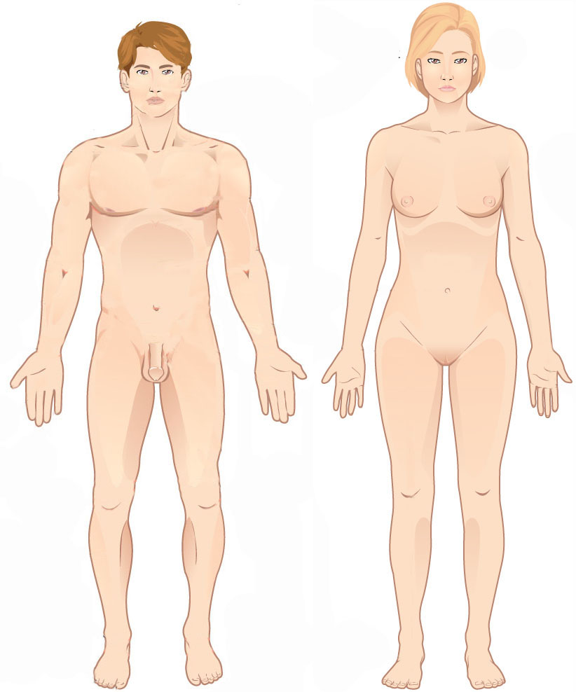
Standard anatomical terms of location are used to unambiguously describe the anatomy of animals, including humans. The terms, typically derived from Latin or Greek roots, describe something in its standard anatomical position. This position provides a definition of what is at the front ("anterior"), behind ("posterior") and so on. As part of defining and describing terms, the body is described through the use of anatomical planes and anatomical axes. The meaning of terms that are used can change depending on whether an organism is bipedal or quadrupedal. Additionally, for some animals such as invertebrates, some terms may not have any meaning at all; for example, an animal that is radially symmetrical will have no anterior surface, but can still have a description that a part is close to the middle ("proximal") or further from the middle ("distal"). International organisations have determined vocabularies that are often used as standard vocabularies for subdisciplines o ... [...More Info...] [...Related Items...] OR: [Wikipedia] [Google] [Baidu] |
Sternum
The sternum or breastbone is a long flat bone located in the central part of the chest. It connects to the ribs via cartilage and forms the front of the rib cage, thus helping to protect the heart, lungs, and major blood vessels from injury. Shaped roughly like a necktie, it is one of the largest and longest flat bones of the body. Its three regions are the manubrium, the body, and the xiphoid process. The word "sternum" originates from the Ancient Greek στέρνον (stérnon), meaning "chest". Structure The sternum is a narrow, flat bone, forming the middle portion of the front of the chest. The top of the sternum supports the clavicles (collarbones) and its edges join with the costal cartilages of the first two pairs of ribs. The inner surface of the sternum is also the attachment of the sternopericardial ligaments. Its top is also connected to the sternocleidomastoid muscle. The sternum consists of three main parts, listed from the top: * Manubrium * Body (gl ... [...More Info...] [...Related Items...] OR: [Wikipedia] [Google] [Baidu] |
Metacarpal Bones
In human anatomy, the metacarpal bones or metacarpus form the intermediate part of the skeletal hand located between the phalanges of the fingers and the carpal bones of the wrist, which forms the connection to the forearm. The metacarpal bones are analogous to the metatarsal bones in the foot. Structure The metacarpals form a transverse arch to which the rigid row of distal carpal bones are fixed. The peripheral metacarpals (those of the thumb and little finger) form the sides of the cup of the palmar gutter and as they are brought together they deepen this concavity. The index metacarpal is the most firmly fixed, while the thumb metacarpal articulates with the trapezium and acts independently from the others. The middle metacarpals are tightly united to the carpus by intrinsic interlocking bone elements at their bases. The ring metacarpal is somewhat more mobile while the fifth metacarpal is semi-independent.Tubiana ''et al'' 1998, p 11 Each metacarpal bone consists of a body ... [...More Info...] [...Related Items...] OR: [Wikipedia] [Google] [Baidu] |
Humerus
The humerus (; ) is a long bone in the arm that runs from the shoulder to the elbow. It connects the scapula and the two bones of the lower arm, the radius and ulna, and consists of three sections. The humeral upper extremity consists of a rounded head, a narrow neck, and two short processes (tubercles, sometimes called tuberosities). The body is cylindrical in its upper portion, and more prismatic below. The lower extremity consists of 2 epicondyles, 2 processes (trochlea & capitulum), and 3 fossae (radial fossa, coronoid fossa, and olecranon fossa). As well as its true anatomical neck, the constriction below the greater and lesser tubercles of the humerus is referred to as its surgical neck due to its tendency to fracture, thus often becoming the focus of surgeons. Etymology The word "humerus" is derived from la, humerus, umerus meaning upper arm, shoulder, and is linguistically related to Gothic ''ams'' shoulder and Greek ''ōmos''. Structure Upper extremity The upper or pr ... [...More Info...] [...Related Items...] OR: [Wikipedia] [Google] [Baidu] |
Clavipectoral Triangle
The clavipectoral triangle (also known as the deltopectoral triangle) is an anatomical region found in humans and other animals. It is bordered by the following structures: * Clavicle (superiorly) * Lateral border of Pectoralis Major (medially) * Medial border of Deltoid muscle (laterally) It contains the cephalic vein, and deltopectoral fascia, which is a layer of deep fascia that invests the three structures that make up the border of the triangle. The deltoid branch of the thoracoacromial artery also passes through this triangle, giving branches to both the deltoid and pectoralis major muscles. The subclavian vein and the subclavian artery may be accessed via this triangle, as they are deep to it. Clinical significance * Palpation of coracoid process of scapula The coracoid process of the scapula is not subcutaneous; It is covered by the anterior border of the deltoid. However, the tip of the coracoid process can be felt on deep palpation on the lateral aspect of the ... [...More Info...] [...Related Items...] OR: [Wikipedia] [Google] [Baidu] |
Maxilla
The maxilla (plural: ''maxillae'' ) in vertebrates is the upper fixed (not fixed in Neopterygii) bone of the jaw formed from the fusion of two maxillary bones. In humans, the upper jaw includes the hard palate in the front of the mouth. The two maxillary bones are fused at the intermaxillary suture, forming the anterior nasal spine. This is similar to the mandible (lower jaw), which is also a fusion of two mandibular bones at the mandibular symphysis. The mandible is the movable part of the jaw. Structure In humans, the maxilla consists of: * The body of the maxilla * Four processes ** the zygomatic process ** the frontal process of maxilla ** the alveolar process ** the palatine process * three surfaces – anterior, posterior, medial * the Infraorbital foramen * the maxillary sinus * the incisive foramen Articulations Each maxilla articulates with nine bones: * two of the cranium: the frontal and ethmoid * seven of the face: the nasal, zygomatic, lacrimal, ... [...More Info...] [...Related Items...] OR: [Wikipedia] [Google] [Baidu] |
Mandibular Symphysis
In human anatomy, the facial skeleton of the skull The skull is a bone protective cavity for the brain. The skull is composed of four types of bone i.e., cranial bones, facial bones, ear ossicles and hyoid bone. However two parts are more prominent: the cranium and the mandible. In humans, t ... the external surface of the mandible is marked in the median line by a faint ridge, indicating the mandibular symphysis (Latin: ''symphysis menti'') or line of junction where the two lateral halves of the mandible typically fuse at an early period of life (1-2 years). It is not a true symphysis as there is no cartilage between the two sides of the mandible. This ridge divides below and encloses a triangular eminence, the mental protuberance, the base of which is depressed in the center but raised on either side to form the mental tubercle. The lowest (most inferior) end of the mandibular symphysis — the point of the chin — is called the "menton". It serves as the origin for ... [...More Info...] [...Related Items...] OR: [Wikipedia] [Google] [Baidu] |
Rostralward
Standard anatomical terms of location are used to unambiguously describe the anatomy of animals, including humans. The terms, typically derived from Latin or Greek roots, describe something in its standard anatomical position. This position provides a definition of what is at the front ("anterior"), behind ("posterior") and so on. As part of defining and describing terms, the body is described through the use of anatomical planes and anatomical axes. The meaning of terms that are used can change depending on whether an organism is bipedal or quadrupedal. Additionally, for some animals such as invertebrates, some terms may not have any meaning at all; for example, an animal that is radially symmetrical will have no anterior surface, but can still have a description that a part is close to the middle ("proximal") or further from the middle ("distal"). International organisations have determined vocabularies that are often used as standard vocabularies for subdisciplines of anatom ... [...More Info...] [...Related Items...] OR: [Wikipedia] [Google] [Baidu] |


_dorsal_view.png)


