|
Cistern Of Lateral Cerebral Fossa
The cistern of lateral cerebral fossa (also cistern of the lateral sulcus, or Sylvian cistern) is an elongated subarachnoid cistern formed by arachnoid mater bridging the lateral sulcus The lateral sulcus (or lateral fissure, also called Sylvian fissure, after Franciscus Sylvius) is the most prominent sulcus (neuroanatomy), sulcus of each cerebral hemisphere in the human brain. The lateral sulcus (neuroanatomy), sulcus is a deep ... between the frontal, temporal, and parietal opercula. The cistern contains the middle cerebral artery (MCA) and its branches, and the two (i.e. superficial and deep) middle cerebral veins (MCVs). The cistern is subdivided into three compartments: the superficial opercular compartment (SOC) (most superficial), deep opercular compartment (DOC) (intermediate), and cisternal compartment (CC) (deepest). The SOC contains the superficial MCV, and distal branches of the MCA; the DOC contains the M3 segment of the MCA; the CC contains the M1 and M2 segment ... [...More Info...] [...Related Items...] OR: [Wikipedia] [Google] [Baidu] |
Subarachnoid Cisterns
The subarachnoid cisterns are spaces formed by openings in the subarachnoid space, an anatomic space in the meninges of the brain. The space is situated between the two meninges, the arachnoid mater and the pia mater. These cisterns are filled with cerebrospinal fluid (CSF). Structure Although the pia mater adheres to the surface of the brain, closely following the contours of its gyri and sulci, the arachnoid mater only covers its superficial surface, bridging across the gyri. This leaves wider spaces between the pia and arachnoid and the cavities are known as the subarachnoid cisterns. Although they are often described as distinct compartments, the subarachnoid cisterns are not truly anatomically distinct. Rather, these subarachnoid cisterns are separated from each other by a trabeculated porous wall with various-sized openings. Cisterns There are many cisterns in the brain with several large ones noted with their own name. At the base of the spinal cord is another subarachno ... [...More Info...] [...Related Items...] OR: [Wikipedia] [Google] [Baidu] |
Arachnoid Mater
The arachnoid mater (or simply arachnoid) is one of the three meninges, the protective membranes that cover the brain and spinal cord. It is so named because of its resemblance to a spider web. The arachnoid mater is a derivative of the neural crest mesoectoderm in the embryo. Structure The arachnoid mater is interposed between the two other meninges, the more superficial (closer to the surface) and much thicker dura mater and the deeper pia mater, from which it is separated by the subarachnoid space. The delicate arachnoid layer is not attached to the inside of the dura but against it, and surrounds the brain and spinal cord. It does not line the brain down into its sulci (folds), as does the pia mater, with the exception of the longitudinal fissure, which divides the left and right cerebral hemispheres. Cerebrospinal fluid (CSF) flows under the arachnoid in the subarachnoid space, within a meshwork of trabeculae which span between the arachnoid and the pia. The arachnoid ma ... [...More Info...] [...Related Items...] OR: [Wikipedia] [Google] [Baidu] |
Lateral Sulcus
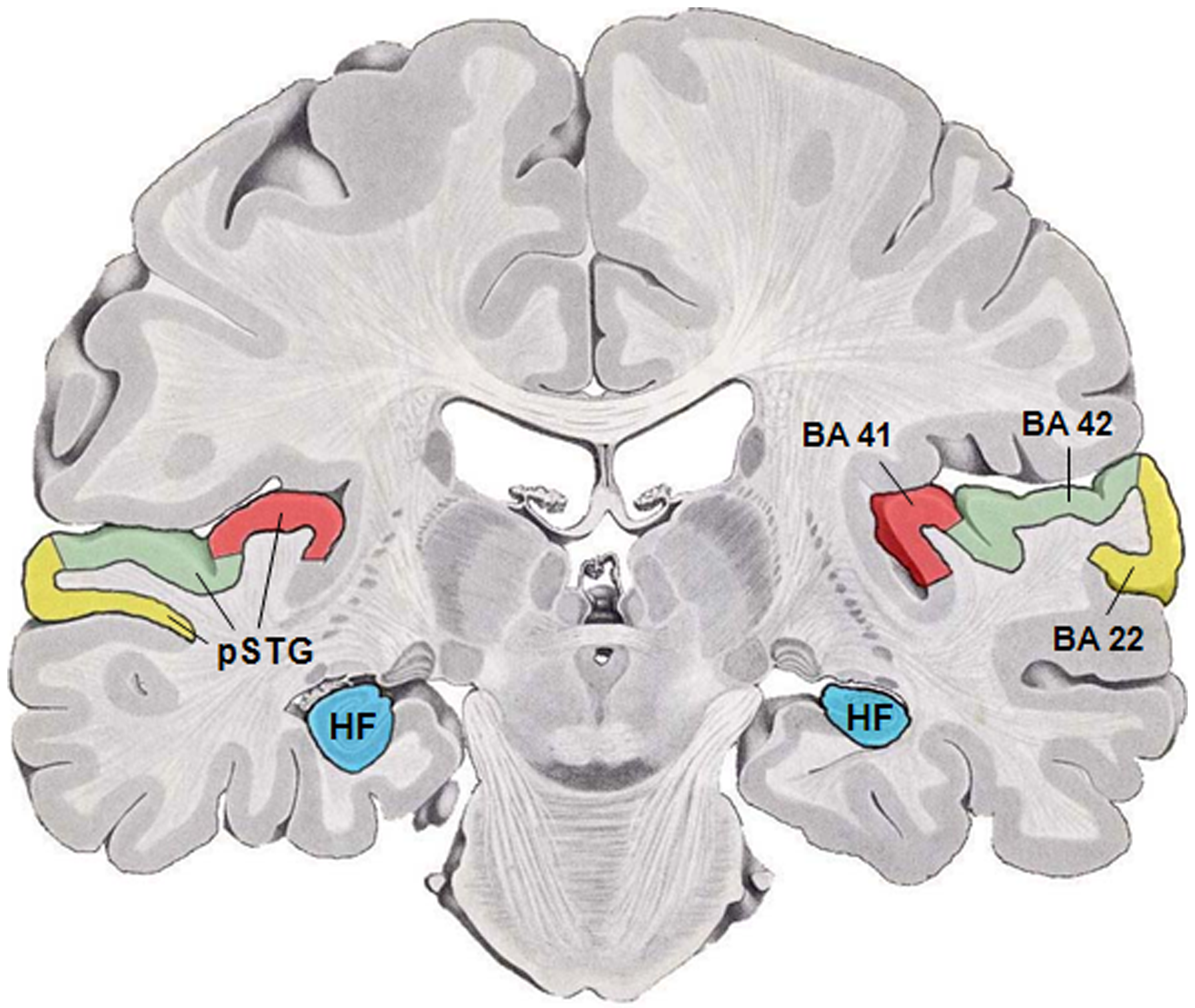
The lateral sulcus (or lateral fissure, also called Sylvian fissure, after Franciscus Sylvius) is the most prominent sulcus (neuroanatomy), sulcus of each cerebral hemisphere in the human brain. The lateral sulcus (neuroanatomy), sulcus is a deep fissure (anatomy), fissure in each hemisphere that separates the frontal lobe, frontal and parietal lobes from the temporal lobe. The insular cortex lies deep within the lateral sulcus. Anatomy The lateral sulcus divides both the frontal lobe and parietal lobe above from the temporal lobe below. It is in both Cerebral hemisphere, hemispheres of the brain. The lateral Sulcus (neuroanatomy), sulcus is one of the earliest-developing sulci of the human brain, appearing around the fourteenth week of gestational age. The insular cortex lies deep within the lateral sulcus. The lateral sulcus has a number of side branches. Two of the most prominent and most regularly found are the ascending (also called vertical) ramus and the horizontal ramus ... [...More Info...] [...Related Items...] OR: [Wikipedia] [Google] [Baidu] |
Operculum (brain)
In human brain anatomy, an operculum (Latin, meaning "little lid") (: opercula), may refer to the frontal, temporal, or parietal operculum, which together cover the insular cortex, insula as the opercula of insula. It can also refer to the occipital operculum, part of the occipital lobe. The insular lobe is a portion of the cerebral cortex that has invaginated to lie deep within the lateral sulcus. It sits like an island (the meaning of ''insular'') almost surrounded by the groove of the Circular sulcus of insula, circular sulcus and covered over and obscured by the insular opercula. A part of the parietal lobe, the frontoparietal operculum, covers the upper part of the insular lobe from the front to the back. The opercula lie on the precentral gyrus, precentral and postcentral gyrus, postcentral gyrus, gyri (on either side of the central sulcus). The part of the parietal operculum that forms the ceiling of the lateral sulcus functions as the secondary somatosensory cortex. De ... [...More Info...] [...Related Items...] OR: [Wikipedia] [Google] [Baidu] |
Middle Cerebral Artery
The middle cerebral artery (MCA) is one of the three major paired cerebral artery, cerebral arteries that supply blood to the cerebrum. The MCA arises from the internal carotid artery and continues into the lateral sulcus where it then branches and projects to many parts of the lateral cerebral cortex. It also supplies blood to the anterior temporal lobes and the insular cortex, insular cortices. The left and right MCAs rise from trifurcations of the internal carotid artery, internal carotid arteries and thus are connected to the anterior cerebral artery, anterior cerebral arteries and the posterior communicating artery, posterior communicating arteries, which connect to the posterior cerebral artery, posterior cerebral arteries. The MCAs are not considered a part of the Circle of Willis. Structure The middle cerebral artery divides into four segments, named by the region they supply as opposed to order of branching as the latter can be somewhat variable: *M1: The ''sphenoidal' ... [...More Info...] [...Related Items...] OR: [Wikipedia] [Google] [Baidu] |
Middle Cerebral Veins
The middle cerebral veins - are the superficial and deep veins - that run along the lateral sulcus. The superficial middle cerebral vein is also known as the superficial Sylvian vein, and the deep middle cerebral vein is also known as the deep Sylvian vein. The lateral sulcus or lateral fissure, is also known as the Sylvian fissure. Superficial middle cerebral vein The superficial middle cerebral vein (superficial Sylvian vein) begins on the lateral surface of the hemisphere. It runs along the lateral sulcus to empty into either the cavernous sinus, or the sphenoparietal sinus. It is adherent to the deep surface of the arachnoid mater bridging the lateral sulcus. It drains the adjacent cortex. Anastomoses At its posterior extremity, the superficial middle cerebral vein is connected with the superior sagittal sinus via the superior anastomotic vein, and with the transverse sinus via the inferior anastomotic vein. Deep middle cerebral vein The deep middle cerebral vein (dee ... [...More Info...] [...Related Items...] OR: [Wikipedia] [Google] [Baidu] |


