|
Cervical Spinal Nerve
A spinal nerve is a mixed nerve, which carries motor, sensory, and autonomic signals between the spinal cord and the body. In the human body there are 31 pairs of spinal nerves, one on each side of the vertebral column. These are grouped into the corresponding cervical, thoracic, lumbar, sacral and coccygeal regions of the spine. There are eight pairs of cervical nerves, twelve pairs of thoracic nerves, five pairs of lumbar nerves, five pairs of sacral nerves, and one pair of coccygeal nerves. The spinal nerves are part of the peripheral nervous system. Structure Each spinal nerve is a mixed nerve, formed from the combination of nerve root fibers from its dorsal and ventral roots. The dorsal root is the afferent sensory root and carries sensory information to the brain. The ventral root is the efferent motor root and carries motor information from the brain. The spinal nerve emerges from the spinal column through an opening (intervertebral foramen) between adjacent ... [...More Info...] [...Related Items...] OR: [Wikipedia] [Google] [Baidu] |
Mixed Nerve
A mixed nerve is any nerve that contains both sensory ( afferent) and motor ( efferent) nerve fibers. All 31 pairs of spinal nerves are mixed nerves. Four of the twelve cranial nerves – V, VII, IX and X are mixed nerves. Examples Spinal nerves The 31 pairs of spinal nerves are mixed. * 8 cervical nerves * 12 thoracic nerves * 5 lumbar nerves * 5 sacral nerves * 1 coccygeal nerve Cranial nerves Four of the cranial nerves Cranial nerves are the nerves that emerge directly from the brain (including the brainstem), of which there are conventionally considered twelve pairs. Cranial nerves relay information between the brain and parts of the body, primarily to and f ... are mixed nerves. * Trigeminal nerve (CN V) * Facial nerve (CN VII) * Glossopharyngeal nerve (CN IX) * Vagus nerve (CN X) References {{Reflist Nerves Spinal nerves Cranial nerves ... [...More Info...] [...Related Items...] OR: [Wikipedia] [Google] [Baidu] |
Axon
An axon (from Greek ἄξων ''áxōn'', axis) or nerve fiber (or nerve fibre: see American and British English spelling differences#-re, -er, spelling differences) is a long, slender cellular extensions, projection of a nerve cell, or neuron, in Vertebrate, vertebrates, that typically conducts electrical impulses known as action potentials away from the Soma (biology), nerve cell body. The function of the axon is to transmit information to different neurons, muscles, and glands. In certain sensory neurons (pseudounipolar neurons), such as those for touch and warmth, the axons are called afferent nerve fibers and the electrical impulse travels along these from the peripheral nervous system, periphery to the cell body and from the cell body to the spinal cord along another branch of the same axon. Axon dysfunction can be the cause of many inherited and acquired neurological disorders that affect both the Peripheral nervous system, peripheral and Central nervous system, central ne ... [...More Info...] [...Related Items...] OR: [Wikipedia] [Google] [Baidu] |
Hypaxial Muscles
In adult vertebrates, trunk muscles can be broadly divided into hypaxial muscles, which lie ventral to the horizontal septum of the vertebrae and epaxial muscles, which lie dorsal to the septum. Hypaxial muscles include some vertebral muscles, the diaphragm, the abdominal muscles, and all limb muscles. The serratus posterior inferior and serratus posterior superior are innervated by the ventral primary ramus and are hypaxial muscles. Epaxial muscles include other (dorsal) muscles associated with the vertebrae, ribs, and base of the skull. In humans, the erector spinae, the transversospinales (including the multifidus, semispinalis and rotatores), the splenius and suboccipital muscles are the only epaxial muscles. Hypaxial and epaxial muscles develop directly from somitic cells. Differentiation of hypaxial and epaxial muscles is postulated to have evolved as a new trait in vertebrate animals. Location The hypaxial muscles are located on the ventral side of the body, ... [...More Info...] [...Related Items...] OR: [Wikipedia] [Google] [Baidu] |
Ventral Ramus
The ventral ramus (: rami) (Latin for 'branch') is the anterior division of a spinal nerve. The ventral rami supply the antero-lateral parts of the trunk and the limbs. They are mainly larger than the dorsal rami. Shortly after a spinal nerve exits the intervertebral foramen, it branches into the dorsal ramus, the ventral ramus, and the ramus communicans. Each of these three structures carries both sensory and motor information. Each spinal nerve is a mixed nerve that carries both sensory and motor information, via efferent and afferent nerve fibers—ultimately via the motor cortex in the frontal lobe and to somatosensory cortex in the parietal lobe—but also through the phenomenon of reflex. In the thoracic region they remain distinct from each other and each innervates a narrow strip of muscle and skin along the sides, chest, ribs, and abdominal wall. These rami are called the intercostal nerves. In regions other than the thoracic, ventral rami converge with each othe ... [...More Info...] [...Related Items...] OR: [Wikipedia] [Google] [Baidu] |
Epaxial Muscles
In adult vertebrates, trunk muscles can be broadly divided into hypaxial muscles, which lie ventral to the horizontal septum of the vertebrae and epaxial muscles, which lie dorsal to the septum. Hypaxial muscles include some vertebral muscles, the diaphragm, the abdominal muscles, and all limb muscles. The serratus posterior inferior and serratus posterior superior are innervated by the ventral primary ramus and are hypaxial muscles. Epaxial muscles include other (dorsal) muscles associated with the vertebrae, ribs, and base of the skull. In humans, the erector spinae, the transversospinales (including the multifidus, semispinalis and rotatores), the splenius and suboccipital muscles are the only epaxial muscles. Hypaxial and epaxial muscles develop directly from somitic cells. Differentiation of hypaxial and epaxial muscles is postulated to have evolved as a new trait in vertebrate animals. Location The hypaxial muscles are located on the ventral side of the body, ... [...More Info...] [...Related Items...] OR: [Wikipedia] [Google] [Baidu] |
Dorsal Ramus Of Spinal Nerve
The dorsal ramus of spinal nerve, posterior ramus of spinal nerve, or posterior primary division is the posterior division of a spinal nerve. The dorsal rami provide motor innervation to the deep (a.k.a. intrinsic or true) muscles of the back, and sensory innervation to the skin of the posterior portion of the head, neck and back. A spinal nerve splits within the intervertebral foramen to form a dorsal ramus and a ventral ramus. The dorsal ramus then turns to course posterior-ward before splitting into a medial branch and a lateral branch. Both these branches provide motor innervation to deep back muscles. In the neck and upper back, the medial branch is also responsible for providing sensory innervation of the skin; in the lower back, the lateral branch does so. All medial branches additionally also provide sensory innervation to the zygapophyseal joints and periosteum of the vertebral column. Structure Ventral root axons join with dorsal root ganglia to form mixed spinal nerves ... [...More Info...] [...Related Items...] OR: [Wikipedia] [Google] [Baidu] |
Lumbar Vertebrae
The lumbar vertebrae are located between the thoracic vertebrae and pelvis. They form the lower part of the back in humans, and the tail end of the back in quadrupeds. In humans, there are five lumbar vertebrae. The term is used to describe the anatomy of humans and quadrupeds, such as horses, pigs, or cattle. These bones are found in particular cuts of meat, including tenderloin or sirloin steak. Human anatomy In human anatomy, the five vertebrae are between the rib cage and the pelvis. They are the largest segments of the vertebral column and are characterized by the absence of the foramen transversarium within the transverse process (since it is only found in the cervical region) and by the absence of facets on the sides of the body (as found only in the thoracic region). They are designated L1 to L5, starting at the top. The lumbar vertebrae help support the weight of the body, and permit movement. General characteristics The adjacent figure depicts the general cha ... [...More Info...] [...Related Items...] OR: [Wikipedia] [Google] [Baidu] |
Atlas (anatomy)
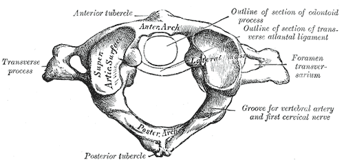
In anatomy, the atlas (C1) is the most superior (first) cervical vertebra of the spine and is located in the neck. The bone is named for Atlas of Greek mythology, just as Atlas bore the weight of the heavens, the first cervical vertebra supports the head. However, the term atlas was first used by the ancient Romans for the seventh cervical vertebra (C7) due to its suitability for supporting burdens. In Greek mythology, Atlas was condemned to bear the weight of the heavens as punishment for rebelling against Zeus. Ancient depictions of Atlas show the globe of the heavens resting at the base of his neck, on C7. Sometime around 1522, anatomists decided to call the first cervical vertebra the atlas. Scholars believe that by switching the designation atlas from the seventh to the first cervical vertebra Renaissance anatomists were commenting that the point of man's burden had shifted from his shoulders to his head—that man's true burden was not a physical load, but rather, his m ... [...More Info...] [...Related Items...] OR: [Wikipedia] [Google] [Baidu] |
Occipital Bone
The occipital bone () is a neurocranium, cranial dermal bone and the main bone of the occiput (back and lower part of the skull). It is trapezoidal in shape and curved on itself like a shallow dish. The occipital bone lies over the occipital lobes of the cerebrum. At the base of the skull in the occipital bone, there is a large oval opening called the foramen magnum, which allows the passage of the spinal cord. Like the other cranial bones, it is classed as a flat bone. Due to its many attachments and features, the occipital bone is described in terms of separate parts. From its front to the back is the basilar part of occipital bone, basilar part, also called the basioccipital, at the sides of the foramen magnum are the lateral parts of occipital bone, lateral parts, also called the exoccipitals, and the back is named as the squamous part of occipital bone, squamous part. The basilar part is a thick, somewhat quadrilateral piece in front of the foramen magnum and directed toward ... [...More Info...] [...Related Items...] OR: [Wikipedia] [Google] [Baidu] |
Intervertebral Foramen
The intervertebral foramen (also neural foramen) (often abbreviated as IV foramen or IVF) is an opening between (the intervertebral notches of) two pedicles (one above and one below) of adjacent vertebra in the articulated spine. Each intervertebral foramen gives passage to a spinal nerve and spinal blood vessels, and lodges a posterior (dorsal) root ganglion. Cervical, thoracic, and lumbar vertebrae all have intervertebral foramina. Anatomy Structure In the thoracic region and lumbar region, each vertebral foramen is additionally bounded anteriorly by (the inferior portion of) the body of vertebra (particularly in the thoracic region) and adjacent intervertebral disc (particularly in the lumbar region). In the cervical region, a small part of the body of vertebra inferior to the intervertebral disc also forms the anterior boundary of the IVF (due to the fact that the junction of the pedicle with the body of vertebra is situated somewhat more inferiorly on the body). ... [...More Info...] [...Related Items...] OR: [Wikipedia] [Google] [Baidu] |
Efferent Nerve Fiber
Efferent nerve fibers are axons (nerve fibers) of efferent neurons that exit a particular region. These terms have a slightly different meaning in the context of the peripheral nervous system (PNS) and central nervous system (CNS). The efferent fiber is a long process projecting far from the neuron's body that carries nerve impulses away from the central nervous system toward the peripheral effector organs (muscles and glands). A bundle of these fibers constitute an efferent nerve. The opposite direction of neural activity is afferent conduction, which carries impulses by way of the afferent nerve fibers of sensory neurons. In the nervous system, there is a "closed loop" system of sensation, decision, and reactions. This process is carried out through the activity of sensory neurons, interneurons, and motor neurons. In the CNS, afferent and efferent projections can be from the perspective of any given brain region. That is, each brain region has its own unique set of affe ... [...More Info...] [...Related Items...] OR: [Wikipedia] [Google] [Baidu] |


